The Scrotum
The scrotum of the stallion is located high in the inguinal region and is slightly pendulous. It forms two distinct pouches that contain, protect, and thermoregulate the testes, epididymides, spermatic cords, and cremaster muscles. The testes are located in the scrotum to maintain testicular temperature at several degrees below core body temperature, a necessity for normal spermatogenesis.1,2 Thermography of scrotal contents of stallions has demonstrated a scrotal skin temperature of 33°C, with testis contents at 30.5° to 32.5°C.1
The wall of the scrotum consists of four layers: (1) skin, (2) tunica dartos, (3) scrotal fascia, and (4) parietal vaginal tunic.3-6 The scrotal skin is thin, generally hairless, and slightly oily, containing numerous sebaceous and sweat glands, which assist in testis thermoregulation.3,7 The tunica dartos layer is adherent to the scrotal skin and consists of muscular and fibroelastic tissue. It lines both scrotal pouches and extends into the median septum, seen externally as the median raphae of the scrotum. The degree of contraction or relaxation of this layer allows alterations in the size, shape, and position of the scrotum in relation to the body wall, thereby aiding testis thermoregulation. The scrotal fascia, a loose connective tissue layer between the tunica dartos and parietal vaginal tunic, allows the testes and associated parietal tunic layer to move freely within the scrotum.5 The innermost layer of the scrotum, the parietal vaginal tunic, is an evagination of the parietal peritoneum through the inguinal rings, which forms during testicular descent. This layer forms a sac that lines the scrotum and is closely apposed to the visceral vaginal tunic, the outer layer of the testis. The vaginal cavity is the space between the parietal and visceral layers of the vaginal tunic. It normally contains a very small amount of viscous fluid to allow some free movement of the testis within. The vaginal cavity is a potential space within which considerable fluid may accumulate as a result of a variety of causes.
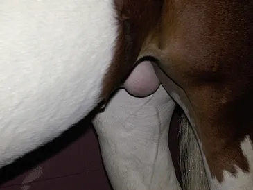
The scrotum of the normal stallion should appear slightly pendulous, globular, and generally symmetric (Fig. 1-1). Normal variations may be observed in the positioning of the testes if one is relatively anterior to or ventral to the other. The skin should have no evidence of trauma, scarring, or skin lesions. Palpation of the scrotum of a normal stallion reveals a thin and pliable covering, which slides loosely and easily over the testicles and epididymides within.
The Testes
Testicular Descent
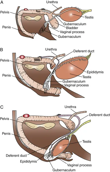
The testicles normally descend into a scrotal position between the last 30 days of gestation and the first 10 days postpartum (Fig. 1-2).7,8-11 In some colts, the testes may descend into the inguinal region and remain there for some time before fully descending into the scrotum. The hormonal factors involved in testicular descent in the stallion are poorly understood. Research in the rat has demonstrated the involvement of androgens and luteinizing hormone (LH) in the process. The timing of testicular descent coincides with significant rises in endogenous gonadotropins. It appears that an intact hypothalamic-pituitary axis, adequate LH levels, and several physical factors must be present for normal testicular descent to occur. In midgestation, the abdominal fetal testis hypertrophies significantly, resulting in the developing gonad resting close to the vaginal ring. The developing gubernaculum and abdominal pressure hold the testes in place until late gestation, despite considerable decrease in the size of the gonads later in gestation. The caudal ligament, which attaches the epididymis to the caudal pole of the testis, initially lengthens faster than the rest of the gubernaculum, resulting in the epididymis being drawn into the vaginal ring and inguinal canal. Dilation of the vaginal ring and inguinal canal, combined with abdominal pressure and traction from the gubernaculum, eventually draws the testes into the ring as well.