- Features more than 1, 000 superb illustrations depicting the full spectrum of retinal diseases using OCT scans, supported by clinical photos and ancillary imaging technologies.- Presents images as large as possible on the page with an abundance of arrows, pointers, and labels to guide you in pattern recognition and eliminate any uncertainty.- Includes the latest high-resolution spectral domain OCT technology and new insights into OCT angiography technology to ensure you have the most up-to-date and highest quality examples available.- Provides key feature points for each disorder giving you the need-to-know OCT essentials for quick comprehension and rapid reference.- An excellent diagnostic companion to Handbook of Retinal OCT: Optical Coherence Tomography, by the same expert author team of Drs. Jay S. Duker, Nadia K. Waheed, and Darin R. Goldman.
eBook - ePub
Atlas of Retinal OCT E-Book
Optical Coherence Tomography
- 400 pages
- English
- ePUB (mobile friendly)
- Available on iOS & Android
eBook - ePub
Atlas of Retinal OCT E-Book
Optical Coherence Tomography
About this book
Trusted by 375,005 students
Access to over 1.5 million titles for a fair monthly price.
Study more efficiently using our study tools.
Information
Topic
MedicineSubtopic
Opthalmology & OptometryPart 1
Normal Optical Coherence Tomography
Section 1
Normal Optic Nerve
1.1
Normal Optic Nerve
Carlos A. Moreira Neto, Carl Rebhun
Spectral domain OCT (SD-OCT) devices have two scan patterns to analyze the optic nerve head (ONH): volume scans and line scans.
Volume Scans
Volume scans acquire a volumetric set of data, centered at the ONH. It delineates the optic disc margin and optic disc surface contour and is segmented to obtain the retinal nerve fiber boundaries. Each device has its own scanning protocol. The Cirrus HD-OCT identifies the center of the optic disc and creates a 3.46-mm circle on this location and calculates the thickness of the retinal nerve fiber layer (RNFL). The Heidelberg Spectralis creates a cylindrical volume with a diameter of 3.4 mm through and around the ONH (Duker, Waheed & Goldman 2014). The Optovue RTVue's protocol for the ONH consists of a grid pattern with circular and radial scans that acquires a 4- × 4-mm volume around the optic nerve. Because different machines use circles of different diameters around the center of the ONH, the measurement of RNFL between machines is not comparable (Duker et al. 2014).
Retinal Nerve Fiber Layer Thickness (RNFL)
OCT devices calculate RNFL thickness as the distance between the internal limiting membrane and the outer aspect of the RNFL (Fig. 1).
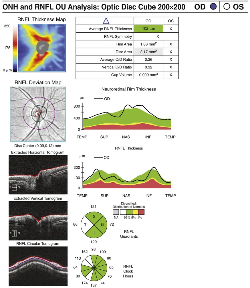
FIG. 1 Normal peripapillary RNFL, neuroretinal rim thickness, and disc area measurements using SD-OCT.
Ganglion Cell Complex
The ganglion cell complex (GCC) consists of the thickness of three inner retinal layers: the NFL, the ganglion cell layer, and the inner plexiform layer. The scan is centered at the fovea, and the software presents the results as a color-coded map, comparing to a normative database (Fig. 2).
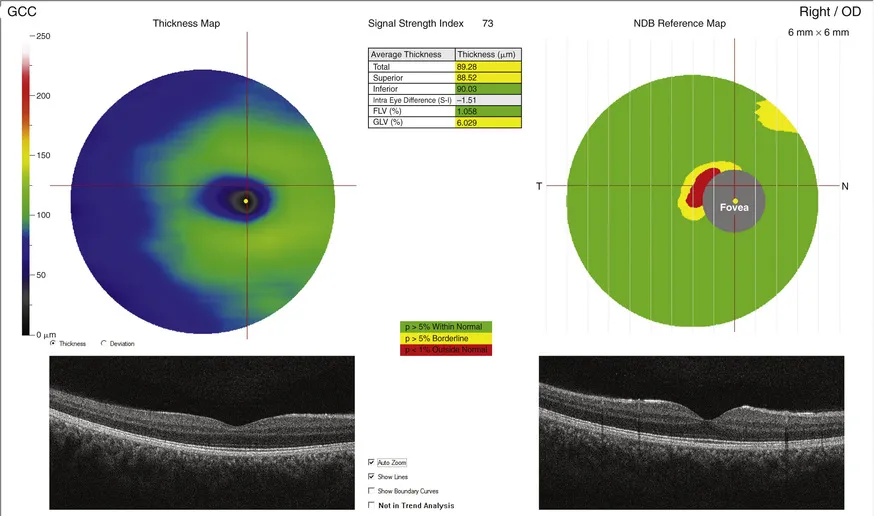
FIG. 2 Normal color-coded ganglion cell complex (GCC) thickness using SD-OCT.
Optic Nerve Morphology
SD-OCT devices also calculate optic nerve diameter, area, cup, and rim measurements (see Fig. 1). Each measurement varies according to age (Cavallotti et al. 2002) and ethnicity (Girkin 2008). According to Budenz et al. (2007), the mean RNFL thickness in a normal population is 100.1 µm. Thinner RNFL measurements were associated with older age. Caucasians had slightly thinner RNFL thickness than Hispanics or Asians. Persons with smaller optic disc areas also have thinner RNFL thickness.
Line Scans
Aiming to obtain a higher resolution visualization of structure and anatomic anomalies at the ONH, line scans provide a single or a series of high-resolution B-scans similar to the scans obtained in the macula (Fig. 3).
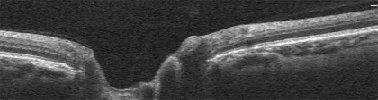
FIG. 3 Line scan of the ONH.
OC...
Table of contents
- Cover image
- Title Page
- Table of Contents
- Copyright
- Preface
- Contributors
- Acknowledgments
- Dedications
- Part 1 Normal Optical Coherence Tomography
- Part 2 Isolated Macular Disorders
- Part 3 Vasoocclusive Disorders
- Part 4 Uveitis and Inflammatory Disorders
- Part 5 Retinal and Choroidal Tumors
- Part 6 Trauma
- Part 7 Inherited Retinal Degenerations
- Part 8 Vitreous Disorders
- Part 9 Miscellaneous Retinal Disorders
- Index
Frequently asked questions
Yes, you can cancel anytime from the Subscription tab in your account settings on the Perlego website. Your subscription will stay active until the end of your current billing period. Learn how to cancel your subscription
No, books cannot be downloaded as external files, such as PDFs, for use outside of Perlego. However, you can download books within the Perlego app for offline reading on mobile or tablet. Learn how to download books offline
We are an online textbook subscription service, where you can get access to an entire online library for less than the price of a single book per month. With over 1.5 million books across 990+ topics, we’ve got you covered! Learn about our mission
Look out for the read-aloud symbol on your next book to see if you can listen to it. The read-aloud tool reads text aloud for you, highlighting the text as it is being read. You can pause it, speed it up and slow it down. Learn more about Read Aloud
Yes! You can use the Perlego app on both iOS and Android devices to read anytime, anywhere — even offline. Perfect for commutes or when you’re on the go.
Please note we cannot support devices running on iOS 13 and Android 7 or earlier. Learn more about using the app
Please note we cannot support devices running on iOS 13 and Android 7 or earlier. Learn more about using the app
Yes, you can access Atlas of Retinal OCT E-Book by Darin Goldman,Nadia K. Waheed,Jay S. Duker,Nadia K Waheed in PDF and/or ePUB format, as well as other popular books in Medicine & Opthalmology & Optometry. We have over 1.5 million books available in our catalogue for you to explore.