Atlas of Normal Radiographic Anatomy and Anatomic Variants in the Dog and Cat - E-Book
eBook - ePub

Atlas of Normal Radiographic Anatomy and Anatomic Variants in the Dog and Cat - E-Book

  1. 320 pages
  2. English
  3. ePUB (mobile friendly)
  4. Available on iOS & Android
eBook - ePub

Atlas of Normal Radiographic Anatomy and Anatomic Variants in the Dog and Cat - E-Book

Book details
Book preview
Table of contents
Citations

About This Book

Equip yourself to make accurate diagnoses and achieve successful treatment outcomes with this highly visual comprehensive atlas. Featuring a substantial number of new high contrast images, Atlas of Normal Radiographic Anatomy and Anatomic Variants in the Dog and Cat, 2nd Edition provides an in-depth look at both normal and non-standard subjects along with demonstrations of proper technique and image interpretations. Expert authors Donald E. Thrall and Ian D. Robertson describe a wider range of "normal" as compared to competing books — not only showing standard dogs and cats, but also non-standard subjects such as overweight and underweight pets and animals with breed-specific variations. Every body part is put into context with a textual description to help explain why a structure appears as it does in radiographs, and enabling practitioners to appreciate variations of normal that are not included, based on an understanding of basic radiographic principles.

  • Radiographic images of normal or standard prototypical animals are supplemented by images of non-standard subjects exhibiting breed-specific differences, physiologic variants, or common congenital malformations.
  • Images that depict a wider range of "normal" — such as images that detail the natural growth and aging characteristics of normal pediatric and senior animals — prevents clinical under- and over-diagnosing.
  • In-depth coverage of patient positioning and radiographic exposure guidelines assist clinicians in producing the very best results.
  • Unlabeled radiographs along side labeled counterparts clarifies important anatomic structures of clinical interest.
  • High-quality digital images provide excellent contrast resolution and better visibility of normal structures to assist clinicians in making accurate diagnoses.
  • Brief descriptive text and explanatory legends accompany all images to help put concepts into the proper context.
  • An overview of radiographic technique includes the effects of patient positioning, respiration, and exposure factors.
  • NEW! Companion website features additional radiographic CT scans and more than 100 questions with answers and rationales.
  • NEW! Additional CT and 3D images have been added to each chapter to help clinicians better evaluate the detail of bony structures.
  • NEW! Breed-specific images of dogs and cats are included throughout the atlas to help clinicians better understand the variances in different breeds.
  • NEW! Updated material on oblique view radiography provides a better understanding of an alternative approach to radiography, particularly in fracture cases.
  • NEW! 8.5" x 11" trim size makes the atlas easy to store.

Frequently asked questions

Simply head over to the account section in settings and click on “Cancel Subscription” - it’s as simple as that. After you cancel, your membership will stay active for the remainder of the time you’ve paid for. Learn more here.
At the moment all of our mobile-responsive ePub books are available to download via the app. Most of our PDFs are also available to download and we're working on making the final remaining ones downloadable now. Learn more here.
Both plans give you full access to the library and all of Perlego’s features. The only differences are the price and subscription period: With the annual plan you’ll save around 30% compared to 12 months on the monthly plan.
We are an online textbook subscription service, where you can get access to an entire online library for less than the price of a single book per month. With over 1 million books across 1000+ topics, we’ve got you covered! Learn more here.
Look out for the read-aloud symbol on your next book to see if you can listen to it. The read-aloud tool reads text aloud for you, highlighting the text as it is being read. You can pause it, speed it up and slow it down. Learn more here.
Yes, you can access Atlas of Normal Radiographic Anatomy and Anatomic Variants in the Dog and Cat - E-Book by Donald E. Thrall, Ian D. Robertson in PDF and/or ePUB format, as well as other popular books in Médecine & Médecine vétérinaire. We have over one million books available in our catalogue for you to explore.

Information

Publisher
Saunders
Year
2015
ISBN
9780323312776
Chapter 1

Basic Imaging Principles and Physeal Closure Time
image

How to Use This Atlas

As described in the Preface, a radiographic atlas is intended to help decide whether any given radiographic finding is normal or abnormal. Determining normal from abnormal is one of the most difficult parts of interpreting a radiograph (if not the most difficult). No atlas will be able to provide a definitive answer to the “What is that, and is it normal?” question in every circumstance, but the information in this atlas can help guide the decision-making process.
The best way to use this atlas is to spend some time with it, and get to know it. Of course, labeled images are provided—every atlas needs these. But, contrary to a pure picture atlas, some of the most valuable information in this atlas is contained in the text. Being familiar with the text, which is brief and focused, and noting the important principles that have been augmented with illustrative examples can help formulate a basis for interpretation that extends beyond structure identification alone.

What Is Normal?

Many dogs and cats have congenital or developmental changes that are apparent radiographically but insignificant clinically. Discussing some of these common variations in this atlas, along with normal structures, is justified even though they are not completely normal because they are often confused with disease. This book demonstrates much of the morphologic diversity currently present in domestic canine and feline companions that has come to be commonly accepted as normal or clinically insignificant.

Why Are Computed Tomography Images Included in This Atlas?

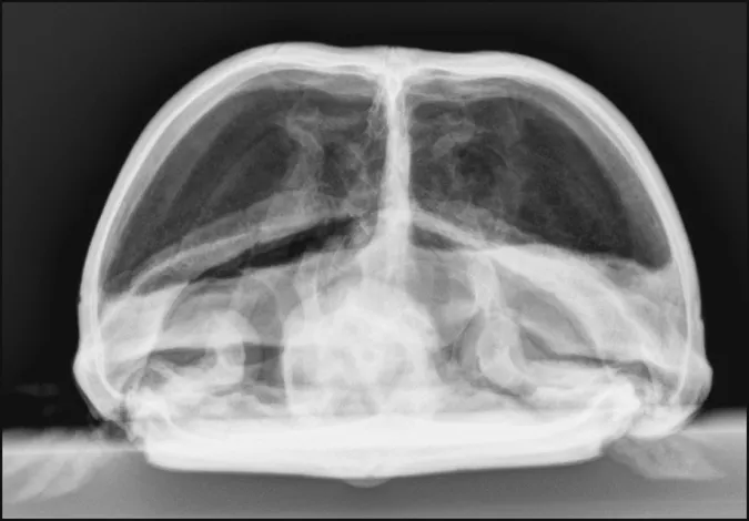
Because this is a radiographic atlas, the majority of images are radiographs. This book is not intended to be an atlas of normal computed tomography (CT) appearances, but some CT images are included to reinforce the appearance of selected structures in radiographs. A radiograph is a 2-dimensional image of a 3-dimensional (3-D) object, and, as a result, spatial localization of structures cannot always be done accurately, even with multiple views of the object or patient. CT images are tomographic, creating multiple image slices of the volume of interest. Slicing the patient solves the problem of spatial localization and can assist with understanding radiographic anatomy more thoroughly. Additionally, CT images, which are typically acquired in a transverse plane, can be reformatted into other planes, typically sagittal and dorsal planes, for clarification or to improve structure visualization. Transverse CT images can even be reformatted into volumetric images for illustration of complex structures. Therefore, a careful selection of planar and volumetric CT images has been included with this goal in mind.

Radiographic Terminology

This book uses the standard method for naming radiographic projections approved by the American College of Veterinary Radiology.1 In general, this naming method is based on anatomic directional terms (as defined by the Nomina Anatomica Veterinaria) combined with the point-of-entrance to point-of-exit of the primary x-ray beam. In other words, any radiograph can be named by knowing the point of entrance and point of exit of the primary x-ray beam. For example, a spinal radiograph made with a dog lying in dorsal recumbency would be cal...

Table of contents

  1. Cover image
  2. Title page
  3. Table of Contents
  4. Copyright
  5. Preface
  6. Acknowledgments
  7. The Images
  8. 1 Basic Imaging Principles and Physeal Closure Time
  9. 2 The Skull
  10. 3 The Spine
  11. 4 The Thoracic Limb
  12. 5 The Pelvic Limb
  13. 6 The Thorax
  14. 7 The Abdomen
  15. Index
  16. Color Plate