- 696 pages
- English
- ePUB (mobile friendly)
- Available on iOS & Android
Small Animal Endoscopy
About this book
The latest edition of the critically acclaimed Small Animal Endoscopy presents informative, practical, and up-to-date guidance on endoscopic indications, instrumentation, patient preparation, and techniques. Todd R. Tams and Clarence A. Rawlings, the foremost experts in veterinary endoscopy, provide the novice as well as the advanced practitioner with the information needed to deliver the safest, high-quality endoscopic services for small animals, including avian and exotics. Chapters are organized consistently and lavishly illustrated to help you easily find and understand key concepts and procedures. This edition includes a companion website with expert demonstrations of techniques.- Enables you to deliver the safest, high quality care and a wider range of services to the pets of increasingly concerned and savvy owners.- Features cutting-edge information on minimally invasive procedures to improve diagnostic accuracy, reduce operating time, improve success, minimize post-operative stress and pain, and promote faster healing.- Helps you recognize the many indications for endoscopy in everyday practice.- Covers a vast range of topics in a clear, concise and readable style.- Describes instrumentation, examination, and sample procurement techniques in detail.- Shows both normal and abnormal findings you may encounter during a procedure in an atlas of images in relevant chapters.- Provides minimally invasive examination and surgical options for veterinarians treating uniquely sensitive avian and exotic patients.- Provides step-by-step instructions on specific techniques.- Helps beginners master endoscopic diagnosis and treatment and more experienced endoscopists utilize their endoscopic equipment to its fullest capacity.
Tools to learn more effectively

Saving Books

Keyword Search

Annotating Text

Listen to it instead
Information
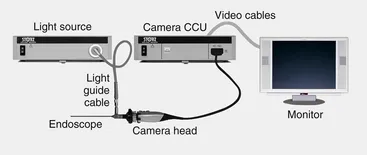
Endoscope System
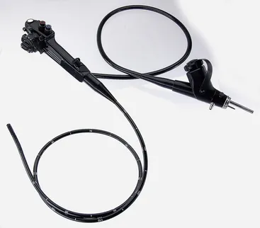
Flexible Endoscopes
| Feature | Fiberscope | Video endoscope |
|---|---|---|
| Image quality | Good | Excellent |
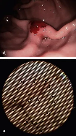
| Broken fibers seen as “black dots” | Likely over time | N/A (image is electronic, not fiberoptic) |
| Cost | Moderate | High |
| Diameters available | Wide range available | Smaller diameters not available∗ |
| Video capability | Requires attachable charge coupled device (CCD) camera | Integral |
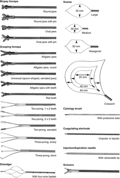
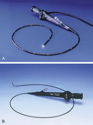
Table of contents
- Cover
- Title Page
- Front Matter
- Copyright
- Contributors
- Preface
- Acknowledgments
- Table of Contents
- Part One: Flexible Endoscopy
- Part Two: Rigid Endoscopy
- Index
Frequently asked questions
- Essential is ideal for learners and professionals who enjoy exploring a wide range of subjects. Access the Essential Library with 800,000+ trusted titles and best-sellers across business, personal growth, and the humanities. Includes unlimited reading time and Standard Read Aloud voice.
- Complete: Perfect for advanced learners and researchers needing full, unrestricted access. Unlock 1.4M+ books across hundreds of subjects, including academic and specialized titles. The Complete Plan also includes advanced features like Premium Read Aloud and Research Assistant.
Please note we cannot support devices running on iOS 13 and Android 7 or earlier. Learn more about using the app