Winner of a HIGHLY COMMENDED AWARD in the Surgical specialties category of the 2011 BMA Medical Book Competition. Key Questions in Cardiac Surgery will systematically cover all the main topics involved in the current practice of a cardiac surgeon. It will incorporate current guidelines for practice (such as from the American Heart Association and European Society of Cardiology) and up-to-date information based on current literature. The data and body of knowledge presented in this book are strictly evidence-based which makes it ideal as a revision aid for residents/registrars undertaking their Cardiothoracic Surgery Board examinations around the world. Although these examinations vary in format in different countries, this book is applicable to all cardiothoracic surgical trainees. Its concise, yet complete coverage of the important topics, make it the ideal guide to answer the key questions in cardiac surgery that are asked within the confines of an examination. Cardiologists, cardiothoracic intensive care unit specialists, nursing staff, physiotherapists and other professions allied to medicine, both surgical and cardiological, will also find the book useful in terms of the indications and surgical management of these patients, as they are integral to the cardiac surgical process.
- English
- ePUB (mobile friendly)
- Available on iOS & Android
eBook - ePub
About this book
Trusted by 375,005 students
Access to over 1 million titles for a fair monthly price.
Study more efficiently using our study tools.
Information
Topic
MedicineChapter 1
Cardiac anatomy
| 1 | Describe the anatomy of the coronary artery system (Figure 1) |
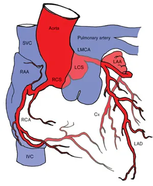
Figure 1. Coronary artery system. SVC = superior vena cava; RAA = right atrial appendage; IVC = inferior vena cava; RCS = right coronary sinus; RCA = right coronary artery; LCS = left coronary sinus; LMCA = left main coronary artery; LAA = left atrial appendage; Cx = circumflex artery; LAD = left anterior descending artery.
| • | The coronary artery system originates from the aortic root and consists of the left and right coronary arteries and their individual branches. |
| • | The left coronary artery originates from the left coronary ostium as the left main stem and divides early into the left anterior descending artery (also known as the anterior interventricular artery) and circumflex artery (see below). |
| • | The right coronary artery originates from the right coronary ostium and eventually terminates as the posterior descending artery (also known as the posterior interventricular artery) and posterior left ventricular artery (see below). |
| 2 | Describe the anatomy of the left main coronary artery (Figure 2) |
| • | The left main coronary artery (left main stem) courses from the left coronary sinus of the aorta in an anterior and inferior direction between the pulmonary trunk and the left atrial appendage. |
| • | It then divides into two major arteries of nearly equal diameter, the left anterior descending artery and the circumflex artery. Typically, no branches are seen before this bifurcation. |
| • | In some patients, the left main coronary artery trifurcates into the intermediate coronary artery (ramus intermedius), left anterior descending artery and circumflex artery. |
| • | The left main coronary artery is typically 10-40mm in length but may be absent in patien... |
Table of contents
- Cover Page
- Title Page
- Copyright Page
- Contents
- Preface
- Foreword
- Acknowledgements
- Abbreviations
- Recommendations and evidence
- Chapter 1: Cardiac anatomy
- Chapter 2: Cardiac physiology
- Chapter 3: Cardiac pharmacology
- Chapter 4: Electrocardiography
- Chapter 5: Echocardiography
- Chapter 6: Cardiac catheterisation
- Chapter 7: Radiological imaging
- Chapter 8: Cardiopulmonary bypass
- Chapter 9: Cardiopulmonary bypass scenarios
- Chapter 10: Adjuncts to cardiopulmonary bypass
- Chapter 11: Myocardial protection
- Chapter 12: Aortic valve disease
- Chapter 13: Mitral valve disease
- Chapter 14: Tricuspid valve disease
- Chapter 15: Infective endocarditis
- Chapter 16: Thoracic aortic disease
- Chapter 17: Coronary artery disease
- Chapter 18: Heart failure
- Chapter 19: Arrhythmia surgery
- Chapter 20: Pericardial disease, cardiac tumours and cardiac trauma
- Chapter 21: Cardiac anaesthesia and intensive care management
- Chapter 22: Postoperative management
- Appendix I: Transoesophageal echocardiographic views
- Appendix II: Transthoracic echocardiographic views
- Appendix III: Normal echocardiographic values
- Appendix IV: Standard coronary angiographic views
- Appendix V: Normal arterial blood gas values
- Appendix VI: Normal cardiac physiological values
- Appendix VII: AHA guidelines for quantifying the severity of valvular disease
- Appendix VIII: EuroSCORE
- Index
Frequently asked questions
Yes, you can cancel anytime from the Subscription tab in your account settings on the Perlego website. Your subscription will stay active until the end of your current billing period. Learn how to cancel your subscription
No, books cannot be downloaded as external files, such as PDFs, for use outside of Perlego. However, you can download books within the Perlego app for offline reading on mobile or tablet. Learn how to download books offline
Perlego offers two plans: Essential and Complete
- Essential is ideal for learners and professionals who enjoy exploring a wide range of subjects. Access the Essential Library with 800,000+ trusted titles and best-sellers across business, personal growth, and the humanities. Includes unlimited reading time and Standard Read Aloud voice.
- Complete: Perfect for advanced learners and researchers needing full, unrestricted access. Unlock 1.4M+ books across hundreds of subjects, including academic and specialized titles. The Complete Plan also includes advanced features like Premium Read Aloud and Research Assistant.
We are an online textbook subscription service, where you can get access to an entire online library for less than the price of a single book per month. With over 1 million books across 990+ topics, we’ve got you covered! Learn about our mission
Look out for the read-aloud symbol on your next book to see if you can listen to it. The read-aloud tool reads text aloud for you, highlighting the text as it is being read. You can pause it, speed it up and slow it down. Learn more about Read Aloud
Yes! You can use the Perlego app on both iOS and Android devices to read anytime, anywhere — even offline. Perfect for commutes or when you’re on the go.
Please note we cannot support devices running on iOS 13 and Android 7 or earlier. Learn more about using the app
Please note we cannot support devices running on iOS 13 and Android 7 or earlier. Learn more about using the app
Yes, you can access Key Questions in Cardiac Surgery by Moorjani, Narain,Viola, Nicola,Ohri, Sunil K. in PDF and/or ePUB format, as well as other popular books in Medicine & Medical Theory, Practice & Reference. We have over one million books available in our catalogue for you to explore.