- 272 pages
- English
- ePUB (mobile friendly)
- Available on iOS & Android
eBook - ePub
About this book
'It gives me immense personal pleasure to see this publication come to fruition - edited by two very committed young teachers, ably supported by several up and coming young surgeons who are setting out on the road to a surgical vocation. 'A picture is equal to a thousand words' - this adage is exemplified by the excellent pictures in this volume generously contributed by many young surgeons. The reader will find the following pages a compelling read. This book is the outcome of the painstaking collection of clinical material over many years by the two editors, aided by colleagues who have contributed much towards this very worthwhile publication.' Pradip K. Datta, in the Foreword from "Book 1".
Information
Section 1
Orthopaedics
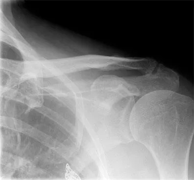
Fracture dislocation of the shoulder (l)
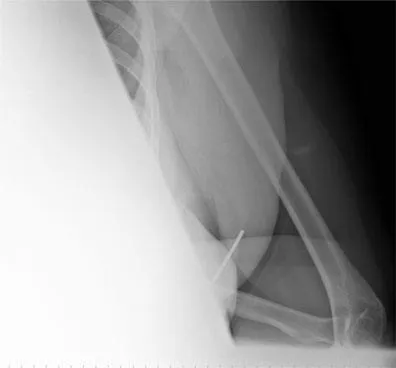
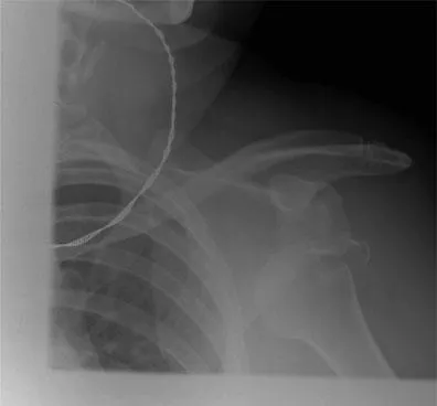
A 45-year-old female patient fell on to her outstretched hand, suffering an injury to her shoulder. These X-rays were taken on her attendance at the accident and emergency department.
Questions
Q1 Describe the injury shown on X-ray.
Q2 Discuss the management of this injury.
Q3 List the complications of this injury and the management of one of the complications.
Q4 What is the likely outcome after such an injury in a patient in her mid-40s?
Answers
A1 The injury is a fracture dislocation of the shoulder. The dislocation is likely to be anterior, but cannot be categorically stated on a single view.
A2 Management is reduction of the fracture dislocation by any of the standard methods described.
A3 Complications are:
1 failure to reduce the fracture fragment such that it remains in the subacromial space or significantly displaced (Figure 1.3)
2 an axillary nerve palsy, or brachial plexus lesion
3 a vascular injury.
Treatments of each of these are:
1 accurate anatomical reduction of the greater tuberosity; this may require surgery and fixation
2 the axillary nerve injury may need exploration and repair
3 angiography and vascular repair.
A4 The outcome of the injury in a lady in her mid-40s is that recovery may take up to 12 months, rotator cuff function after union of the greater tuberosity fracture may not be entirely satisfactory, and the incidence of recurrent instability is low. In the event that the axillary nerve palsy does not recover, very poor active elevation of the arm, and shoulder function, can be expected.
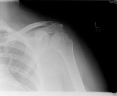
Acromioclavicular joint osteoarthrosis
A 60-year-old patient presented with pain in the shoulder affecting primarily overhead activity, and pain on lying on the left side at night which interfered with sleep. X-rays are as shown.
Questions
Q1 What radiological diagnosis is present on this X-ray which would account for the patient’s symptoms?
Q2 In the presence of reduced shoulder strength, what investigations would be appropriate, and why?
Q3 Describe the treatment of:
• osteoarthrosis of the AC joint
• subacromial impingement
• rotator cuff tear.
Answers
A1 The radiological diagnosis is osteoarthrosis of the AC joint and irregularity of the undersurface of the acromion, a normal glenohumeral joint and slight narrowing of the subacromial space.
A2 Further investigations would be an ultrasound to determine the integrity of the cuff, and/or an MRI scan to detect the presence of a rotator cuff tear, particularly if there is loss of strength.
A3 Treatment of AC joint arthrosis is arthroscopic or open excision of the acromioclavicular joint; treatment of subacromial impingement is acromioplasty (either open or arthroscopic); and treatment of a rotator cuff tear is by arthroscopic or open means, if it is symptomatic – namely if there is shoulder weakness. Conservative treatment in the event that the rotator cuff tear is asymptomatic is to be recommended. Other forms of treatment include conservative treatment with anti-inflammatories and analgesics, physiotherapy, local steroid and local anaesthetic injection into the AC joint and subacromial space.
Ankle fracture
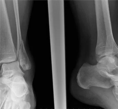
A male patient aged 30 years suffered a twisting injury to his right ankle in a football match, and was unable to weight-bear thereafter. X-rays of the right ankle are shown in Figure 1.5.
Questions
Q1 Describe the appearances of the X-ray indicating the nature of any bony or ligamentous injury that is apparent.
Q2 Name two ankle fracture classifications that you know of, and describe one in detail.
Q3 What treatment options are available to a patient with this injury?
Answers
A1 There is a spiral fracture of the distal fibular shaft at the level of the ankle joint, without significant displacement. There is slight lateral displacement of the talus on the tibia, opening up the medial side of the ankle, and suggesting a stretch or tear of the medial collateral ligament of the ankle.
A2 The two classifications that you should know are:
• the Lauge–Hansen classification...
Table of contents
- Cover
- Title Page
- Copyright Page
- Table of Contents
- Foreword
- Preface
- List of contributors
- Acknowledgements
- Abbreviations
- Dedication
- Section 1: Orthopaedics
- Section 2: Critical care and trauma
- Section 3: Transplant and vascular access
- Section 4: Paediatric surgery
- Section 5: Breast and endocrine surgery
- Index
Frequently asked questions
Yes, you can cancel anytime from the Subscription tab in your account settings on the Perlego website. Your subscription will stay active until the end of your current billing period. Learn how to cancel your subscription
No, books cannot be downloaded as external files, such as PDFs, for use outside of Perlego. However, you can download books within the Perlego app for offline reading on mobile or tablet. Learn how to download books offline
Perlego offers two plans: Essential and Complete
- Essential is ideal for learners and professionals who enjoy exploring a wide range of subjects. Access the Essential Library with 800,000+ trusted titles and best-sellers across business, personal growth, and the humanities. Includes unlimited reading time and Standard Read Aloud voice.
- Complete: Perfect for advanced learners and researchers needing full, unrestricted access. Unlock 1.4M+ books across hundreds of subjects, including academic and specialized titles. The Complete Plan also includes advanced features like Premium Read Aloud and Research Assistant.
We are an online textbook subscription service, where you can get access to an entire online library for less than the price of a single book per month. With over 1 million books across 990+ topics, we’ve got you covered! Learn about our mission
Look out for the read-aloud symbol on your next book to see if you can listen to it. The read-aloud tool reads text aloud for you, highlighting the text as it is being read. You can pause it, speed it up and slow it down. Learn more about Read Aloud
Yes! You can use the Perlego app on both iOS and Android devices to read anytime, anywhere — even offline. Perfect for commutes or when you’re on the go.
Please note we cannot support devices running on iOS 13 and Android 7 or earlier. Learn more about using the app
Please note we cannot support devices running on iOS 13 and Android 7 or earlier. Learn more about using the app
Yes, you can access MRCS Picture Questions by Tjun Tang,BV Praveen in PDF and/or ePUB format, as well as other popular books in Medicine & Medical Theory, Practice & Reference. We have over one million books available in our catalogue for you to explore.