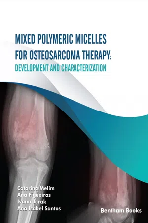
Mixed Polymeric Micelles for Osteosarcoma Therapy
eBook - ePub

Mixed Polymeric Micelles for Osteosarcoma Therapy

Development and Characterization

  1. English
  2. ePUB (mobile friendly)
  3. Available on iOS & Android
eBook - ePub

Mixed Polymeric Micelles for Osteosarcoma Therapy

Development and Characterization

Book details
Book preview
Table of contents
Citations

About This Book

Osteosarcoma is a rare bone tumor that has a high incidence amongchildren and young adults. Despite recent therapeutic developments, osteosarcomastill presents major hurdles to achieving successful results, mainly due to thepresence of multi-drug-resistant cells.This monograph primarily aims to provide information about the basicscience behind the treatment of osteosarcoma along with experimental resultsfor a novel formulation that overcomes multidrug resistance, and therefore, mayserve as a viable treatment option. The book starts with an updated and conciseguide to the pathophysiology of the disease, while also introducing the readerto new therapies and materials (specifically chitosan, polyethyleneimine, poloxamers, poloxamines, and PluronicsĀ®) used in the treatment process along with the aimsof the experiments present subsequently. Next, the book documents the materialsand methods used in developing polymeric micelles for delivering drugs toosteosarcoma sites. By explaining the basics of nanomedicine as a startingpoint, readers will understand how polymeric micelles act as facilitators ofdrug transport to cancer cells, and how one can synthesize a small stablemicelle (by creating derivatives of base nanomaterials), capable of activelytargeting osteosarcoma cells and overcoming multi-drug resistance. The chapterexplains the synthesis and characterization techniques of the materials used todevelop polymeric micelles.The results, a reflection of the conjugation of different experimentalsolutions initiated here, point to a modern route towards the search for a therapeutic solution for osteosarcoma.The simple, structured presentation coupled with relevant informationon the subject of micelle-based nanotherapeutic drug delivery make thismonograph an essential handbook for pharmaceutical scientists involved in thefield of nanomedicine, drug delivery, cancer therapy and any researchersassisting specialists in clinical oncology for the treatment of osteosarcoma.

Frequently asked questions

Simply head over to the account section in settings and click on ā€œCancel Subscriptionā€ - itā€™s as simple as that. After you cancel, your membership will stay active for the remainder of the time youā€™ve paid for. Learn more here.
At the moment all of our mobile-responsive ePub books are available to download via the app. Most of our PDFs are also available to download and we're working on making the final remaining ones downloadable now. Learn more here.
Both plans give you full access to the library and all of Perlegoā€™s features. The only differences are the price and subscription period: With the annual plan youā€™ll save around 30% compared to 12 months on the monthly plan.
We are an online textbook subscription service, where you can get access to an entire online library for less than the price of a single book per month. With over 1 million books across 1000+ topics, weā€™ve got you covered! Learn more here.
Look out for the read-aloud symbol on your next book to see if you can listen to it. The read-aloud tool reads text aloud for you, highlighting the text as it is being read. You can pause it, speed it up and slow it down. Learn more here.
Yes, you can access Mixed Polymeric Micelles for Osteosarcoma Therapy by Catarina Melim, Ana Figueiras, Ivana Jarak, Ana Isabel Santos in PDF and/or ePUB format, as well as other popular books in Medicine & Nursing. We have over one million books available in our catalogue for you to explore.

Information

Year
2022
ISBN
9789815040371
Subtopic
Nursing

Introduction


1. Osteosarcoma

Osteosarcoma (OS) is a rare condition, with a yearly worldwide incidence of 3.4 per million people. It is, however, one of the most common cancers in adolescents, behind lymphoma and brain tumors (Misaghi et al., 2018). The defining feature that identifies the disease is the observation of osteoid matrix production by cancerous cells (Abarrategi et al., 2016). OS metastasis spreads via the hematogenous route in the same way as mesenchymal tumors and, typically, patients perish due to lung metastasis (Kansara & Thomas, 2007).
OS is characterized by a biphasic pattern, showing an incidence peak during adolescence and after the age of 60, with the first peak associated with the growth spurt during puberty. In addition, since OS development in adolescents mainly occurs in the more active areas of growth, a link between carcinogenesis and osteoblast activity was proposed (Fletcher et al., 2013; Kansara & Thomas, 2007). In the elderly population, the appearance of OS is of a secondary nature attributed to other diseases such as Pagetā€™s disease of bone. In these patients, tumors develop in the axial part of the bone or in locations that were irradiated beforehand (Mirabello, Troisi & Savage, 2009).
OSā€™ patients often present swelling as well as pain in the metaphyseal bone of the distal femur, the proximal tibia, and proximal humerus. About 10% of cases involve the axial skeleton, mostly affecting the pelvis. Pain is mostly associated with the performance of active tasks and gradually starts appearing at rest (Cottrell, 2018; Ritter & Bielack, 2010). The painā€™s onset is usually in adolescence and is associated with hospitalization, reduced survival, and poor quality of life of the patient (Smeester, Moriarity & Beitz, 2017).
In the past two decades, there has been little advancement regarding the prognosis of this disease, despite numerous research attempts. Children and adolescents present the worst prognostic. One of the most common problems of OS is the low patient survival rate, which has remained practically unchanged for 15 years, especially in those with metastatic tumors or in an advanced stage locally at the
time of diagnosis. Additionally, for those patients who experience disease relapse, treatment will depend on whether the tumor is removable, on the prior chemotherapy regimen and the time to relapse (Oā€™Day & Gorlick, 2009). As it is assumed that changes in the current chemotherapy scenario will not provide an improvement in the OS landscape, there has been an increasing effort in the discovery of new therapeutic agents (Sampson et al., 2015).
According to the World Health Organization, the primary malignant bone tumor can be classified into seven types (see Table 1). In order to categorize the tumor, it is important to examine the microscopic, histological, and radiographic findings (Fletcher et al., 2013).
Table 1 Classification of OS subtypes according to their histological appearance(Hang & Chen, 2014; Kumar, Barwar, & Khan, 2014; Malhas et al., 2012; Misaghi et al., 2018; Yin et al., 2018).
Osteosarcoma Subtype Histological Characteristics
Low-grade central Fibroblastic stroma of variable cellularity; osteoids arranged in parallel seams resembling parosteal osteosarcoma
Conventional Subdivided into osteoblastic, fibroblastic, and chondroblastic subtypes; bone or osteoid production by tumor cells
Telangiectatic Dilated blood-filled cavities; high-grade sarcomatous cells on the septae and peripheral rim
Small-cell Small-cell production; round hypochromatic nuclei with slight nuclear polymorphism
Parosteal Fibroblastic in appearance; streams of bone trabeculae arranged in a parallel orientation
Periosteal Chondroblastic in appearance; matrix component mainly cartilaginous
High-grade surface Malignant spindle cells; high degree of atypical cells

The tumorā€™s microenvironment (TME) is an essential feature to be regarded. In OS, the local TME has been linked to the induction and development of the disease, further contributing to a poor prognosis. Amid the non-tumor cells that compose the TME are mesenchymal stem cells, or MSCs. These non-hematopoietic precursor cells derived from the bone marrow are thought to be the origin of OS cells given the diseaseā€™s varied histological subtypes (Zheng et al., 2018). In fact, MSCs have the ability to self-differentiate and renew into multiple skeletal mesodermal lineages, including adipocytes, chondrocytes and osteoblasts (Tsukamoto et al., 2012). Tumor tissue MSCs, when recruited to the lesion, can obtain a cancer-associated fibroblast-like phenotype and promote tumor growth and progression (Bonuccelli et al., 2014; Zheng et al., 2018).

1.1. Pathophysiology

The majority of reported cases are sporadic in origin. OS develops in rapidly growing bones, preferentially during puberty and in the knee area (Choong et al., 2011). This disease is more prominent in males, with a male to female ratio of 1.5/1. Also, several environmental factors have been connected to the emergence of OS. UV light and ionizing radiation are the best described agents causative of OS. Exposure to radiation is responsible for 2% of the cases observed (Cottrell, 2018).
Chemical agents can be behind the development of OS. In 1938, Brunschwig injected 3-methylcholanthrene (MC) in mice, which resulted in the formation of an ossifying sarcoma in the tibia (Brunschwig, 1938). The combination of MC with chromium salts and the treatment with chromium compounds alone were explored regarding th...

Table of contents

  1. Welcome
  2. Table of Content
  3. Title
  4. BENTHAM SCIENCE PUBLISHERS LTD.
  5. FOREWORD
  6. PREFACE
  7. Abstract
  8. Introduction
  9. Materials and Methods
  10. Results and Discussion
  11. Conclusion
  12. Annex
  13. ABBREVIATIONS
  14. REFERENCES