- 52 pages
- English
- ePUB (mobile friendly)
- Available on iOS & Android
Genetic Testing
About this book
It is hard to avoid hearing about genetic testing. It is advertised, discussed, debated, and offered to patients. Some are over the counter, such as paternity testing, testing for risk for diabetes and others. Others are offered by private companies and still others by drug companies, These tests may or may not show a distinct answer, so it important for patients to understand these results. Early in 1920s a Eugenics movement began in the United States, courts decided which person had undesirable traits and would be sterilized so they could not pass these traits to their children. The idea here was to create a population with better genes (therefore healthier and richer). Families who were chosen received awards and people began to see the importance of genetics. But little did they know how it would EXPLODE! This book will look at genetic testing as it applies today and how the serious decisions that it demands, cannot be ignored.
Tools to learn more effectively

Saving Books

Keyword Search

Annotating Text

Listen to it instead
Information
At meiosis |
During meiosis the chromosomes can break
By external change
Free radicals attach to the DNA (epigenetics)
External exposure when in the mother uterus
Disrupting the sequences that turn the gene on or off Inherit a mutation from a parent |
Outside influences |
As we all know things that mother injests or breathes in can be harmful to the fetus |
When genes are turned on and off |
Epigenetics examines what happens within the mother between conception and birth.
Free radicals can make changes by holding the DNA |
Huntington |
Dominant |
A child with a parent with HD has a 50% chance of inheriting the gene and therefore the condition |
Sickle cell anemia |
Recessive |
If both parents have the gene, there is a 25% chance that the fetus will get both |
Color blindness |
Sex linked |
The gene for color blindness is on the X chromosome and two genes are needed to show the condition, a male can only have one (and therefore has normal vision) |
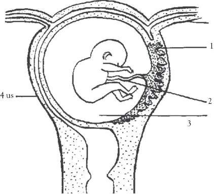
(1) placenta; (2) umbilcal cord; (3) amniotic fluid; (4) uterus
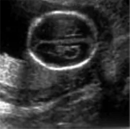
This ultrasound is taken from the top of the skull. The bright white circle is the skull. This type of view is used to determine if the brain is growing correctly
Table of contents
- Cover
- Half-title Page
- Title Page
- Copyright
- Contents
- The Basics
- Chapter 1 Introduction
- Chapter 2 Prenatal Testing
- Chapter 3 Adult Testing
- Chapter 4 Screening
- Chapter 5 Ancestry DNA
- Chapter 6 Other Tests
- Chapter 7 Laboratory Methods
- Chapter 8 Landmark Legal Cases
- Chapter 9 Other Interesting Cases
- Chapter 10 Some Interesting Problems
- Epilogue
- Appendix A
- Decision Making Model
- References
- Index
- Backcover
Frequently asked questions
- Essential is ideal for learners and professionals who enjoy exploring a wide range of subjects. Access the Essential Library with 800,000+ trusted titles and best-sellers across business, personal growth, and the humanities. Includes unlimited reading time and Standard Read Aloud voice.
- Complete: Perfect for advanced learners and researchers needing full, unrestricted access. Unlock 1.4M+ books across hundreds of subjects, including academic and specialized titles. The Complete Plan also includes advanced features like Premium Read Aloud and Research Assistant.
Please note we cannot support devices running on iOS 13 and Android 7 or earlier. Learn more about using the app