Stem Cells, Tissue Engineering And Regenerative Medicine
eBook - ePub

Stem Cells, Tissue Engineering And Regenerative Medicine

  1. 552 pages
  2. English
  3. ePUB (mobile friendly)
  4. Available on iOS & Android
eBook - ePub

Stem Cells, Tissue Engineering And Regenerative Medicine

Book details
Book preview
Table of contents
Citations

About This Book

Stem cells, tissue engineering and regenerative medicine are fast moving fields with vastly transformative implications for the future of health care and capital markets. This book will show the state of the art in the translational fields of stem cell biology, tissue engineering and regenerative medicine. The state of developments in specific organ systems, where novel solutions to organ failure are badly needed such as the lungs, kidney and so forth, are discussed in various chapters. These present and future advances are placed in the context of the overall field, offering a comprehensive and quick up-to-date drink from the fountain of knowledge in this rapidly emerging field.

This book provides an investigator-level overview of the current field accessible to the educated scientific generalist as well as a college educated readership, undergraduates and science writers, educators and professionals of all kinds.

Contents:

  • Developmental Biology, Regenerative Medicine and Stem Cells: The Hope Machine is Justified (David Warburton)
  • Towards Broader Approaches to Stem Cell Signaling and Therapeutics (Edwin Jesudason)
  • Pluripotent Stem Cells from the Early Embryo (Claire E Cuddy and Martin F Pera)
  • The First Cell Fate Decision During Mammalian Development (Melanie D White and Nicolas Plachta)
  • Asymmetric Cell Divisions of Stem/Progenitor Cells (Ahmed HK El-Hashash)
  • Microenvironmental Modulation of Stem Cell Differentiation with Focus on the Lung (Shimon Lecht, Collin T Stabler, Seda Karamil, Athanasios Mantalaris, Ali Samadikuchaksaraei, Julia M Polak and Peter I Lelkes)
  • Smart Matrices for Distal Lung Tissue Engineering (Mark J Mondrinos and Peter I Lelkes)
  • Skin Stem Cells and Their Roles in Skin Regeneration and Disorders (Chao-Kai Hsu, Chao-Chun Yang and Shyh-Jou Shieh)
  • Stem Cell Recruitment and Impact in Skin Repair and Regeneration (Tim Hsu, Tai-Lan Tuan and Yun-Shain Lee)
  • Epigenetic and Environmental Regulation of Skin Appendage Regeneration (Ting-Xin Jiang, Chih-Chiang Chen, Michael W Hughes, Cheng-Ming Chuong and Randall Widelitz)
  • Cranial Neural Crest: An Extraordinarily Migratory and Multipotent Embryonic Cell Population (Samuel G Cox and J Gage Crump)
  • Modeling Neurodegenerative Diseases and Neurodevelopmental Disorders with Reprogrammed Cells (Kate E Galloway and Justin K Ichida)
  • Cytokine Regulation of Intestinal Stem Cells (Philip E Dubé, Unice J K Soh and D Brent Polk)
  • The Intestinal Stem Cell Niche and Its Regulation by ErbB Growth Factor Receptors (Dana Almohazey and Mark R Frey)
  • Tissue Engineering: Intestine (Avafia Y Dossa, Kathy A Schall, Tracy C Grikscheit and Christopher P Gayer)
  • Liver Stem and Progenitor Cells in Development, Disease and Regenerative Medicine (Nirmala Mavila and Kasper S Wang)
  • Lung Mesenchymal Stem Cells (Wei Shi)
  • FGF Signaling in Lung Stem and Progenitor Cells (Soula Danopoulos and Denise Al Alam)
  • Bioengineering Distal Airways (Christine Finck and Todd Jensen)
  • The Isolation and Molecular Characterization of Cancer Stem Cells (Aggressive Endophenotypes) in Individual Lung Cancers (Raj K Batra, Scott Oh and Saroj Basak)
  • Mesenchymal Stromal Cell-Based Therapies for Lung Diseases and Critical Illnesses (Fernanda Cruz, Patricia RM Rocco and Daniel J Weiss)
  • Heart Regeneration and Repair: What We Have Learned from Model Organisms (Laurent Gamba, Michael R Harrison and Ching-Ling Lien)
  • Leveraging Structure-Based Rational Drug Design and Nanotechnology to Destroy Leukemic Stem Cells (Fatih M Uckun, Jianjun Cheng, Cheney Mao and Sanjive Qazi)
  • Placenta-Derived Stem Cells: Development and Preclinical Applications for Regenerative Medicine (Jennifer Izumi Divine, Hee Kyung Jung and Toshio Miki)
  • Stem Cells in the Real World: Environmental Impacts (Theresa M Bastain, Lu Gao and Frank D Gilliland)
  • Establishing a Research Grade Human Pluripotent Stem Cell Laboratory (Laura-Marie Nucho and Victoria Fox)


Readership: Stem cell and tissue engineering scientists, patient advocates, educated laypeople, high school science students, undergraduate students, graduate students, physicians and surgeons.
Key Features:

  • This book presents up-to-date latest breakthroughs and near future applications
  • Bench to bedside
  • This book features potential cures

Frequently asked questions

Simply head over to the account section in settings and click on ā€œCancel Subscriptionā€ - itā€™s as simple as that. After you cancel, your membership will stay active for the remainder of the time youā€™ve paid for. Learn more here.
At the moment all of our mobile-responsive ePub books are available to download via the app. Most of our PDFs are also available to download and we're working on making the final remaining ones downloadable now. Learn more here.
Both plans give you full access to the library and all of Perlegoā€™s features. The only differences are the price and subscription period: With the annual plan youā€™ll save around 30% compared to 12 months on the monthly plan.
We are an online textbook subscription service, where you can get access to an entire online library for less than the price of a single book per month. With over 1 million books across 1000+ topics, weā€™ve got you covered! Learn more here.
Look out for the read-aloud symbol on your next book to see if you can listen to it. The read-aloud tool reads text aloud for you, highlighting the text as it is being read. You can pause it, speed it up and slow it down. Learn more here.
Yes, you can access Stem Cells, Tissue Engineering And Regenerative Medicine by David Warburton in PDF and/or ePUB format, as well as other popular books in Biological Sciences & Science General. We have over one million books available in our catalogue for you to explore.

Information

Publisher
WSPC
Year
2014
ISBN
9789814612791

1

Pluripotent Stem Cells from the Early Embryo

Claire E. Cuddyā€  and Martin F. Pera*ā€ ,ā€”,Ā§

ā€ University of Melbourne
ā€”Florey Neuroscience and Mental Health Institute
Ā§Walter and Eliza Hall Institute of Medical Research

Introduction

Pluripotent stem cells (PSC) have the unique ability to give rise to any cell in the body. Our understanding of these remarkable cells has grown exponentially over the past two decades, and indeed has progressed to the point where today, cellular therapeutics made from pluripotent cells are undergoing clinical trials in man.
However, the concept of pluripotency dates back many years, to early pioneering work on teratocarcinomas, unusual tumors consisting of primitive undifferentiated stem cells, alongside a wide array of mature tissues such as muscle, skin and gut.
The identification, characterization, and propagation in vitro of teratocarcinoma stem cells, coupled with the realization that they were similar in phenotype and developmental potential to the cells of the early embryo, led to the isolation of embryonic stem (ES) cells in the mouse in 1981, and ultimately in the human in 1998. The discovery of mouse ES cells brought about a revolution in mammalian genetics, by enabling the creation of transgenic strains of animals with desired targeted genetic modifications. The isolation of PSC from the human blastocyst marked the beginning of the second stem cell revolution, which has given us powerful new tools for the study of human biology and disease, and is opening up bright new prospects for regenerative medicine. In this chapter, we discuss the key features of PSC derived from the early mammalian embryo.

Pluripotent Stem Cells have Unique Properties

The two key properties that define a stem cell are the capacity to self-renew, or to divide to produce new stem cells, and the ability to give rise to multiple specialised cell types. Multipotent stem cells can give rise to a restricted subset of tissue-specific cell types. For example, hematopoietic stem cells, the best-characterized stem cell population in adult tissues, give rise to all elements of the blood (white cells, red cells, and platelets) but no other cell types. ES cells are pluripotent. PSC can give rise to derivatives of all three embryonic germ layers; the ectoderm, mesoderm and endoderm. The three germ layers of the embryo form just after implantation of the embryo into the womb, and each gives rise to distinct lineages. The nervous system and skin are derived from the ectoderm, the mesoderm becomes the musculoskeletal, circulatory, and urogenital systems and the endoderm will form most of the major internal organ systems. PSC can form derivatives of all of these lineages.
We reserve the term totipotent to refer to PSC that, on their own, can give rise to a new individual given appropriate maternal support. Under this definition, ES cells are not totipotent, because in the absence of appropriate signals, they cannot undergo the morphogenetic patterning to generate the body plan, even though they can form all its constituent tissues. This limitation may relate to a requirement in mammals for maternally derived patterning factors which would be lost during the establishment of a stem cell line.1 Totipotency is a property displayed only by the zygote and blastomeres of the embryo up to the eight-cell stage.

Mouse Embryonic Development Provides the Conceptual Basis for Understanding PSC

An understanding of the origin and properties of PSC requires familiarity with the basic features of mammalian preimplantation embryonic development. Most of our knowledge of this process stems from studies performed in the mouse, or in humans from research into in vitro fertilisation procedures. Because we are able to study postimplantation embryonic development only in the mouse, we will focus on this model of development here. However, it is important to note that whilst many fundamental developmental mechanisms are strongly conserved, there are also important differences in the details of embryonic development between mammalian species. In the early human embryo, the timing of key developmental milestones, the architecture of the conceptus, the development of the extraembryonic tissues like the placenta and yolk sac that support the embryo properly, and the signalling mechanisms that control growth and differentiation, vary considerably from their counterparts in the mouse.2
In all placental mammals, as the fertilised egg travels down the oviduct to the uterus, both the uterus and the conceptus are preparing for the event of implantation.3 During this time, the cells of the embryo are steadily dividing.4 Three distinct cell lineages emerge during preimplantation development. Up until the eight cell stage, all the cells or blastomeres of the embryo are totipotent. An event called compaction occurs around the eight cell stage, and two lineages become segregated, with the trophectoderm to develop on the outside of the embryo, and the precursors of the inner cell mass in the interior. Later, just before implantation, a third lineage emerges below the inner cell mass, called the primitive endoderm. Once the primitive endoderm has formed, the tissue above it is called the epiblast. The inner cell mass and the epiblast will give rise to all the tissues of the body. The trophectoderm and primitive endoderm, which go on to comprise parts of the placenta and the yolk sac respectively, are known as extraembryonic tissues (tissues derived from the conceptus that play a supporting role in development and are discarded at or before birth). The placenta functions in fetal-maternal exchange. The extraembryonic endoderm has the function of a primitive placenta very early in development, but also serves a source of signals that help to pattern the embryo.
After implantation at day 4.5, the embryo grows at a tremendous rate and the basic cell lineages and body plan are determined within days 5ā€“10 post-fertilisation. Following implantation, the primitive ectoderm layer is incorporated into a bilaminar cylinder of tissue made up of the epiblast and hypoblast or primitive endoderm. At around day 6, this epiblast undergoes a process known as gastrulation. A cleft known as the primitive streak forms, and the cells of the epiblast migrate toward and through this cleft. The process of gastrulation produces three cell layers, which form the three distinct germ layer lineages; the ectoderm, mesoderm and endoderm. By the end of gastrulation, the cells of the embryo have become committed to particular fates, and, in normal development, are no longer pluripotent. Organogenesis occurs from day 10 to the end of gestation, at around day 19/20.

Normal Pluripotent Stem Cells Were First Derived From the Epiblast

ES cell lines are derived from the blastocyst stage embryo (Fig. 1). The blastocyst consists of an outer single cell layer (the trophoblast) encompassing a fluid-filled cavity. Within this cavity and anchored to the trophoblast is the inner cell mass. The inner cell mass gives rise to the epiblast, which is the tissue of origin of mouse ES cells.5 In this species, the inner cell mass is isolated and explanted into culture at around day four of development, often with a layer of mitotically inactivated embryo fibroblasts for support. The cells begin to divide and ultimately give rise to ES cell lines that have properties very similar to the epiblast itself.
In the human, embryos donated by couples undergoing infertility treatment and surplus to clinical requirements are cultured to blastocyst stage (day 5/6) usually in sequential media mimicking the changes in the in vivo environment as the embryo travels through the oviduct. The highest quality embryos (based on morphology) are picked for establishing a cell line. Often, in deriving mouse or human ES cells, at the blastocyst stage, the inner cell mass is isolated from the trophoblast. This can be done using immunosurgery whereby complement binds animal-sourced antibodies directed against trophoblast antigens and destroys the trophoblast cells.6 More recently, in the human, isolation of the inner cell mass has been performed by dissection with needles or lasers, eliminating the use of xenomaterials. As in the mouse, the inner cell mass is explanted onto a layer of feeder cells, or into one of the defined culture systems described below. There is an intermediate cell type, different in phenotype to the inner cell mass or ES cells, that emerges transiently during the early phases of human ES cell establishment.7 However, the precise embryonic counterpart of human ES cells remains undetermined.
Image
Figure 1. Derivation of pluripotent stem cells from the early mammalian embryo. The egg is a source of reprogramming factors for the development of somatic cell nuclear transfer ES cell lines, and, following activation, can also give rise to parthenogenetic ES cells, in mouse or human. The inner cell mass of the preimplantation embryo gives rise to the epiblast, from which mouse, rat, and human ES cells are derived. The postimplantation epiblast is the tissue of origin of epiblast stem cells in the mouse. Primordial germ cells arise directly from the epiblast and can be converted into EG cells with properties similar to ES cells in the mouse and rat. EG cell lines have been isolated from human primordial germ cells, but are difficult to propagate long term as pluripotent cells.

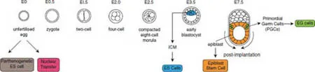
Pluripotent Stem Cells can be Generated From Different Stages of Development and by Cellular Reprogramming

Mammalian PSC were first isolated from mouse teratocarcinomas and then from the preimplantation mouse embryo. However, in the past two decades, it has been found that PSC lines can be isolated from germ cells, or from mouse embryos at later stages of embryonic development, or by cellular reprogramming, either through somatic cell nuclear transfer or by introduction of defined factors. In addition to these PSC lines, in the mouse, stem cell lines representing the major extraembryonic lineages have also been isolated. The varieties of stem cells corresponding to early stages of mammalian development (Fig. 1) are listed in Table 1.8
Though ES cells were originally isolated from preimplantation embryos, it has been known for many years that pluripotent stem cells persist in the mammalian embryo up until the end of gastrulation. Using different culture techniques to those used in ES cell isolation, it is possible to derive stem cells from the postimplantation epiblast.9 These cells, called epiblast stem cells, are pluripotent but have different growth requirements and different behavior to mouse embryonic stem cells. Specifically, they are unable to generate germ line chimeras using conventional in vivo assays, though they can contribute extensively to development when placed into postimplantation embryos cultured in vitro.10
Table 1. Types and sources of cultured stem cell lines from the early embryo.
Type of Pluripotent...

Table of contents

  1. Cover
  2. Halftitle
  3. Title Page
  4. Copyright Page
  5. Contents
  6. Introduction: Developmental Biology, Regenerative Medicine and Stem Cells: The Hope Machine is Justified
  7. Foreword: Towards Broader Approaches to Stem Cell Signaling and Therapeutics
  8. Chapter 1 Pluripotent Stem Cells from the Early Embryo
  9. Chapter 2 The First Cell Fate Decision During Mammalian Development
  10. Chapter 3 Asymmetric Cell Divisions of Stem/Progenitor Cells
  11. Chapter 4 Microenvironmental Modulation of Stem Cell Differentiation with Focus on the Lung
  12. Chapter 5 Smart Matrices for Distal Lung Tissue Engineering
  13. Chapter 6 Skin Stem Cells and Their Roles in Skin Regeneration and Disorders
  14. Chapter 7 Stem Cell Recruitment and Impact in Skin Repair and Regeneration
  15. Chapter 8 Epigenetic and Environmental Regulation of Skin Appendage Regeneration
  16. Chapter 9 Cranial Neural Crest: An Extraordinarily Migratory and Multipotent Embryonic Cell Population
  17. Chapter 10 Modeling Neurodegenerative Diseases and Neurodevelopmental Disorders with Reprogrammed Cells
  18. Chapter 11 Cytokine Regulation of Intestinal Stem Cells
  19. Chapter 12 The Intestinal Stem Cell Niche and Its Regulation by ErbB Growth Factor Receptors
  20. Chapter 13 Tissue Engineering: Intestine
  21. Chapter 14 Liver Stem and Progenitor Cells in Development, Disease and Regenerative Medicine
  22. Chapter 15 Lung Mesenchymal Stem Cells
  23. Chapter 16 FGF Signaling in Lung Stem and Progenitor Cells
  24. Chapter 17 Bioengineering Distal Airways
  25. Chapter 18 The Isolation and Molecular Characterization of Cancer Stem Cells (Aggressive Endophenotypes) in Individual Lung Cancers
  26. Chapter 19 Mesenchymal Stromal Cell-Based Therapies for Lung Diseases and Critical Illnesses
  27. Chapter 20 Heart Regeneration and Repair: What We Have Learned From Model Organisms
  28. Chapter 21 Leveraging Structure-Based Rational Drug Design and Nanotechnology to Destroy Leukemic Stem Cells
  29. Chapter 22 Placenta-Derived Stem Cells: Development and Preclinical Applications for Regenerative Medicine
  30. Chapter 23 Stem Cells in the Real World: Environmental Impacts
  31. Chapter 24 Establishing a Research Grade Human Pluripotent Stem Cell Laboratory
  32. Index