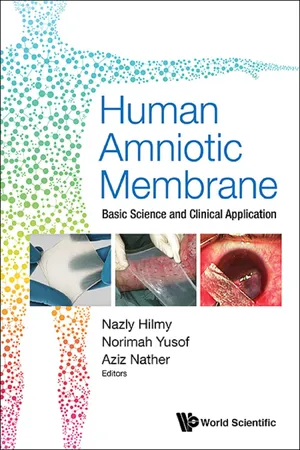
Human Amniotic Membrane: Basic Science And Clinical Application
eBook - ePub

Human Amniotic Membrane: Basic Science And Clinical Application

Basic Science and Clinical Application

  1. 380 pages
  2. English
  3. ePUB (mobile friendly)
  4. Available on iOS & Android
eBook - ePub

Human Amniotic Membrane: Basic Science And Clinical Application

Basic Science and Clinical Application

Book details
Book preview
Table of contents
Citations

About This Book

-->

This book is a comprehensive guide for all tissue bank operators to screen, procure and process amniotic membrane for clinical application.

The amnion comes close to being the ideal biological membrane or dressing — readily available, inexpensive to procure and process. Its basic science is discussed in detail — anatomy, biological and biomechanical properties.

It can be procured from the placenta in normal vaginal deliveries and from Caesarean Sections. Processing is by freeze-drying or by air-drying process with sterilisation using gamma irradiation.

The product has low antigenicity, has anti-microbial properties with ability to enhance epithelisation with marked relief of pain. It is useful as a dressing for wounds — flap wounds, burn wounds, injury wounds, diabetic ulcers, leprous ulcers and post-surgery wounds and post-radiation wounds. It is also used as a biological scaffold for cells in tissue engineering. Its ophthalmic applications include treatment of corneal ulcers and conjunctival tumours. Oral uses include gingiva depigmentation and periodontal regeneration.

--> Contents:

  • Section I: Introduction:
    • IAEA Programmes in Tissue Banking in Asia-Pacific Region (Nazly Hilmy and Norimah Yusof)
    • Tissue Banking in Asia Pacific Region: Past, Present, Future (Aziz Nather, Foong Shi Yun Mandy, Tan Ning and Wang Kaiying)
    • Training Tissue Bank Operators — 20 Years of Experience by IAEA/NUS Regional Training Centre (Aziz Nather and Wo Yu Jun)
  • Section II: Historical Development and Basic Science:
    • Historical Development of Amnion (Norimah Yusof and Nazly Hilmy)
    • Anatomy and Histology of Amnion (Nazly Hilmy and Norimah Yusof)
    • Biological Properties and Functions of Amnion (Paramita Pandansari, Retno Dwijartini Tantin, Basril Abbas and Nazly Hilmy)
    • Physical Properties of Amnion (Norimah Yusof and Nazly Hilmy)
  • Section III: Screening, Procurement and Processing:
    • Transmissible Diseases (Aziz Nather, Sherilyn Leong Li Juan and Wo Yu Jun)
    • Donor Suitability (Aziz Nather, Wo Yu Jun, Sherilyn Leong Li Juan, Norimah Yusof and Nazly Hilmy)
    • Procurement and Processing of Amniotic Membrane (Nazly Hilmy and Norimah Yusof)
  • Section IV: Sterilisation and Quality Control:
    • Sterilisation of Amnion (Norimah Yusof and Nazly Hilmy)
    • Routine Quality Control of Processed Amniotic Membrane (Norimah Yusof and Nazly Hilmy)
  • Section V: Clinical Applications:
    • Management of Wounds:
      • Use of Amnion in Plastic Surgery (Ahmad Sukari Halim, Leow Aik Ming, Aravazhi Ananda Dorai and Wan Azman Wan Sulaiman)
      • Role of Amnion for Treating Burns (Hasim Mohamad)
      • Role of Amnion for Healing of Wounds (Menkher Manjas, Petrus Tarusaraya and Nazly Hilmy)
      • Amnion Dressing in the Management of Radiation Skin Reaction Following Post Radiotherapy (Menkher Manjas)
    • Ophthalmic Applications:
      • Freeze-Dried Irradiated Amnion in Ophthalmic Surgery (Nazly Hilmy, Paramita Pandansari, Getry Sukmawati Ibrahim, S Indira, S Bambang, Radiah Sunarti and Susi Heryati)
      • Multi Layer Amniotic Membrane Transplantation (MLAMT) for Ocular Reconstruction (Getry Sukmawati Ibrahim and Havriza Vitresia)
    • Oral Cavity Applications:
      • Gingiva Depigmentation Using Amniotic Membrane (Retno Dwijartini Tantin, Basril Abbas, Paramita Pandansari and Nazly Hilmy)
      • Amniotic Membrane in Periodontal Regeneration (Shaila Kothiwale)

-->
--> Readership: This book will serve as a useful guide to tissue bank operators in the Asia Pacific region including China, Hong Kong, Korea, Japan, Bangladesh, India, Indonesia, Malaysia, Myanmar, Pakistan, Philippines, Sri Lanka, Thailand, Vietnam and Singapore. -->
Keywords:Human Amniotic Membrane;Basic Science;Clinical Application;Radiation SterilisedReview: Key Features:

  • First book on Amnion Membrane Allograft in the world
  • Part of tissue banking curriculum of Diploma Course in Tissue Banking run by IAEA and NUS annually since 1997
  • Will become textbook for all diploma course students

Frequently asked questions

Simply head over to the account section in settings and click on “Cancel Subscription” - it’s as simple as that. After you cancel, your membership will stay active for the remainder of the time you’ve paid for. Learn more here.
At the moment all of our mobile-responsive ePub books are available to download via the app. Most of our PDFs are also available to download and we're working on making the final remaining ones downloadable now. Learn more here.
Both plans give you full access to the library and all of Perlego’s features. The only differences are the price and subscription period: With the annual plan you’ll save around 30% compared to 12 months on the monthly plan.
We are an online textbook subscription service, where you can get access to an entire online library for less than the price of a single book per month. With over 1 million books across 1000+ topics, we’ve got you covered! Learn more here.
Look out for the read-aloud symbol on your next book to see if you can listen to it. The read-aloud tool reads text aloud for you, highlighting the text as it is being read. You can pause it, speed it up and slow it down. Learn more here.
Yes, you can access Human Amniotic Membrane: Basic Science And Clinical Application by Nazly Hilmy, Norimah Yusof;Aziz Nather;; in PDF and/or ePUB format, as well as other popular books in Biological Sciences & Cell Biology. We have over one million books available in our catalogue for you to explore.

Information

Publisher
WSPC
Year
2017
ISBN
9789813226364
Section V: Clinical Applications

Management of Wounds

13

Use of Amnion in Plastic Surgery

Ahmad Sukari Halim∗, Leow Aik Ming,
Aravazhi Ananda Dorai and
Wan Azman Wan Sulaiman
Reconstructive Sciences Department,
School of Medical Sciences
Health Campus
Universiti Sains Malaysia
16150 Kubang Kerian, Kelantan,
Malaysia
∗Email: [email protected]
Introduction
A plethora of synthetic dressings are available, most of which are beneficial, accounts for a substantial portion of any health budget. One of the natural occurring products, the human afterbirth, is constantly available worldwide but is almost always discarded despite its membranes’ established efficacy as a true biological dressing (Trelford and Trelford-Sauder, 1979; Matthews et al., 1982).
In 1910, William Thornton who was a final year medical student at Johns Hopkins Hospital has suggested to Dr. Staige Davis that fetal membranes might be useful for wound healing. Although the membrane used at that time did not provide permanent cover on the only recorded case in which Davis applied, he was enthusiastic about the concept and recommended further investigations (Davis, 1910).
The term “amniotic membrane” and “amnion” are used loosely in literature. Failure to recognize the appropriate terminology has bedeviled interpretation of clinical results and their outcomes as highlighted by Trelford. Thus, these terminologies become important for accurate definition of clinical practice and subsequent assessment of results. The term “extra-embryonic tissue” (EET) is referred to placenta, amnion and chorion. “Extra-embryonic membranes” (EEM) or amniotic membrane denotes a general name encompassing an inner membrane, the amnion and an outer membrane, the chorion, which is in contact with the maternal decidua during gestation. Hence, the term “amnion” is reserved for the inner layer of EEM as obtained when the amnion epithelium and its underlying layers are stripped from the chorion without using any elaborate techniques of separation, although this does not result in absolutely anatomical separation in histologic terms (Matthews et al., 1982).
Procurement of Amnion for Clinical Usage
The real possibilities of transmitting diseases via human blood, sera and tissue products in the modern era cannot be ignored. Therefore, all the potential donors of placenta should be stringently screened for the possibility of blood borne diseases such as HIV, HbSAg, HCV, Syphilis and Gonorrhoea.
To obtain the membranes, EEM is cut from the edge of placenta and the reflected portion of the membrane is stripped from the placenta surface to the base of the umbilical cord. Strict aseptic technique should be exercised throughout. Ideally, only membranes obtained at Caesarean Sections are used, but vaginally delivered tissues have been widely applied without complications. For the vaginal delivered tissue, the amniotic membrane should be ruptured less than 12 hours before delivery. There are various conditions where the membranes are not suitable such as meconium-stained or dysmorphic, those from the mothers with seropositive blood screening or a history of venereal disease, endometriosis, prolonged labour, or toxemia. The membranes obtained are thoroughly rinsed in a normal saline to remove all blood traces, once in 0.025% sodium hypochlorite, and again several times in normal saline, before refrigeration at 4°C (Robson and Krizek, 1973) the addition of antibiotics being optional (Shun and Ramsey-Stewart, 1983).
Specific Features of Amnion Relevant to Plastic Surgical Practice
Skin substitutes and biological dressings play a major role in plastic and reconstructive surgery. The histological characteristic of amnion is quite similar to skin. The normal amnion is 0.02–0.05 mm thick and it has the thickness of only one third the thickness of the stratum corneum epidermis (Rao and Chandrasekharam, 1981). Among the various types of biological dressings, human amnion membrane play a vital role in facilitating wound healing. Amnion seems to satisfy all the criteria of an ideal biological dressing. It is thin, elastic and adherence to the wound surface. It provides an effective vapor barrier, a durable cover for raw surfaces, substantial pain relief with potential bacteriostatic properties and facilitates rapid re-epithelialisation (Rao and Chandrasekharam, 1981; Colocho et al., 1974; Talmi et al., 1990; Unger and Roberts, 1976). One of the most important principles in plastic surgery is to achieve rapid and complete wound healing in the shortest possible time. Any method that promotes and enhances rapid wound healing has an enormous potential for research and clinical use. Human amnion membrane has an inherent angiogenic property to increase the rate of healing and epithelialisation. Human amnion membrane has long been used for various clinical applications in the field of plastic surgery. Human amnion membrane has been widely used to treat superficial and partial thickness burns in many centers worldwide (Subrahmanyam, 1995). Amnion can also be used as a biological dressing in the treatment of chronic ulcers and pressure sores (Ward et al., 1989). Skin graft donor sites can also be effectively treated by using human amnion membrane. Recently, radiation induced ulcers have been effectively treated using human amnion membrane (Gajiwala and Sharma, 2003). Human amnion thus has the potential to become an attractive biomaterial which is inexpensive and easily obtainable.
Type of Amnion Preparations Available to Clinical Applications
Amnion is mostly used as fresh, but dried (Rao and Chandrasekharam, 1981) frozen (Colocho et al., 1974) irradiated (Rao and Chandrasekharam, 1981) or lyophilised (Burgos and Sergeant, 1983) preparations have also been used.
Factors that Contribute to the Success of Amnion in Plastic Surgical Practice
A. Anti-bacterial Properties
Theories of mechanism of the anti-bacterial effect observed in EEM and amnion are plentiful. Many authors have concurred that EEM and amnion are effective biological dressings since both membranes fall intimately into the contour of the wound to lie in close apposition to its surface (Trelford et al., 1973; Gruss and Jirsch, 1978; Bose, 1979). It has been noted that the degree of bacterial contamination was inversely proportional to the number of leucocytes beneath a graft (Gruss and Jirsch, 1978). Saymen et al. found that leucocytes migrate toward the surface of a covered wound and away from it if it is uncovered (Saymen et al., 1973). Thus the precise elimination of dead space is considered important. Amnion alone, being thinner and even pliable than EEM, is probably the most potentially effective dressing available in this context. Simko et al. added the surface pH fell after covering with amnion and that the temperature in the underlying granulation tissue rose, increasing phagocytosis and reducing the exudation of interstitial fluid.
Robson and Krizek has found that EEM was superior to allograft (homograft) and to xenograft (heterograft) skin and as effective as autograft skin in lowering the bacterial count when applied to 20% rat scalds topically inoculated with Pseudomonas aeruginosa (Burgos and Sergeant, 1983). They concluded that no such anti-bacterial substance existed. However, a substance that enhanced leucotaxis or angiogenesis, with subsequent leucotaxis and promotion of host defense mechanisms, would be important (Robson and Krizek, 1973). Similarly, a substance depressing the normal host rejection to a “foreign” surface dressing and thus permitting continued close surface adherence with at least the aforementioned mechanical benefits, would achieve an additional antibacterial effect. The presence of a weak antibiotic-like substance in amniotic fluid has also been demonstrated, and the possibility of its elaboration in EEM epithelium remains strong (Galask and Synder, 1970; Sachs and Stern, 1970).
B. Effect of Amnion on Vessel Development
The vascular response of amnion on the wound bed has been extensively studied using standard histologic staining and immunohistologic techniques (Faulk et al., 1980) It was found that not only was there a considerable proliferation of capillary vessels towards the surface of leg ulcers following 5 days of tissue-culture-preserved EEM application, but also found that the blood vessel walls became thinner and more regular and their lamina were observed to be more consistently patent. Increased vascularity in burns to which EEM was applied has also been noted microscopically (Kirschbaum and Hernandez, 1963).
Proteins elaborated by amniotic epithelial cells have angiogenic effects, but the precise mode of action remains unknown (Burgos, 1983).
C. Alterations in Connective Tissues
Studies have shown that Reticulin Stain was able to reveal a marked loosening of the thick, tightly-woven tissue pattern often seen in the bed of chronic refractory skin ulcers (Faulk et al., 1980). W...

Table of contents

  1. Cover
  2. Halftitle
  3. Title
  4. Copyright
  5. Dedication
  6. Lifetime Achievement Awards
  7. Preface
  8. About the Editors
  9. List of Contributors
  10. Contents
  11. Section I: Introduction
  12. Section II: Historical Development and Basic Science
  13. Section III: Screening, Procurement and Processing
  14. Section IV: Sterilisation and Quality Control
  15. Section V: Clinical Applications