Quenched-phosphorescence Detection of Molecular Oxygen
eBook - ePub

Quenched-phosphorescence Detection of Molecular Oxygen

Applications in Life Sciences

  1. 368 pages
  2. English
  3. ePUB (mobile friendly)
  4. Available on iOS & Android
eBook - ePub

Quenched-phosphorescence Detection of Molecular Oxygen

Applications in Life Sciences

Book details
Book preview
Table of contents
Citations

About This Book

Significant progress has been made in recent years in quenched-phosphorescence oxygen sensing, particularly in the materials and applications of this detection technologythat are open to commercialization, like uses in brain imaging and food packaging. Prompted by this, the editors have delivered a dedicated bookthat brings together these developments, provides a comprehensive overview of the different detection methodologies, and representative examples and applications.

This book is intended to attract new researchers from various disciplines such as chemistry, physics, biology and medicine, stimulate further progress in the field and assist in developing new applications. Providing a concise summary at the cutting edge, this practical guide for current experts and new potential users will increase awareness of this versatile sensing technology.

Frequently asked questions

Simply head over to the account section in settings and click on ā€œCancel Subscriptionā€ - itā€™s as simple as that. After you cancel, your membership will stay active for the remainder of the time youā€™ve paid for. Learn more here.
At the moment all of our mobile-responsive ePub books are available to download via the app. Most of our PDFs are also available to download and we're working on making the final remaining ones downloadable now. Learn more here.
Both plans give you full access to the library and all of Perlegoā€™s features. The only differences are the price and subscription period: With the annual plan youā€™ll save around 30% compared to 12 months on the monthly plan.
We are an online textbook subscription service, where you can get access to an entire online library for less than the price of a single book per month. With over 1 million books across 1000+ topics, weā€™ve got you covered! Learn more here.
Look out for the read-aloud symbol on your next book to see if you can listen to it. The read-aloud tool reads text aloud for you, highlighting the text as it is being read. You can pause it, speed it up and slow it down. Learn more here.
Yes, you can access Quenched-phosphorescence Detection of Molecular Oxygen by Dmitri B Papkovsky, Ruslan I Dmitriev, Dmitri B Papkovsky, Ruslan I Dmitriev in PDF and/or ePUB format, as well as other popular books in Physical Sciences & Analytic Chemistry. We have over one million books available in our catalogue for you to explore.

Information

Year
2018
ISBN
9781788014557
CHAPTER 1
Fundamentals of Quenched Phosphorescence O2 Sensing and Rational Design of Sensor Materials
Sergey M. Borisova
a Graz University of Technology, Stremayrgasse 9, 8010, Graz, Austria
*E-mail: [email protected]

1.1 Introduction

In the last decades, optical oxygen sensors (oxygen optodes) became indispensable analytical tools, which are nowadays widely applied in academia and industry. Their popularity is explained by the numerous advantages offered by the optical detection method such as the absence of electromagnetic interferences, minimally invasive character (measurement though a transparent wall of a reactor), versatility of sensor formats varying from planar foils and fiber-optic sensors to nanoparticles, suitability for imaging etc. Optical oxygen sensors do not consume the analyte, which favourably distinguishes them from electrochemical sensors such as Clark electrode or galvanic cells. Optodes allow for oxygen measurement in gases and solutions with dynamic ranges, which can be adjusted over many orders of magnitude. Finally, optical oxygen sensors are very useful for measurement of air pressure on surfaces (pressure sensitive paints) or as transducers for enzymatic sensors making use of oxygen consumption such as glucose or lactate sensors.

1.2 Mechanism of Oxygen Quenching

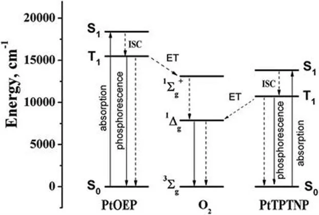
Oxygen is one of the most powerful luminescence quenchers. Quenching of fluorescent dyes (excited singlet state, S1) and phosphorescent dyes (excited triplet state, T1) is spin-allowed. Moreover, the energies of excited states of oxygen (1āˆ‘g+ and 1Ī”g) are lower than the energies of the excited states of most organic dyes and metal complexes (Figure 1.1), which makes quenching via energy transfer favourable.
image
Figure 1.1 Energy diagram for two phosphorescent oxygen indicators: platinum(ii) octaethylporphyrin (PtOEP) and platinum(ii) tetraphenyltetranaphthoporphyrin (PtTPTNP).
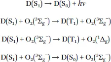
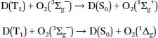
The mechanism of oxygen quenching is rather complex and the exact pathways and formed products depend on many factors.1 Electron-exchange Dexter-type energy transfer is the predominant mechanism of oxygen quenching. Quenching of fluorescent dyes (D) can result in the formation of the dye in the triplet excited state or in the ground state:1
image
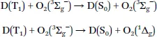
The triplet state of the dye is deactivated to the ground state:
image
For the quenching of phosphorescence, the dye is deactivated into the ground state and singlet oxygen is formed:
image
Depending on the triplet energy of the dye, formation of singlet oxygen either only in the 1Ī”g state (e.g. for PtTPTNP, Figure 1.1) or in both 1Ī”g and 1āˆ‘g+ states (e.g. for PtOEP) is possible. Notably, O2(1āˆ‘g+) deactivates very fast into O2(1Ī”g) state.
Apart from the energy transfer, electron transfer leading to superoxide is also possible:
image
This process can play a significant role for metal complexes with strong reducing properties (particularly in the excited state), for instance Ir(iii) cyclometalated complexes.2 Rapid back electron transfer can result in the formation of singlet oxygen and the sensitizer in the ground state.
Importantly for all these processes, singlet oxygen represents one of the main products. Since its deactivation to the triplet state regenerates the analyte, optical oxygen sensors do not consume the analyte in theory. However, the lifetime of singlet oxygen in polymers can be much longer compared to that in the aqueous phase (āˆ¼3 Āµs), which can be sufficient for it to react with the sensor components (dye or polymer), see Chapter 1.7.
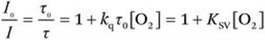
Independent of the quenching mechanism, the quenching behavior for dissolved dyes is described by the Sternā€“Volmer equation:
image
(1.1)
where I0(Ļ„0) and I(Ļ„) are the luminescence intensity (decay time) in the absence and in the presence of oxygen, respectively, kq is the bimolecular quenching constant and KSV is the Sternā€“Volmer constant.
From eqn (1.1) it is evident that the efficiency of quenching depends on both the bimolecular quenching constant and the decay time of the luminophore Ļ„0. The kq constant is determined mostly by oxygen diffusion since the diffusion of the much larger dye is significantly slower already in solution and is virtually non-existant for immobilized dyes. The kq constant often approaches the diffusion-controlled limit kdiff for quenching of fluorescence1 but is lower for quenching of phosphorescence. For common phosphorescent indicators such as Pt(ii) porphyrins or Ru(ii) polypyridyl complexes, it is usually close to 1/9 of kdiff,2 where 1/9 is the spin statistical factor accounting for the formation of both products in the singlet state. However, kq is sometimes higher than this value even in the case of purely energy transfer-based quenching1 and may be even higher if electron transfer is involved.2
Clearly, the Ļ„0 has a much stronger influence on the Sternā€“Volmer constant than kq. In fact, assuming a kq = kdiff = 2.1 Ɨ 1010 Māˆ’1 sāˆ’1 for an air-saturated toluene solution (C(O2) ā‰ˆ 1.8 mM) of a typical fluorescent dye with Ļ„0 of 4 ns, the I0/I value calculated with eqn (1.1) is only 1.15. On the other hand, for a phosphorescent indicator such as PtOEP (Ļ„0 = 85 Āµs) even with much lower kq 2.4 Ɨ 109 Māˆ’1 sāˆ’1 (1/9kdiff) the luminescence intensity and decay time decrease 367-fold in the same conditions. Since the diffusion of oxygen is significantly slower in the polymers compared to the solution, it is evident that only phosphorescent indicators will provide the required resolution when embedded in common polymeric matrices. Additionally, whereas the tunability of fluorescence decay times is usually limited by 1ā€“2 orders of magnitude, the phosphorescence decay time can vary from several microseconds to hundreds of milliseconds. This provides virtually unlimited flexibility in designing oxygen-sensing materials for very different applications.

1.3 Requirements for Phosphorescent Indicators

In order to navigate among hundreds of reported oxygen indicators it is useful to define the important parameters, which should be considered when the indicators are selected. These include:
  • (i) Spectral properties (absorption and emission maxima). In contrast to fluorescent dyes, phosphorescent indicators possess large Stokes shifts, which simplifies signal separation and reduces interferences caused by scattering and autofluorescence. Nevertheless, indicators excitable and emitting at longer wavelength are preferable for the same reasons. In photosynthetic systems, however, such excitation can result in much higher levels of autofluorescence and dyes with other spectral properties can be a better choice. Clearly, autofluorescence can be completely eliminated in the time domain measurement, but it does interfere with the measurement in the frequency domain unless multi-frequency measurement is performed.
    Compatibility of the indicator with the light sources, photodetectors and other optical components should also be considered. Whereas a wide range of light sources is available for the whole spectral range, the detectors are mainly limited to avalanche photodiodes, CCD-arrays and photomultipliers. Although the sensitivity of PMTs is generally very high, it deteriorates fast in the NIR part of the spectrum. In the case of fiber-optic sensors, the quartz glass fibers are compatible with all oxygen indicators. In contrast, much cheaper plastic fibers show strong absorption in the NIR part of the spectrum, which limits the practically useful length to 1ā€“2 meters.
  • (ii) Brightness of luminescence. Brightness can be defined as the product of molar absorption coefficient Īµ and luminescence quantum yield Ļ•. Clearly, in case of phosphorescent indicators high efficiency of inter-system crossing (ISC, S1 ā†’ T1 transition, Figure 1.1) is one of the prerequisites for bright phosphorescence. Bright indicators allow for thinner sensing layers and therefore for faster response times. In the case of very bright but less photostable indicators the oper...

Table of contents

  1. Cover
  2. Title
  3. Preface
  4. Contents
  5. Chapter 1 Fundamentals of Quenched Phosphorescence O2 Sensing and Rational Design of Sensor Materials
  6. Chapter 2 New Polymer-based Sensor Materials and Fabrication Technologies for Large-scale Applications
  7. Chapter 3 Evolution of Cell-penetrating Phosphorescent O2 Probes
  8. Chapter 4 Hydrophilic Ir(iii) Complexes for In vitro and In vivo Oxygen Imaging
  9. Chapter 5 Protection of Triplet Excited State Materials from Oxygen Quenching and Photooxidation in Optical Sensing Applications
  10. Chapter 6 Progress in Phosphorescence Lifetime Measurement Instrumentation for Oxygen Sensing
  11. Chapter 7 Optical O2 Sensing in Aquatic Systems and Organisms
  12. Chapter 8 Monitoring of Extracellular and Intracellular O2 on a Time-resolved Fluorescence Plate Reader
  13. Chapter 9 Monitoring Parameters of Oxygen Transport to Cells in the Microcirculation
  14. Chapter 10 Photoacoustic Imaging of Oxygen
  15. Chapter 11 Imaging of Tissue Oxygen Ex vivo
  16. Chapter 12 Tracking of Hypoxia and Cancer Metastasis with Iridium(iii)-based O2 Probes
  17. Chapter 13 Probing Tissue Oxygenation by Delayed Fluorescence of Protoporphyrin IX
  18. Chapter 14 Microfluidic Systems and Optical Oxygen Sensors: A Perfect Match for Advancing Bioprocessing and Microbiology
  19. Chapter 15 pO2 Measurements in Biological Tissues by Luminescence Lifetime Spectroscopy: Strategies to Exploit or Minimize Phototoxic Effects in Tumors
  20. Chapter 16 In vivo Brain Functional Imaging Using Oxygenation-related Optical Signal
  21. Chapter 17 Applications of Phosphorescent O2 Sensors in Food and Beverage Packaging Systems
  22. Subject Index