![]()
Chapter 1
ANATOMY AND STRUCTURE OF THE VISUAL SYSTEM
Although it is beyond the scope of this book to provide a comprehensive technical description of the visual system, it is important to have at least a basic understanding of the structures involved and their relationship to perception and to learning. Structurally, the visual system can be thought of as having three distinct parts: 1. the organs of sensation, which are the eyes; 2. the optic nerves, which transmit visual images from the eye and transport it to the brain; and 3. the visual cortex, which is the part of the brain responsible for interpreting information received via the optic nerves. Problems occurring in any part of the visual system can impact the child’s perception, and must be considered when selecting appropriate intervention strategies. These three parts of the visual system are interdependent, so that problems in one area can impact another. Clearly, if there is damage to the eyeball that prevents it from taking in an accurate picture, or if the optic nerves fail to transmit images correctly, the brain will not have the information it needs to accurately interpret the picture. Similarly, if the brain receives an accurate picture but does not know how to make sense of the picture because of cognitive or perceptual interferences, the child may not learn what types of visual information he or she needs to pay attention to, and may lose the ability to attend to relevant visual details needed for learning. For this reason, it is important to consider all aspects of vision when attempting to understand a child’s visual perceptual skills.
The eye and extraocular muscles
Every sensory system in the human body begins with a peripheral, or distant, organ of sensation. The eye is the peripheral organ for sight, just as the nose is the organ for smell, the skin is the organ for touch, the ear is the organ for hearing, and the tongue is the organ for taste. Figure 1.1 presents a simplified illustration of the structure of the eye.
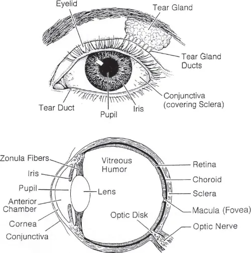
Figure 1.1: Structure of the eye. Reproduced from Miller, Menacker and Batshaw (2002), p.166 with permission from Mark L. Batshaw.
In many ways, the eye functions like a camera. It contains a convex lens system that helps to aim and focus the picture, a variable opening system that allows light to enter, and a structure that records the picture in much the same way as a camera’s film. The eyelid helps to keep the eye moist and provides some protection from injury or foreign objects. On the inside of the eyelids lies a thin layer of tissue that contains many tiny blood vessels, called the conjunctiva. Many people are familiar with conjunctivitis, which is an inflammation of this lining due to a viral or bacterial infection, allergy, or other source of irritation. This conjunctival tissue covers most of the white part of the eyeball, which is called the sclera. At the very front of the sclera, conjunctival tissue is replaced by the cornea, which is a transparent structure responsible for allowing light to enter the eye. The cornea covers the iris, which is the colored portion of the eye. The function of the iris is to adjust the amount of light entering the eye by opening and closing its central opening. This central opening is known as the pupil, which is the dark spot in the center of the eyeball. The process of the iris opening and closing to allow light to enter the eye is what makes a person’s pupils appear smaller or larger under different conditions. The pupil appears larger under dark conditions, because it is in an open position to allow as much light as possible to enter the eye. Under bright light conditions, the pupil constricts and becomes smaller. When light passes through the cornea, it projects through the lens, which lies directly behind the pupil. The lens further focuses light for projection onto the retina, which is a thin layer of tissue lying in the innermost part of the eye. The retina records the images it receives in an upside down and reversed format, and then transmits the image over the optic nerves to the brain.
The retina contains two types of receptors that change light into nerve impulses for transmission to the brain: the rods and the cones. The cones are concentrated in a small area of the retina called the fovea, or macula, and are the only receptors capable of perceiving color. The cones are responsible for central vision, needed for appreciating fine detail in visual images. Whenever a person needs to have precise vision of a moving target, the eye moves in an effort to keep the image directly focused on the macular portion of the retina. The rods, on the other hand, are scattered throughout the retina, and are responsible for peripheral vision.
Six muscles, called the extraocular muscles, attach to each eye and allow the eye to move in all directions in order to take in visual information from a large field as well as from moving targets. They also ensure that the eyes work together in a well-coordinated fashion, a process known as binocular (two-eyed) vision. Weakness or misalignment of any of these muscles makes it difficult for the eyes to work together in a coordinated manner, and can impact vision significantly. Poor binocular vision can lead to such problems as crossed eyes, double vision, and other problems, which will be discussed further in Chapter 3.
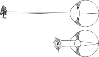
Accommodation describes the process used by the eye to project images clearly onto the retina. Attached to the lens are a number of small muscles, called ciliary muscles. These muscles serve to change the shape of the lens to allow the eye to focus on objects close up or far away. The ability to accommodate vision to changes in distance is at its best in early childhood and gradually declines with age. This is why many adults need glasses for reading as they get older. Their eyes have lost the flexibility to accommodate to view objects at a close range. Figure 1.2 illustrates the process of accommodation.
Figure 1.2: Process of accommodation. The lens changes its shape to focus on a near or far object. Reproduced from Miller, Menacker and Batshaw (2002), p.172 with permission from Mark L. Batshaw.
The optic pathways
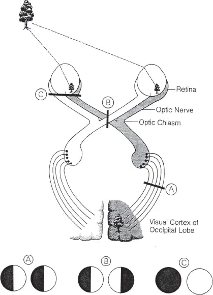
Each eye contains one optic nerve that projects from behind the retina on its way to the brain. Just before the optic nerves enter the brain, some of the fibers from each nerve cross over to the other side in a structure known as the optic chiasm, illustrated in Figure 1.3. Each optic nerve now contains some fibers projecting images from the right eye, and some fibers projecting images from the left eye, as they continue their pathway towards the brain. As the two optic nerves reach the brain, they finally each project their images onto one of the hemispheres of the brain, in the area known as the visual cortex located in the occipital lobe of the brain. Here, visual information is decoded and sent to the temporal and parietal lobes of the brain, where perceptual interpretation of the information takes place.
In this way, each eye is able to send information to both the right and left sides of the brain. That is why, if someone loses sight in one eye, the brain continues to receive a whole picture, although some aspects of vision controlled by binocular eye movements, especially depth perception, may be impacted. However, this also means that damage to either optic nerve can cause visual disturbances to each hemisphere of the brain, which may result in a loss of some part of the child’s field of vision. Problems affecting the optic pathways are not common among children with AD/HD, autism, or learning disabilities, but may be found in some children who have had injuries or illnesses causing brain damage.
Figure 1.3: Visual pathways. One nerve emerges from behind each eye. A portion of the fibers from each nerve cross at the optic chiasm. An abnormality at various points along the route (upper figure) will lead to different patterns of visual loss (shown as black areas in the lower figures). These are illustrated: A) damage to the cortical pathway; B) damage to the optic chiasm; C) damage to the retina. Reproduced from Miller, Menacker and Batshaw (2002), p.176 with permission from Mark L. Batshaw.
![]()
Chapter 2
THE EARLY DEVELOPMENT OF VISUAL SKILLS
Vision plays a major role in helping the young child to develop cognitive, motor and social skills. As early as six months before birth, the eyes begin preparing for their role, practicing the muscular movements that will allow the eyes to focus on their world after birth. The visual system is anatomically mature at birth. However, the newborn infant does not see things in the same way as an older child or adult, because the visual system must proceed through tremendous developmental maturation as the child experiences and learns from visual input. Vision plays a critical role in all aspects of development, and children born without vision demonstrate characteristic delays impacting motor skills, concept formation, language, and social skills. By the time a child enters school, as much as 75% of classroom teaching uses visual media as a primary instructional tool. Even when learning language, children need visual references and models to understand the meaning and contexts of the words used for teaching.
Visual skills in the first year of life
At birth, the infant recognizes patterns of light and dark, and has reflexive closing of the eyes to bright light. At this early age, the infant’s brain responds to any stimulus within its field of vision. However, the eyes can only focus clearly on objects about 8–12 inches away, and the infant is most attracted to objects within this range. Because this is approximately the distance between the infant’s eyes and a caregiver’s eyes when holding the baby, this mechanism sets the tone for developing the critical eye contact necessary for bonding between infant and caregiver. By about two-and-a-half months of age, the infant will smile in response to making eye contact with a caregiver. The newborn prefers to look at the human face as opposed to other visual stimuli, and this is gradually replaced by interest in black and white designs that are large and that contain clear boundaries, especially with horizontal and vertical markings. At this early age, the infant is more attracted to the edges or boundaries of visual patterns than with the internal portions of a pattern.
Gradually, by about three to four months of age, the infant begins to show interest in all parts of a pattern, and can track visual targets as they move sideways or up and down. At this time, the infant also develops the ability to use the two eyes together (binocular control) to adjust visual focus as objects are moved closer or farther away, a process known as convergence (moving eyes inward towards the nose to look at something close up) and divergence (moving eyes outward to look at objects far away). The development of these movements is the precursor for eye hand coordination, so that by three to four months of age, the infant begins to reach purposefully towards objects. By four months of age, the baby can also see the full range of colors.
As the baby begins to see and to manipulate objects within reach, visual input becomes closely associated with the tactile input to develop an inner language of basic concepts, such as hardness/softness, heavy/light, and round/square. The infant develops an awareness of these basic concepts long before he or she has the language to express an understanding of these concepts. This is the beginning of early visual perceptual development, especially the perception of form, shape, color, size, and other simple attributes.
Between five to seven months of age, the infant’s ability to use coordinated binocular eye movements greatly increases. This allows the infant to better judge the location of things in space, and provides motivation to move around and to seek contact with more distant objects. Thus begins the development of such perceptual skills as depth perception and understanding the spatial relationship of objects, which underlies the development of gross and fine motor skills as the infant moves within the environment and contacts objects based on perceptual judgement of their location. At first, the infant must move his or her head or trunk to help aim the eyes towards a desired target, but gradually, eye movement becomes independent of head and trunk movement. Eye movement independent of head and trunk movement does not truly develop until the infant has mature walking and locomotor skills. By about six months of age, the infant usually uses both eyes together when looking at objects. Because this gives an element of depth to the visual image, infants at this age become more interested in looking at three-dimensional than two-dimensional objects.
Gradually, the infant begins to understand that an object can be viewed from a number of angles, or used in different ways, and still be the same object. He or she learns to recognize familiar objects for what they are, even if partially hidden from view, such as a bottle hidden under a blanket with only the nipple protruding. Objects take on a life of their own, appearing more ‘real’ to the infant than when viewed as a two-dimensional image, and are missed when they are out of view (the beginning of simple visual memory for form or shape). The infant begins to understand similarities in shape and form, and can begin to place simple shapes in holes.
Other perceptual skills are also emerging at this time, especially form constancy (the ability to recognize an object even if it looks a little different than the way it usually looks) and figure–ground separation (the ability to recognize relevant details even when there are distracting visual elements nearby). These skills develop and start to work in concert with one another, enhancing the infant’s understanding of his or her world. At approximately 12 months of age, the infant understands these perceptual concepts well enough to start to use imagery in the form of imitating simple movements or patterns. This ability to imitate represents continued development of visual memory and visual sequencing memory skills. The ability to use imagery is tremendously important in the development of language and cognitive skills. At this age, most infants have also developed visual skills to the extent that they have normal or near-normal visual acuity (the ability to focus clearly on visual targets).
Toddlers and preschoolers
By the time the child is 18 months of age, he or she has much more physical control of the body as needed to negotiate the environment. This is a period of great physical energy and physical exploration of the child’s world. The child demonstrates an almost insatiable desire to explore what he or she sees, but has enough physical control of the body that the eyes do not need to guide and direct every movement. For example, the eyes can remain focused on a distant target or destination during walking. The child has enough sense of body awareness that he or she can walk without having to watch their feet during every step. The child gradually begins to be able to focus on objects farther and farther away. Depth perception reaches 4 to 10 feet by 18 months, 10 to 16 feet by 3 years, 16 to 20 feet by 4 years, and 20 feet or greater by the time the child enters kindergarten. Peripheral vision also increases greatly during the preschool years, so the child becomes more aware of visual images outside of his or her direct line of vision. These ...