This is a test
- English
- ePUB (mobile friendly)
- Available on iOS & Android
eBook - ePub
Book details
Book preview
Table of contents
Citations
About This Book
This book is written for sonographers, sonologists, other ultrasound practitioners and students of diagnostic medical ultrasound. The book provides a detailed and clinician-focused overview of the main grayscale artifacts with accompanying descriptions, diagrams, strategies for artifact avoidance and countless examples of clinical images. This book represents the largest collection of ultrasound artifact images ever assembled in a single volume.
Frequently asked questions
At the moment all of our mobile-responsive ePub books are available to download via the app. Most of our PDFs are also available to download and we're working on making the final remaining ones downloadable now. Learn more here.
Both plans give you full access to the library and all of Perlego’s features. The only differences are the price and subscription period: With the annual plan you’ll save around 30% compared to 12 months on the monthly plan.
We are an online textbook subscription service, where you can get access to an entire online library for less than the price of a single book per month. With over 1 million books across 1000+ topics, we’ve got you covered! Learn more here.
Look out for the read-aloud symbol on your next book to see if you can listen to it. The read-aloud tool reads text aloud for you, highlighting the text as it is being read. You can pause it, speed it up and slow it down. Learn more here.
Yes, you can access Artifacts in Diagnostic Medical Ultrasound by Martin Necas in PDF and/or ePUB format, as well as other popular books in Médecine & Technologie et fournitures médicales. We have over one million books available in our catalogue for you to explore.
Information
Publisher
High Frequency PublishingISBN
9780987292179
Topic
MédecineChapter 1: Introduction to Artifacts
Ultrasound artifacts represent a wide range of misleading ultrasound appearances that do not accurately represent anatomical structures or physiologic events. In this chapter, we will discover various types of artifacts and the root causes of their existence.
1.1 What is an artifact?
An ultrasound system sends acoustic pulses into the patient and receives echoes that are generated by tissue reflectors. In B-mode imaging, the depth of the reflector is calculated from the time taken between pulse generation and echo reception using the average speed of sound (1540m/s) in soft tissue as a constant. Echo strength (amplitude) is encoded as brightness. In color, power and spectral Doppler, the returning echoes are further subjected to analysis for Doppler shifts.
In order for the ultrasound imaging system to analyze returning echoes and display them as images, a number of basic assumptions are made:
- Ultrasound pulse travels in a straight line during transmission and echoes follow the same straight path.
- Speed of sound in the body is a constant (1540m/s).
- Attenuation rate is uniform and predictable.
- Two comparable tissue reflectors in a similar location will generate a comparable echo amplitude.
- The beam dimensions are small in height (axial), width (lateral) and thickness (slice thickness).
- Echoes originate only from the central axis of the beam.
- Each reflector only generates one echo.
- The arriving echo was generated by the last emitted ultrasound pulse, not by any preceding pulses.
- The rate of image acquisition exceeds the rate of physiologic events and the rate of transducer movement.
- The operator is utilizing the system appropriately.
- All system controls have been adjusted correctly.
- The transducer elements and electronic system components are functioning normally and without interference from surrounding equipment.
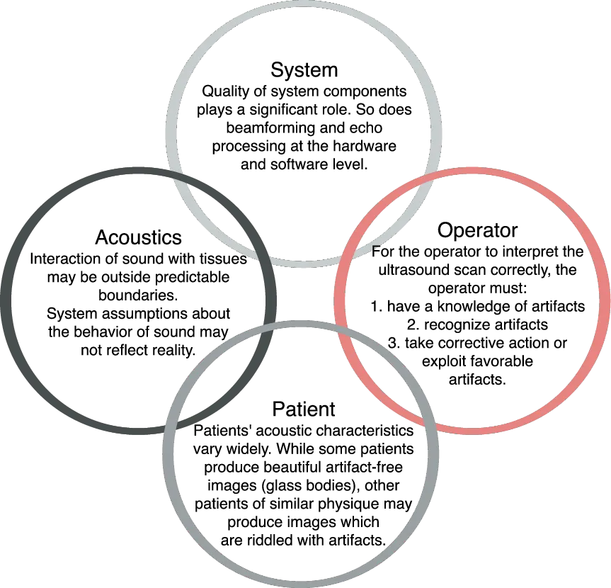
Unfortunately sound (or in this case ultrasound) often fails to obey these convenient rules of pulse propagation, echo generation, signal reception and image processing. Furthermore, the image acquisition rate may sometimes be too slow for rapid physiologic events, leading to a range of temporal display problems. Finally, the operator (sonographer, sonologist) may sometimes play a significant role in creating circumstances for artifacts to occur. An example is shown below.

Figure 1.1 (a): The sonographer used a non-uniform window (falciform ligament and ligamentum teres) to visualize the head of the pancreas. The appearance is highly worrying and suggests the presence of a pancreatic head mass (m).

Figure 1.1 (b): Review of the same area from a different angle revealed a normal pancreatic head.
1.2 General causes of artifacts
1.3 Sorting out the “truth”
While ultrasound artifacts do not accurately reflect reality, to the ultrasound system artifacts appear perfectly “real”. Artifacts represent real echoes or the real absence of echoes, however, they do not represent real structures the way the operator would expect these to appear. Alternatively, the operator may misinterpret artifacts as real structures.
It is the operator’s responsibility to determine whether the image accurately represents reality or not. It is useful to know:
- what types of artifacts exist;
- how each artifact arises;
- the causative agent(s) for each type of artifact;
- the system assumptions that have been violated;
- the range of appearance of each artifact;
- diagnostic uses of artifacts;
- how to accentuate and use diagnostically useful artifacts;
- how to reduce, eliminate or circumvent unhelpful or diagnostically detrimental artifacts.
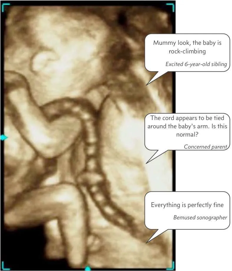
Figure 1.2: Determining what is real requires technical as well as clinical expertise.
1.4 Optical artifacts
Reverberation is caused by bodies of a bright nature with a flat and semi opaque surface which, when the light strikes upon them, throw it back again, like the rebound of a ball, to the former object. (The Notebooks of Leonardo Da Vinci, 1452-1529)
The above quote is a testament to Leonardo Da Vinci’s astonishing insights into optical reverberation. While light and sound behave quite differently, there are a number of parallels between ultrasound imaging and photography which may help explain many acoustic concepts. Some examples of optical artifacts are included on the following pages.

Figure 1.3: Shadowing can be observed both in optical and acoustic images.
Attenuation and scattering
The photograph below was obtained in dense fog and the image of the building appears hazy. Light reflecting from the building is attenuated by the fog.
Another principle that can be seen in this image is that of scatter. Each particle of fog scatters light in multiple directions. In ultrasound imaging, the vast majority of soft tissue reflectors behave as scatterers.
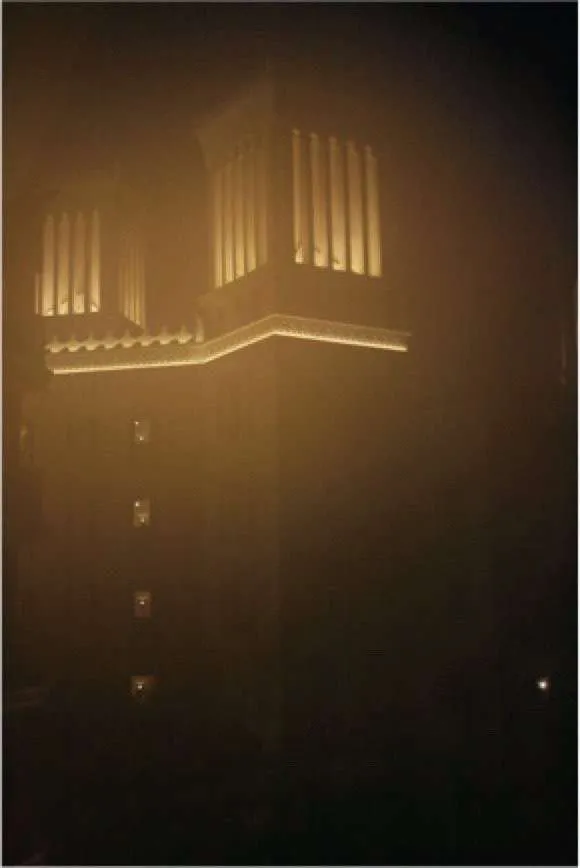
Figure 1.4: Dense fog scatters and attenuates light in a similar way to the scattering and attenuation of sound by soft tissues.
Curved mirror
Light reflects from a mirror at the same angle at which it strikes the mirror. The mirror’s geometry therefore determines the way in which light will reflect and the appearance of the mirrored object. A straight mirror will preserve the size and proportionality of the reflected objects. A concave mirror (see Figure 1.5) has a ‘slimming’ effect. A convex mirror has the opposite (widening) effect. An irregular mirror (see Figure 1.6) will cause irregularity in the reflected image.
All of these scenarios can be encountered with acoustic mirrors as well. An entire chapter will be dedicated to mirror image artifacts....
Table of contents
- Foreword
- Acknowledgements
- About this book
- About the author
- Chapter 1: Introduction to Artifacts
- Chapter 2: Acoustic Speckle
- Chapter 3: Attenuation Artifacts
- Chapter 4: Beam Dimension Artifacts
- Chapter 5: Refraction
- Chapter 6: Reverberation
- Chapter 7: Ringdown
- Chapter 8: Mirror Image Artifacts
- Chapter 9: Reflection Artifact
- Chapter 10: Propagation Speed Artifact
- Chapter 11: Range Ambiguity
- Chapter 12: Motion Artifact
- Chapter 13: Acoustic Window and Angle of Approach Effects
- Chapter 14: System Artifacts and Malfunctions
- Chapter 15: Artifact Avoidance Strategies