eBook - ePub
Cardiology Board Review
ECG, Hemodynamic and Angiographic Unknowns
This is a test
- English
- ePUB (mobile friendly)
- Available on iOS & Android
eBook - ePub
Cardiology Board Review
ECG, Hemodynamic and Angiographic Unknowns
Book details
Book preview
Table of contents
Citations
About This Book
An accessible and engaging review of board exam essentials
Cardiology Board Review lays the groundwork for board exam success with its instructive and easy-to-read explanations of the pathophysiology, diagnosis, and treatment of patients with cardiovascular disease. Breaking topics down into case unknowns, this innovative revision aid provides examples of everyday cardiological issues and then explains how best to address the problem at hand. All content is complemented by clinical images and illustrations, as well as helpful summaries and key points.
Featuring 56 different cases, this essential text:
- Places learning in a practical context. Information about disease states is presented in case-based format which leads to better retention.
- Covers topics including congenital heart disease, coronary artery disease, cardiomyopathies, valvular heart disease, arrhythmias, heart failure, peripheral vascular disease, and more
- Designed to present important concepts and information in a unique way to complement textbook learning
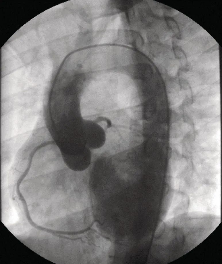
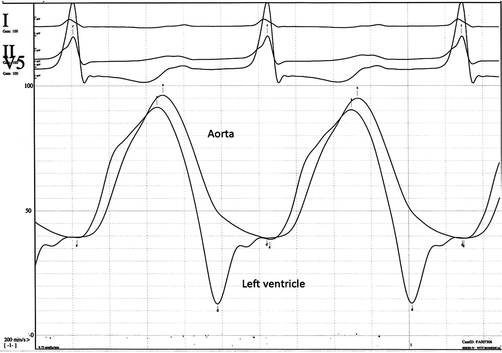
- Features electrocardiograms, angiograms, and pressure tracings
- Is applicable to those working towards certification in Cardiovascular Disease from the American Board of Internal Medicine or preparing for board examinations in other countries
- Is also suitable for those requiring MOC recertification
- Features cases on aortic insufficiency, atrial fibrillation, Brugada syndrome, carotid artery disease, myocardial bridging, congenital heart disease, electrolyte abnormalities, apical HCM, mitral regurgitation, RV outflow tract tachycardia, pulmonary hypertension, arrhythmogenic right ventricular dysplasia, aortic stenosis, atrial myxoma, atrial tachycardia, pulmonic insufficiency, Takotsubo, tricuspid regurgitation, Wolfe-Parkinson-White syndrome, pulmonic stenosis, coronary anomalies, ECG changes of hypothermia, endocarditis, pulmonary embolus, ventricular septal defect, hemodynamics of hypertrophic cardiomyopathy, complete heart block, heart failure, coronary artery disease, atrial septal defect, constrictive pericarditis, fractional flow reserve, dextrocardia, STEMI, early repolarization, giant cell myocarditis, peripheral arterial disease, pericardial tamponade, peripheral arterial disease, pericarditis, myocarditis, long QT syndrome, mitral stenosis, tetralogy of Fallot, and supraventricular tachycardia among others.
Cardiology Board Review offers fellows a fresh and engaging approach to the information required to achieve success in board examinations.
Frequently asked questions
At the moment all of our mobile-responsive ePub books are available to download via the app. Most of our PDFs are also available to download and we're working on making the final remaining ones downloadable now. Learn more here.
Both plans give you full access to the library and all of Perlego’s features. The only differences are the price and subscription period: With the annual plan you’ll save around 30% compared to 12 months on the monthly plan.
We are an online textbook subscription service, where you can get access to an entire online library for less than the price of a single book per month. With over 1 million books across 1000+ topics, we’ve got you covered! Learn more here.
Look out for the read-aloud symbol on your next book to see if you can listen to it. The read-aloud tool reads text aloud for you, highlighting the text as it is being read. You can pause it, speed it up and slow it down. Learn more here.
Yes, you can access Cardiology Board Review by George A. Stouffer, George A. Stouffer in PDF and/or ePUB format, as well as other popular books in Medicine & Cardiology. We have over one million books available in our catalogue for you to explore.
Information
Case 1: A 31‐Year‐Old Man with Fever and Rapidly Progressive Dyspnea
A 31‐year‐old man with a history of ankylosing spondylitis is brought to the Emergency Department by a friend. He reports one week of low‐grade fevers (100.5 °F), cough, progressive shortness of breath, nausea, diarrhea, and occasional vomiting. He has noticed a rapid decline in his exercise tolerance in the past week and states he can't even walk more than 10–20 ft without getting very short of breath. The patient also complains of a frothy cough sometimes laced with blood, paroxysmal nocturnal dyspnea, and orthopnea. He denies any chest pain.
On physical examination, blood pressure is 110/40 mmHg, pulse rate is 110 bpm, and oxygen saturation is 98%. His lungs are clear and he has a diastolic murmur along the right upper sternal border. After an echocardiogram, he is referred for cardiac catheterization. What is the diagnosis based on the aortogram below (Figure 1.1)?
Aortic Regurgitation
The pigtail catheter is in the aortic root and a power injection of a high volume of contrast is performed. This results in filling of the left ventricle, consistent with aortic regurgitation (AR). Angiographic classification of severity of AR is based on the amount of contrast dye entering the left ventricle during an aortogram:
- 1+ some dye enters ventricle but clears with every systole
- 2+ the ventricle becomes completely opacified after several heartbeats and remains opacified throughout the cardiac cycle
- 3+ after several heartbeats – the ventricle becomes as dark as the aorta
- 4+ after several heartbeats – the ventricle becomes darker than the aorta
Note that in the 2014 AHA/ACC Valvular Heart Disease Guideline, transthoracic echocardiography is the first line diagnostic modality for AR as it provides information on the cause of regurgitation, regurgitant severity, and LV (left ventricular) size and systolic function. Cardiac magnetic resonance imaging is indicated in patients with AR in whom echocardiographic images are suboptimal. Cardiac catheterization was performed in this patient to evaluate coronary anatomy prior to surgery.
AR is a condition of increased afterload with associated hemodynamic changes varying depending on the time–course of the valve dysfunction. If AR develops rapidly (i.e. acute or subacute AR), the left ventricle is unable to handle the pressure and volume overload causing a rapid increase in left ventricular pressures during diastole, markedly elevated pressures at end diastole, and premature closure of the mitral valve. Systemic diastolic pressures may be low but generally there is a minimal increase in pulse pressure; in very severe cases of acute AR, cardiac output may fall leading to a decrease in pulse pressure and/or hypotension. In chronic AR, stroke volume increases to maintain effective forward flow. This leads to dilation of the left ventricle, leading in some patients to the development of a massively dilated left ventricle, termed cor bovinum.
In this patient, an echocardiogram showed severely decreased left ventricular contraction with left atrial dilation. The aortic valve was trileaflet with inadequate coaptation and severe AR (Table 1.1). Right ventricular contractile performance was decreased. Hemodynamic changes of severe AR were found at cardiac catheterization including elevated left ventricular end diastolic pressure (38 mmHg), equalization of aortic and left ventricular pressures during diastole (Figure 1.2), and premature closure of the mitral valve during diastole. There was moderate pulmonary hypertension (50/33 mmHg) and reduced cardiac output (2.3 l/min by assumed Fick Method). The patient underwent successful aortic valve replacement with a 25 mm St. Jude prosthesis.
Table 1.1 Echo criteria for grading severity of aortic regurgitation (AR).
Echo criteria | Mild AR | Moderate AR | Severe AR |
Doppler jet width | ≤25% of LVOT | 25–64% of LVOT | ≥65% of LVOT |
Vena Contracta | <0.3 cm | 0.3–0.6 cm | >0.6 cm |
Regurgitant volume | ≤30 ml/beat | 30–59 ml/beat | ≥60 ml/beat |
Regurgitant fraction | ≤30% | 30–49% | ≥50% |
Effective regurgitant orifice (ERO) | <0.1 cm2 | 0.1–0.29 cm2 | ≥0.3 cm2 |
Miscellaneous | Holodiastolic flow reversal in the proximal abdominal aorta |
Table of contents
- Cover
- Table of Contents
- Cases compiled with contributions from
- Preface
- Case 1: A 31-Year-Old Man with Fever and Rapidly Progressive Dyspnea
- Case 2: A Young Man with Palpitations After a Party
- Case 3: A 45-Year-Old Man with Chest Pain After an Automobile Accident
- Case 4: A 67-Year-Old Man with Left-Sided Weakness
- Case 5: A 54-Year-Old Woman with Exertional Angina But No Atherosclerotic Coronary Artery Disease
- Case 6: A 34-Year-Old Woman with Fatigue
- Case 7: An Elderly Woman with a Loud Murmur
- Case 8: A Middle-Aged Woman Who Passes out While Running after her Grandchildren
- Case 9: A 31-Year-Old Man with Palpitations and Dizziness
- Case 10: An Unexpected Finding on a Coronary Angiogram
- Case 11: A 26-Year-Old Man Who Collapses While Talking with Friends
- Case 12: An Elderly Gentleman Who Passes Out While Working on His Farm
- Case 13: A 46-Year-Old Woman with Dyspnea on Exertion and Daily Emesis
- Case 14: A Pregnant Woman with Palpitations
- Case 15: Is this a Positive Brockenbrough Sign?
- Case 16: Dyspnea in a Woman Who Is Five Months Postpartum
- Case 17: Can you Identify This Coronary Anomaly?
- Case 18: Why Is this Patient Short of Breath?
- Case 19: A Clinical Application of Coronary Physiology
- Case 20: An Asymptomatic Patient with a Very Unusual ECG
- Case 21: Is This a STEMI?
- Case 22: Rapidly Progressive Dyspnea, Abdominal Fullness, and Nausea
- Case 23: A 40-Year-Old Man with Dyspnea on Exertion
- Case 24: A Recent Immigrant from Mexico with Complaints of Dyspnea
- Case 25: New Onset Hypertension with Dyspnea and ECG Changes
- Case 26: A 52-Year-Old Woman on Hemodialysis Who Presents with Shortness of Breath and New ECG Changes
- Case 27: A 42-Year-Old Man with Hypotension, Diarrhea, Vomiting, and ECG Changes
- Case 28: A 58-Year-Old Male with Worsening Dyspnea
- Case 29: A 68-Year-Old Woman with Chest Pain and a Normal Stress Test
- Case 30: A 29-Year-Old Woman with Shortness of Breath and Leg Swelling
- Case 31: An ECG Finding You Don't Want to Miss
- Case 32: An Unusual ECG in a Homeless Man Who Is Unconscious
- Case 33: An 18-Year-Old Student with Fever, Chest Pain, and ST Elevation
- Case 34: Recurrent Endocarditis in a 26-Year-Old Woman
- Case 35: A 23-Year-Old Man with a Loud Systolic Murmur
- Case 36: A 66-Year-Old Woman with Dyspnea for Two Weeks Which Has Now Abruptly Worsened
- Case 37: An Unusual Ventriculogram
- Case 38: A 68-Year-Old Male with Generalized Weakness and Dyspnea
- Case 39: A 66-Year-Old Woman with Chest Pain During a Hurricane
- Case 40: A 39-Year-Old Woman Who is Found Unconscious
- Case 41: An Unusual Right Atrial Pressure Tracing
- Case 42: A 20-Year-Old Man with a Heart Rate of 250 bpm
- Case 43: A 46-Year-Old Man with Heart Failure and New Onset Palpitations
- Case 44: Palpitations and Dizziness in a Young Adult with a Very Abnormal ECG
- Case 45: A 36-Year-Old Man with Long-Standing Hypertension and an Abnormal ECG
- Case 46: A 46-Year-Old Man with Fatigue
- Case 47: A 28-Year-Old Runner with Exercise-Induced Palpitations
- Case 48: A 43-Year-Old Man with Chest Pain and an Episode of Syncope
- Case 49: A 50-Year-Old Man with Worsening Cough and Dyspnea
- Case 50: Unsuspected Congenital Heart Disease in a 26-Year-Old Woman
- Case 51: How Likely Is This Patient to Have a Bad Outcome?
- Case 52: A 55-Year-Old Man with Leg Pain
- Case 53: An 18-Year-Old High School Student with an Abnormal ECG and a Nervous Parent
- Case 54: Could This Cardiac Arrest Have Been Prevented?
- Case 55: Why Is This Patient Tachycardic?
- Case 56: A 31-Year-Old Woman with Palpitations While at Work
- Index
- End User License Agreement