- English
- ePUB (mobile friendly)
- Available on iOS & Android
eBook - ePub
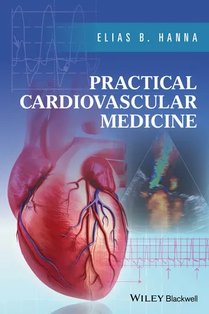
Practical Cardiovascular Medicine
About this book
Prepare yourself for success with this unique cardiology primer which distils the core information you require and presents it in an easily digestible format.
- Provides cardiologists with a thorough and up-to-date review of cardiology, from pathophysiology to practical, evidence-based management
- Ably synthesizes pathophysiology fundamentals and evidence based approaches to prepare a physician for a subspecialty career in cardiology
- Clinical chapters cover coronary artery disease, heart failure, arrhythmias, valvular disorders, pericardial disorders, and peripheral arterial disease
- Practical chapters address ECG, coronary angiography, catheterization techniques, ecnocardiography, hemodynamics, and electrophysiological testing
- Includes over 650 figures, key notes boxes, references for further study, and coverage of clinical trials
- Review questions at the end of each chapter help clarify topics and can be used for Board preparation - over 375 questions in all!
Tools to learn more effectively

Saving Books

Keyword Search

Annotating Text

Listen to it instead
Information
Part 1
CORONARY ARTERY DISEASE
1
Non-ST-Segment Elevation Acute Coronary Syndrome
- I. Types of acute coronary syndrome (ACS)
- II. Mechanisms of ACS
- III. ECG, cardiac biomarkers, and echocardiography in ACS
- IV. Approach to chest pain, likelihood of ACS, risk stratification of ACS
- V. Management of high-risk NSTE-ACS
- VI. General procedural management after coronary angiography: PCI, CABG, or medical therapy only
- VII. Management of low-risk NSTE-ACS and low-probability NSTE-ACS
- VIII. Discharge medications
- IX. Prognosis
- Appendix 1. Complex angiographic disease, moderate disease
- Appendix 2. Women and ACS, elderly patients and ACS, CKD
- Appendix 3. Bleeding, transfusion, prior warfarin therapy, gastrointestinal bleed
- Appendix 4. Antiplatelet and anticoagulant therapy
- Appendix 5. Differences between plaque rupture, plaque erosion, and spontaneous coronary dissection
- Appendix 6. Harmful effects of NSAIDs and cyclooxygenase-2 inhibitors in CAD
- Questions and answers
I. Types of acute coronary syndrome (ACS)
A. Unstable angina
Unstable angina is defined as any of the following clinical presentations, with or without ECG evidence of ischemia and with a normal troponin:
- Crescendo angina: angina that increases in frequency, intensity, or duration, often requiring a more frequent use of nitroglycerin
- New-onset (<2 months) severe angina, occurring during normal activities performed at a normal pace
- Rest angina
- Angina occurring within 2 weeks after a myocardial infarction (post-infarction angina)
B. Non-ST-segment elevation myocardial infarction (NSTEMI)
A rise in troponin, per se, is diagnostic of myocardial necrosis but is not sufficient to define myocardial infarction (MI), which is myocardial necrosis secondary to myocardial ischemia. Additional clinical, ECG, or echocardiographic evidence of ischemia is needed to define MI.
In fact, MI is defined as a troponin elevation above the 99th percentile of the reference limit (~0.03 ng/ml, depending on the assay) with a rise and/or fall pattern, along with any one of the following four features: (i) angina; (ii) ST-T abnormalities, new LBBB, or new Q waves on ECG; (iii) new wall motion abnormality on imaging; (iv) intracoronary thrombus on angiography.1 NSTEMI is defined as MI without persistent (>20 min) ST-segment elevation.
Isolated myocardial necrosis is common in critically ill patients and manifests as a troponin rise, sometimes with a rise and fall pattern, but frequently no other MI features. Also, troponin I usually remains <1 ng/ml in the absence of underlying CAD.2,3
A rise or fall in troponin is necessary to define MI. A fluctuating troponin or a mild, chronically elevated but stable troponin may be seen in chronic heart failure, myocarditis, severe left ventricular hypertrophy, or advanced kidney disease. While having a prognostic value, this stable troponin rise is not diagnostic of MI. Different cutoffs have been used to define a relevant troponin change, but, in general, a troponin that rises above the 99th percentile with a rise or fall of >50–80% is characteristic of MI (ACC guidelines use a less specific cutoff of 20%; 50–80% cutoff is more applicable to low troponin levels <0.1 ng/ml).4
C. ST-segment elevation myocardial infarction (STEMI)
STEMI is defined as a combination of ischemic symptoms and persistent, ischemic ST-segment elevation.1,5 For practical purposes, ischemic symptoms with ongoing ST-segment elevation of any duration are considered STEMI and treated as such. The diagnosis may be retrospectively changed to NSTEMI if ST elevation quickly resolves without reperfusion therapy, in <20 minutes.
Unstable angina and NSTEMI are grouped together as non-ST-segment elevation ACS (NSTE-ACS). However, it must be noted that unstable angina has a much better prognosis than NSTEMI, and particularly that many patients labeled as unstable angina do not actually have ACS.6 In fact, in the current era of highly sensitive troponin assays, a true ACS is often accompanied by a troponin rise. Unstable angina is, thus, a “vanishing” entity.7
II. Mechanisms of ACS
A. True ACS is usually due to plaque rupture or erosion that promotes platelet aggregation (spontaneous or type 1 MI). This is followed by thrombus formation and microembolization of platelet aggregates. In NSTEMI, the thrombus is most often a platelet-rich non-occlusive thrombus. This contrasts with STEMI, which is due to an occlusive thrombus rich in platelets and fibrin. Also, NSTEMI usually has greater collateral flow to the infarct zone than STEMI.
As a result of the diffuse inflammation and alteration of platelet aggregability, multiple plaque ruptures are seen in ~30–80% of ACS cases, although only one is usually considered the culprit in ACS.8 This shows the importance of medical therapy to “cool down” the diffuse process, and explains the high risk of ACS recurrence within the following year even if the culprit plaque is stented.8
Occasionally, a ruptured plaque or, more commonly, an eroded plaque may lead to microembolization of platelets and thrombi and impaired coronary flow without any residual, angiographically significant lesion or thrombus.
B. Secondary unstable angina and NSTEMI (type 2 MI). In this case, ischemia is related to severely increased O2 demands (demand/supply mismatch). The patient may have underlying CAD but the coronary plaques are stable without acute rupture or thrombosis. Conversely, the patient may not have any underlying CAD, in which case troponin I usually remains <0.5–1 ng/ml.2,3 Acute antithrombotic therapy is not warranted.
In the absence of clinical or ECG features of MI, the troponin rise is not even called MI.
Cardiac causes of secondary unstable angina/NSTEMI include: severe hypertension, acute HF, aortic stenosis/hypertrophic cardiomyopathy, tachyarrhythmias. Non-cardiac causes of secondary unstable angina/NSTEMI include: gastrointestinal bleed, severe anemia, hypoxia, sepsis.
While acute HF often leads to troponin elevation, ACS with severe diffuse ischemia may lead to acute HF, and in fact 30% of acute HF presentations are triggered by ACS.9 HF presentation associated with crescendo angina, ischemic ST changes, or severe troponin rise (>0.5–1 ng/ml) should be considered ACS until CAD is addressed with a coronary angiogram.
Acute bleed, severe anemia, or tachyarrhythmia destabilizes a stable angina. Treating the anemia or the arrhythmia is a first priority in these patients, taking precedence over treating CAD.
While acute, malignant hypertension may lead to secondary ACS and troponin rise, ACS with severe angina may lead to hypertension (catecholamine surge). In ACS, hypertension drastically improves with angina relief and nitroglycerin, whereas in malignant hypertension, hypertension is persistent and difficult to control despite multiple antihypertensive therapies, nitroglycerin only having a minor effect. Nitroglycerin has a mild and transient antihypertensive effect, and thus a sustained drop in BP with nitroglycerin often implies that hypertension was secondary to ACS.
C. Coronary vasospasm
It was initially hypothesized by Prinzmetal and then demonstrated in a large series that vasospasm and vasospast...
Table of contents
- Cover
- Title Page
- Table of Contents
- Preface
- Abbreviations
- Part 1: CORONARY ARTERY DISEASE
- Part 2: HEART FAILURE (CHRONIC AND ACUTE HEART FAILURE, SPECIFIC CARDIOMYOPATHIES, AND PATHOPHYSIOLOGY)
- Part 3: VALVULAR DISORDERS
- Part 4: HYPERTROPHIC CARDIOMYOPATHY
- Part 5: ARRHYTHMIAS AND ELECTROPHYSIOLOGY
- Part 6: PERICARDIAL DISORDERS
- Part 7: CONGENITAL HEART DISEASE
- Part 8: PERIPHERAL ARTERIAL DISEASE
- Part 9: OTHER CARDIOVASCULAR DISEASE STATES
- Part 10: CARDIAC TESTS
- Part 11: CARDIAC TESTS: INVASIVE CORONARY AND CARDIAC PROCEDURES
- Index
- End User License Agreement
Frequently asked questions
Yes, you can cancel anytime from the Subscription tab in your account settings on the Perlego website. Your subscription will stay active until the end of your current billing period. Learn how to cancel your subscription
No, books cannot be downloaded as external files, such as PDFs, for use outside of Perlego. However, you can download books within the Perlego app for offline reading on mobile or tablet. Learn how to download books offline
Perlego offers two plans: Essential and Complete
- Essential is ideal for learners and professionals who enjoy exploring a wide range of subjects. Access the Essential Library with 800,000+ trusted titles and best-sellers across business, personal growth, and the humanities. Includes unlimited reading time and Standard Read Aloud voice.
- Complete: Perfect for advanced learners and researchers needing full, unrestricted access. Unlock 1.4M+ books across hundreds of subjects, including academic and specialized titles. The Complete Plan also includes advanced features like Premium Read Aloud and Research Assistant.
We are an online textbook subscription service, where you can get access to an entire online library for less than the price of a single book per month. With over 1 million books across 990+ topics, we’ve got you covered! Learn about our mission
Look out for the read-aloud symbol on your next book to see if you can listen to it. The read-aloud tool reads text aloud for you, highlighting the text as it is being read. You can pause it, speed it up and slow it down. Learn more about Read Aloud
Yes! You can use the Perlego app on both iOS and Android devices to read anytime, anywhere — even offline. Perfect for commutes or when you’re on the go.
Please note we cannot support devices running on iOS 13 and Android 7 or earlier. Learn more about using the app
Please note we cannot support devices running on iOS 13 and Android 7 or earlier. Learn more about using the app
Yes, you can access Practical Cardiovascular Medicine by Elias B. Hanna in PDF and/or ePUB format, as well as other popular books in Medicine & Cardiology. We have over one million books available in our catalogue for you to explore.