- Unique lesion-based problem solving chapter makes this an easy-to-use reference in a clinical setting
- Includes 2D intraoral radiography, the panoramic radiograph, cone beam CT, multidetector CT and MRI
- Multiple cases are presented in order to demonstrate the variation in the radiological appearances of conditions affecting the jaws and teeth
- Special focus on conditions where diagnostic imaging may substantially contribute to diagnosis
- Features a useful chapter covering the temporomandibular joint
- English
- ePUB (mobile friendly)
- Available on iOS & Android
eBook - ePub
Atlas of Oral and Maxillofacial Radiology
About this book
The Atlas of Oral and Maxillofacial Radiology presents an extensive case collection of both common and less common conditions of the jaws and teeth. Focusing on the essentials of radiologic interpretation, this is a go-to companion for clinicians in everyday practice who have radiologically identified a potential abnormality, as well as a comprehensive study guide for students at all levels of dentistry, surgery and radiology.
Trusted by 375,005 students
Access to over 1.5 million titles for a fair monthly price.
Study more efficiently using our study tools.
Information
CHAPTER 1
Problem Solving Diagrams
1.1 Opaque and largely opaque conditions related to the jaws
For conditions affecting the temporomandibular joint (TMJ), nasal cavity, paranasal sinuses, upper airway morphology, skull base and cervical spine, please refer to the dedicated chapters.
On plain films, including panoramic and cephalometric radiographs, soft tissue calcifications may be projected over the jaws (see Chapter 16).
Common conditions
- Reactive sclerosis related to a periapical inflammatory lesion (see section 5.1)
- Bone island (see section 7.4)
- Exostoses (see section 7.3)
- Torus palatinus (see section 7.1)
- Torus mandibularis (see section 7.2)
- Ectopic teeth (see section 3.4)
- Chronic pericoronitis (see section 5.3)
- Supernumerary teeth (see section 3.1)
- Cemento‐osseous dysplasia including periapical osseous dysplasia (see section 9.2)
- Pulp stones (see section 3.21)
- Hypercementosis (see section 3.22)
- Odontoma (see section 10.3)
- Dens invaginatus (see section 3.11)
- Fibrous dysplasia (see section 9.1)
- Enamel pearl (see section 3.9)
- Talon cusp (see section 3.10)
Less common conditions
- Osteoma (see section 10.10)
- Malignant lesions including metastatic disease (see sections 11.1–11.3)
- Chronic osteomyelitis (see section 5.4)
- Ossifying fibroma (see section 9.3)
- Cementoblastoma (see section 10.9)
- Osteoblastoma (see section 10.14)
- Osteoid osteoma (see section 10.15)
- Paget disease of bone (see section 13.5)
- Osteopetrosis (see section 15.2)
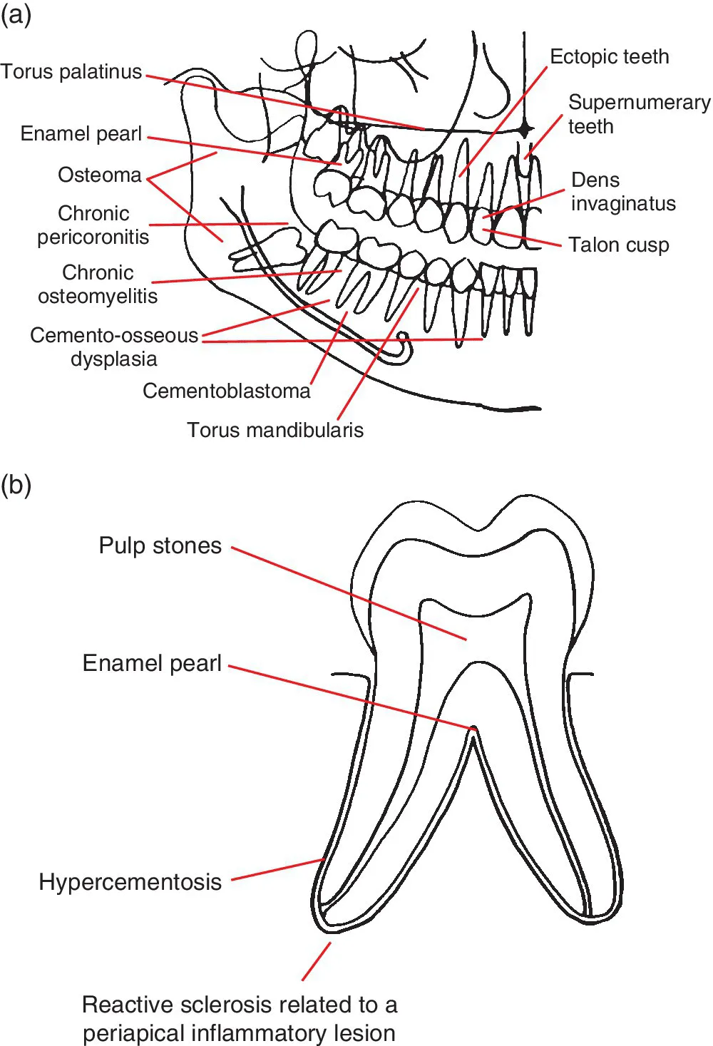
Figure 1.1 (a) Representation of the jaws and teeth and (b) larger representation of the fully erupted tooth. Conditions that have a predilection for certain regions of the jaws and teeth are shown. Note: (1) These lesions are not necessarily more common than other conditions. See the text for lists of common and less common conditions. (2) Most of these lesions also occur elsewhere within the jaws. (3) The pointers identify a region, not a specific site.
1.2 Lucent lesions of the jaws
For conditions affecting the TMJ, nasal cavity, paranasal sinuses, upper airway morphology, skull base and cervical spine, please refer to the dedicated chapters.
Common conditions
- Caries (see section 4.1)
- Periodontal bone loss (see section 5....
Table of contents
- Cover
- Title Page
- Table of Contents
- List of Contributors
- Preface
- Acknowledgements
- How to Use This Atlas
- CHAPTER 1: Problem Solving Diagrams
- CHAPTER 2: Radiological Anatomy
- CHAPTER 3: Anomalies Related to the Teeth
- CHAPTER 4: Conditions Related to Loss of Tooth Structure
- CHAPTER 5: Inflammatory Lesions of the Jaws
- CHAPTER 6: Osteoradionecrosis and Osteonecrosis of the Jaws
- CHAPTER 7: Hamartomatous/Hyperplastic Bony Opacities and Prominences Involving the Jaws
- CHAPTER 8: Cysts and Cyst‐like Lesions Involving the Jaws
- CHAPTER 9: Fibro‐osseous Lesions of the Jaws
- CHAPTER 10: Benign Tumours Involving the Jaws
- CHAPTER 11: Malignant Tumours Involving the Jaws
- CHAPTER 12: Vascular Anomalies of the Mid‐ and Lower Face
- CHAPTER 13: Other Diseases Affecting the Jaws
- CHAPTER 14: Other Morphological Anomalies Involving the Jaws
- CHAPTER 15: Other Systemic Disorders that may Involve the Jaws
- CHAPTER 16: Common Opacities in the Orofacial Soft Tissues
- CHAPTER 17: Trauma and Fractures
- CHAPTER 18: Temporomandibular Joints
- CHAPTER 19: Nasal Cavity, Paranasal Sinuses and Upper Aerodigestive Tract Impressions
- CHAPTER 20: The Skull Base
- CHAPTER 21: The Cervical Spine
- Index
- End User License Agreement
Frequently asked questions
Yes, you can cancel anytime from the Subscription tab in your account settings on the Perlego website. Your subscription will stay active until the end of your current billing period. Learn how to cancel your subscription
No, books cannot be downloaded as external files, such as PDFs, for use outside of Perlego. However, you can download books within the Perlego app for offline reading on mobile or tablet. Learn how to download books offline
Perlego offers two plans: Essential and Complete
- Essential is ideal for learners and professionals who enjoy exploring a wide range of subjects. Access the Essential Library with 800,000+ trusted titles and best-sellers across business, personal growth, and the humanities. Includes unlimited reading time and Standard Read Aloud voice.
- Complete: Perfect for advanced learners and researchers needing full, unrestricted access. Unlock 1.5M+ books across hundreds of subjects, including academic and specialized titles. The Complete Plan also includes advanced features like Premium Read Aloud and Research Assistant.
We are an online textbook subscription service, where you can get access to an entire online library for less than the price of a single book per month. With over 1.5 million books across 990+ topics, we’ve got you covered! Learn about our mission
Look out for the read-aloud symbol on your next book to see if you can listen to it. The read-aloud tool reads text aloud for you, highlighting the text as it is being read. You can pause it, speed it up and slow it down. Learn more about Read Aloud
Yes! You can use the Perlego app on both iOS and Android devices to read anytime, anywhere — even offline. Perfect for commutes or when you’re on the go.
Please note we cannot support devices running on iOS 13 and Android 7 or earlier. Learn more about using the app
Please note we cannot support devices running on iOS 13 and Android 7 or earlier. Learn more about using the app
Yes, you can access Atlas of Oral and Maxillofacial Radiology by Bernard Koong in PDF and/or ePUB format, as well as other popular books in Medicine & Dentistry. We have over 1.5 million books available in our catalogue for you to explore.