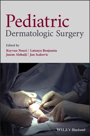
Pediatric Dermatologic Surgery
  1. English
  2. ePUB (mobile friendly)
  3. Available on iOS & Android
eBook - ePub
Book details
Book preview
Table of contents
Citations

About This Book

A complete guide to the surgical techniques used to treat childhood skin conditions

Recent advances have expanded the role of pediatric dermatologic surgery in both specialist and primary care settings. However, such surgeries can pose unique challenges to trainees and experienced practitioners alike. Procedures are carried out under local anesthesia and can be a source of distress and concern among young patients. Moreover, child's skin poses its own set of complicating factors, making the business of performing these procedures especially delicate and precise.

This bookprovides a step-by-step primer on invasive and non-invasive treatments of childhood skin disorders, offering concise and clearly illustrated guidance on current methods and best practices. Addressing conditions' effects, the impact of recent developments in their treatment, the ethics of operative procedures on children, and multiple treatment options for childhood dermatologic disease, Pediatric Dermatologic Surgery is an indispensable resource for trainee dermatologists and pediatricians, as well as practicing specialists.

Frequently asked questions

Simply head over to the account section in settings and click on “Cancel Subscription” - it’s as simple as that. After you cancel, your membership will stay active for the remainder of the time you’ve paid for. Learn more here.
At the moment all of our mobile-responsive ePub books are available to download via the app. Most of our PDFs are also available to download and we're working on making the final remaining ones downloadable now. Learn more here.
Both plans give you full access to the library and all of Perlego’s features. The only differences are the price and subscription period: With the annual plan you’ll save around 30% compared to 12 months on the monthly plan.
We are an online textbook subscription service, where you can get access to an entire online library for less than the price of a single book per month. With over 1 million books across 1000+ topics, we’ve got you covered! Learn more here.
Look out for the read-aloud symbol on your next book to see if you can listen to it. The read-aloud tool reads text aloud for you, highlighting the text as it is being read. You can pause it, speed it up and slow it down. Learn more here.
Yes, you can access Pediatric Dermatologic Surgery by Keyvan Nouri, Latanya Benjamin, Jasem Alshaiji, Jan Izakovic, Keyvan Nouri, Latanya Benjamin, Jasem Alshaiji, Jan Izakovic in PDF and/or ePUB format, as well as other popular books in Medicine & Dermatology. We have over one million books available in our catalogue for you to explore.

Information

Year
2019
ISBN
9781118769782
Edition
1
Subtopic
Dermatology

1
The Embryogenesis of the Skin

Jasem Alshaiji
Department of Dermatology, Amiri Hospital, Kuwait City, Kuwait

1.1 Introduction

The skin is complex tissue and is the biggest constantly renewable organ in the body. It has many different cell types and specialized structures (such as hair, nails, and glands) derived from both embryonic ectoderm and mesoderm. Skin cell types that originate from ectoderm include keratinocytes, neural crest‐derived melanocytes, Merkel cells, and neurons, whereas skin cell types that originate from mesoderm include fibroblasts, endothelial cells, adipocytes and bone marrow‐derived Langerhans cells [1].
There are two distinct dating systems, one refers to estimated gestational age (EGA) and is used in textbooks and by researchers, in which fertilization occurs on day 1 (first day of the last menstrual period (LMP)), whereas menstrual age, used by obstetricians and most clinicians, has fertilization occurring on day 14 [2, 3].
Fetal skin development can be divided into three temporally overlapping stages: organogenesis (specification), histogenesis (morphogenesis) and maturation (differentiation) [4]. These stages roughly correspond to the embryonic period (0–60 days), the early fetal period (2–5 months), and the late fetal period (5–9 months) of development. The first stage involves the specification of ectoderm lateral to the neural plate to become epidermis and the allocation of subsets of mesenchymal and neural crest cells to become dermis. The second stage is the process by which these committed tissues begin to form their specialized structures, including epidermal stratification, epidermal appendage formation, subdivision between the dermis and subcutis, and vascular formation. The third stage refers to the process by which these newly specialized tissues further differentiate and assume their mature forms [1].
Understanding the stages of normal human skin development allows the definition of critical periods when the skin may be more vulnerable to developmental errors; it provides an opportunity to study the evolution of skin function, establishing a background for understanding the natural history of expression of genetic skin disease in its earliest form; and it provides the essential information for the evaluation of skin samples used in the prenatal diagnosis of genodermatoses for which molecular methods are still not adaptable [5].
The timing at which sampling of the skin for various diagnostic procedures are performed should be recognized. Chorionic villus sampling for fetal DNA is sampled around 10 weeks EGA, amniotic fluid cells (amniocentesis) can be obtained at around 14–16 weeks EGA, whereas fetal skin biopsy is performed typically at 19–21 weeks EGA [5–8].

1.2 Stages of Skin Development

1.2.1 Embryonic Stage (Specification)

During the third week after fertilization, the human embryo undergoes gastrulation, a complex process of involution and cell redistribution that results in the formation of the three primary embryonic germ layers: ectoderm, mesoderm, and endoderm. Shortly after gastrulation, ectoderm further subdivides into neuroectoderm and presumptive epidermis [1].

1.2.1.1 Epidermis

At six weeks EGA, the epidermis that covers most of the embryo are two‐layered epithelium consisting of basal cells and periderm cells [9–11]. The basal cells are more columnar and have intercellular attachment mediated by E‐ and P‐cadherin and they express keratins K5, K14, K8 and K19 [12–14]. The periderm cells are larger and flatter than underlying basal cells and they express K5, K14, K8, K18 and K19 [13–16]. Their apical surfaces are studded with microvilli and tight junctions attach them at their lateral surfaces [11]. Two of the immigrant cells, melanocytes and Langerhans cells, are present in the embryonic epidermis among basal cells. Melanocytes are dendritic as early as 50 days EGA in general body skin but there is no evidence of melanosomes in the cytoplasm [17]. Langerhans cells are recognized in embryonic skin as early as 42 days EGA and they have dendritic morphology and probably derived from yolk sac or fetal liver at this age [18–20]. The third immigrant cells, Merkel cells, can be seen in embryonic palmar skin as early as 55–60 days EGA and they express K8, K18, K10 and K20 [21–25]. K20 is the only keratin found exclusively in Merkel cells. They are distributed randomly and in a suprabasal position. They are neuroendocrine cells acting as slow‐adapting mechanoreceptors. It is generally accepted that Merkel cells are derived from keratinocytes in situ [21, 23, 25–27].

1.2.1.2 Dermis

The embryonic dermis is highly cellular and amorphous, consisting of a loose network of mesenchymal cells (fibroblasts, mast cells and skin‐derived precursor‐SKP‐cells) with little intervening fibrous connective tissue matrix, and its origin varies depend on the body site [28]. Dermal mesenchyme of the face and anterior scalp is derived from neural crest ectoderm; the limb and ventral body wall mesenchyme is derived from the lateral plate mesoderm, whereas the dorsal body wall mesenchyme derives from the dermomyotomes of the embryonic somite [29].
The cell migration is promoted through the high water content and hyaluronic‐acid‐rich environment of the dermal mesenchyme, whereas the exchange of signals between epidermis and dermis is achieved through the compact mesenchyme and is very important in stimulating the onset of appendage formation [5, 30].
Types I, III and VI collagen are distributed uniformly throughout the dermis whereas type V collagen is concentrated primarily along basement membranes and surrounding cells. The ratio of collagen III to collagen I is 3:1, the opposite what it is in the adult [31].
The embryonic dermis does not contain elastic fibers yet, but fibrillin and elastin proteins of the elastic fibers can be identified by immunohistochemistry and microfibrils can be seen by electron microscopy (EM) [11]. Blood vessels have been identified in fetal skin through the process of vasculogenesis (de novo) as early as nine weeks EGA and its pattern varies among different regions of the body [32]. Development of the cutaneous innervation closely parallels that of the vascular system in term of its pattern, rate of maturation, and organization. Cutaneous nerves are composed of somatic sensory fibers, which can be identified at seven weeks EGA, and sympathetic autonomic fibers, which are not yet recognized [33].

1.2.1.3 Dermoepidermal Junction (DEJ)

The Dermoepidermal Junction (DEJ) connects the developing epidermis (the basal cells) to the dermis and it provides resistance against shearing forces ...

Table of contents

  1. Cover
  2. Table of Contents
  3. List of Contributors
  4. 1 The Embryogenesis of the Skin
  5. 2 Basic Structure and Function of the Neonatal, Infantile, and Childhood Skin
  6. 3 Approach to the Child as a Patient
  7. 4 Preoperative and Postoperative Care of Children Undergoing Pediatric Dermatology Procedures
  8. 5 The Pediatric Surgical Tray
  9. 6 Anesthesia for Children
  10. 7 Skin Biopsy Techniques
  11. 8 Common Pediatric Dermatologic Surgery Procedures
  12. 9 Suturing Techniques
  13. 10 Wound Closure Technique
  14. 11 Wound Closure Material
  15. 12 Dressings
  16. 13 Nail Surgery
  17. 14 Complications of Surgery and Invasive Procedures
  18. 15 Improving Scars
  19. 16 Special Dermatologic Surgery
  20. 17 Lasers for Vascular Lesions
  21. 18 Lasers for Scars and Striae
  22. 19 Lasers for Acne
  23. 20 Laser for Verrucae
  24. 21 Lasers and Lights for Onychomycosis
  25. 22 Lasers for Pigmented Lesions
  26. 23 Lasers for Tattoos
  27. 24 Lasers for Hair Removal
  28. 25 Lasers for Other Specific Dermatologic Disorders
  29. 26 Medical Ethics and Bioethics in Pediatric Dermatology
  30. Index
  31. End User License Agreement