Topographical and Pathotopographical Medical Atlas of the Chest, Abdomen, Lumbar Region, and Retroperitoneal Space
eBook - ePub

Topographical and Pathotopographical Medical Atlas of the Chest, Abdomen, Lumbar Region, and Retroperitoneal Space

  1. English
  2. ePUB (mobile friendly)
  3. Available on iOS & Android
eBook - ePub

Topographical and Pathotopographical Medical Atlas of the Chest, Abdomen, Lumbar Region, and Retroperitoneal Space

Book details
Book preview
Table of contents
Citations

About This Book

The third medical atlas in this new series on the human body and filled with detailed pictures, this atlas details the topographical and pathotopographical anatomy of the chest, abdomen, lumbar region, and retroperitoneal space, a useful reference for medical professionals and students alike.

Written by an experienced and well-respected physician and professor, this new volume, building on the previous volume, Ultrasonic Topographical and Pathotopographical Anatomy, and its sequel, Topographical and Pathotopographical Medical Atlas of the Head and Neck, also available from Wiley-Scrivener, presents the ultrasonic topographical and pathotopographical anatomy of the chest, abdomen, lumbar region, and retroperitoneal space, offering further detail into these important areas for use by medical professionals.

This series of atlases of topographic and pathotopographic human anatomy is a fundamental and practically important series designed for doctors of all specializations and students of medical schools. Here you can find almost everything that is connected with the topographic and pathotopographic human anatomy, including original graphs of logical structures of topographic anatomy and development of congenital abnormalities, topography of different areas in layers, pathotopography, and computer and magnetic resonance imaging (MRI) of topographic and pathotopographic anatomy. Also you can find here new theoretical and practical sections of topographic anatomy developed by the author himself which are published for the first time. They are practically important for mastering the technique of operative interventions and denying the possibility of iatrogenic complications during operations.

This important new volume will be valuable to physicians, junior physicians, medical residents, lecturers in medicine, and medical students alike, either as a textbook or as a reference. It is a must-have for any physician's library.

Frequently asked questions

Simply head over to the account section in settings and click on “Cancel Subscription” - it’s as simple as that. After you cancel, your membership will stay active for the remainder of the time you’ve paid for. Learn more here.
At the moment all of our mobile-responsive ePub books are available to download via the app. Most of our PDFs are also available to download and we're working on making the final remaining ones downloadable now. Learn more here.
Both plans give you full access to the library and all of Perlego’s features. The only differences are the price and subscription period: With the annual plan you’ll save around 30% compared to 12 months on the monthly plan.
We are an online textbook subscription service, where you can get access to an entire online library for less than the price of a single book per month. With over 1 million books across 1000+ topics, we’ve got you covered! Learn more here.
Look out for the read-aloud symbol on your next book to see if you can listen to it. The read-aloud tool reads text aloud for you, highlighting the text as it is being read. You can pause it, speed it up and slow it down. Learn more here.
Yes, you can access Topographical and Pathotopographical Medical Atlas of the Chest, Abdomen, Lumbar Region, and Retroperitoneal Space by Z. M. Seagal in PDF and/or ePUB format, as well as other popular books in Medicine & Anatomy. We have over one million books available in our catalogue for you to explore.

Information

Year
2018
ISBN
9781119526711
Edition
1
Subtopic
Anatomy

Part 1
The Chest

Topographic Anatomy of the Chest

Chest borders. The chest walls (paries thoracis) and chest cavity (cavum thoracis) together compose the chest (thorax). The superior chest border runs along the upper edge of the clavicle and the manubrium of sternum, and on the back — along the horizontal line drawn through the spinous process of the 7th cervical vertebra. The lower border goes down obliquely from the xiphoid process along the costal arches and on the back along the 12th rib and the spinous process of the 12th thoracic vertebra. The muscular-fascial layer of the chest is presented at the back with the latissimus dorsi muscle, on the sides with the serratus anterior muscles, and in front with the major and minor pectoral muscles. External and internal intercostal muscles are located in the chest itself; the space between these muscles is filled with cellular tissue with intercostal arteries, veins and nerves. The superior chest aperture (apertura thoracis superior) is bounded by the posterior surface of the manubrium of the sternum, the inner edges of the first ribs and the first thoracic vertebra. The inferior chest aperture (aperture thoracis inferior) is bounded by the posterior surface of the xiphoid process, the lower margins of the costal arches and the 10th thoracic vertebra anteriorly.
The prethoracic, thoracic, inframammary, scapular, subscapular and vertebral regions are identified.

Chest Cavity Organs Projection and Layers of Chest

Pleura projection (Figure 1). Lower pleural margins go on the midclavicular line — along the 7th rib; on the anterior axillary line — along the 8th rib; on the midaxillary line — along the 10th rib; on the scapular line — along the 11th rib; on the paraspinal line — until the 12th thoracic vertebra. Posterior margins correspond to costovertebral joints. The cervical pleura overhang the collar bone and correspond to the level of the spinous process of the 7th clervical vertebra posteriorly and anteriorly it is projected 2-3 cm above the collar bone.
Figure 1 Transverse section of the chest. Diaphragm.
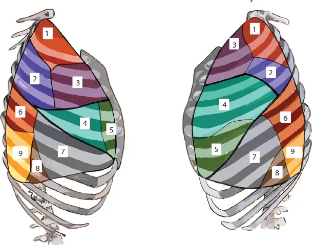
Lung projection (Figure 2). The anterior margin of the left lung starts from the 4th costal cartilage. Then, because of the cardiac notch, it slants to the left midclavicular line. The lower margins of the lungs correspond to the 6th costal cartilage on the right sternal line and on the left parasternal line: on the midclavicular line — to the upper margin of the 7th rib; on the anterior axillary line — to the lower margin of the 7th rib; on the midaxillary line — to the 8th rib; on the scapular line — to the 10th rib, and on the parasinal line — to the 11th rib. The lung margin moves down in inhale. The lung apex is identified 3-4 cm above the collar bone.
Figure 2 Lung segments.
Lobus superior: 1 – seg. apicale; 2 – seg. posterius; 3 – seg. anterius. Lobus medius: 4 – seg. laterale (правого легкого) et seg. lingulare superius (left lung); 5 – seg. mediale (right lung) et seg. lingulare inferius (left lung). Lobus inferior: 6 – seg. apicale; 7 – seg. basale anterius; 8 – seg. basale laterale; 9 – seg. basale posterius
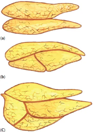
Thymus (Figures 3, 4) is located in the superior interpleural space. Superiorly it borders on the jugular notch of the sternum, above the level of the 2nd rib; on the sides — with the parietal pleura margins.
Figure 3 Thymus gland and its connection with the thyroid gland.
Figure 4 Differences in the shape and number of thymus glands.
a – two lobes, b – three lobes, c – four lobes.
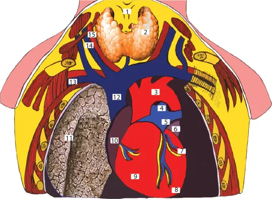
Heart projection (Figure 5). Upper margin of the heart matches a horizontal line, drawn at the level of the 3rd costal cartilage insertion to the breast bone. The right margin is a line, connecting the upper edge of the 2nd rib on the right with the upper edge of the 3rd rib 1 cm to the right of the breast bone; then it continues in the form of the arch from the 3rd to the 5th ribs, as a bulge, heading to the right, at a distance of 1.5 cm from the right edge of the breast bone. The lower margin starts from the place where the 5th rib is attached on the right, through the metasternum base to the fifth intercostal space on the left, stopping short 1.5 cm from the midclavicular line.
Figure 5 Topography of blood vessels, heart, right lung.
1 – larynx; 2 – gl. thyroidea; 3 – arcus aortae; 4 – truncus arteriosus; 5 – truncus pulmonalis; 6 – auricula sinistra; 7 – ventriculus sinister; 8 – apex cordis; 9 – ventriculus dexter; 10 – atrium dextrum; 11 – pulmo dextrum; 12 – v. cava superior; 13 – v. subclavia dextra; 14 – v. jugularis interna; 15 – a. carotis communis dextra.
The left margin is a line connecting the lower edge of the 1st rib on the left and the upper edge of the 2nd rib where they are attached to the breast bone, at the level of the 2nd intercostal space 2.5 cm to the left of the breast bone edge, then up to the point, placed 1.5-2 cm inwards the midclavicular line. The apex of the heart is projected on the left in the 5th intercostal sp...

Table of contents

  1. Cover
  2. Title page
  3. Copyright page
  4. Preface
  5. Part 1: The Chest
  6. Part 2: Abdomen
  7. Part 3: Lumbar Region and Retroperitoneal Space
  8. Part 4: Pathotography Chest
  9. About the Author
  10. End User License Agreement