- English
- ePUB (mobile friendly)
- Available on iOS & Android
Dermatology
About this book
"A very well written introductory dermatology text with excellent clinical photographs and diagrams. We would highly recommend this for those wishing to grasp the basic concepts in dermatology."
—British Journal of Dermatology
Dermatology Lecture Notes presents an accessible overview of skin structure and function, along with the practical aspects of disease management. Now in its 11th edition, it has been thoroughly updated to focus on recent advances in the knowledge of skin diseases and their treatment. It combines readability with high quality illustrations, and is the ideal guide for new comers to the specialty as well as those more advanced in their studies.
Key features include:
- An overview of the basics of skin structure and function, as well as practical aspects of disease management
- Excellent clinical photographs, diagrams and histological images
- Newly expanded and updated sections on benign skin tumours, viruses, emergency dermatology (skin failure in particular) and vascular disorders
- Includes a companion website at www.lecturenoteseries.com/dermatology featuring self-assessment and case studies
With beautiful colour artwork throughout, Dermatology Lecture Notes includes a glossary of dermatological terms, and provides an excellent balance between theory and clinical relevance.
Tools to learn more effectively

Saving Books

Keyword Search

Annotating Text

Listen to it instead
Information
1
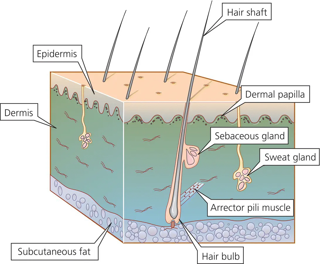
Structure and function of the skin, hair and nails
Skin structure
The epidermis
Keratinocytes
Basal layer
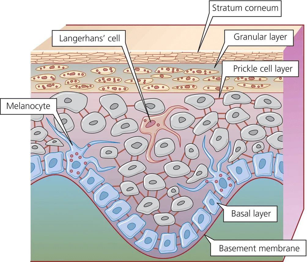
Melanocytes
Prickle cell layer
Granular cell layer
Table of contents
- Cover
- Title Page
- Table of Contents
- Preface
- Acknowledgements
- About the companion website
- 1 Structure and function of the skin, hair and nails
- 2 Approach to the diagnosis of dermatological disease
- 3 Emergency dermatology
- 4 Bacterial and viral infections
- 5 Fungal infections
- 6 Ectoparasite infections
- 7 Acne, acneiform eruptions and rosacea
- 8 Eczema
- 9 Psoriasis
- 10 Benign and malignant skin tumours
- 11 Naevi
- 12 Inherited disorders
- 13 Pigmentary disorders
- 14 Disorders of the hair and nails
- 15 Bullous disorders
- 16 Miscellaneous erythematous and papulosquamous disorders, and light-induced skin diseases
- 17 Vascular disorders
- 18 Connective tissue diseases
- 19 Pruritus
- 20 Systemic disease and the skin
- 21 Skin and the psyche
- 22 Cutaneous drug reactions
- 23 Treatment of skin disease
- Glossary of dermatological terms
- Index
- End User License Agreement
Frequently asked questions
- Essential is ideal for learners and professionals who enjoy exploring a wide range of subjects. Access the Essential Library with 800,000+ trusted titles and best-sellers across business, personal growth, and the humanities. Includes unlimited reading time and Standard Read Aloud voice.
- Complete: Perfect for advanced learners and researchers needing full, unrestricted access. Unlock 1.4M+ books across hundreds of subjects, including academic and specialized titles. The Complete Plan also includes advanced features like Premium Read Aloud and Research Assistant.
Please note we cannot support devices running on iOS 13 and Android 7 or earlier. Learn more about using the app