A unique reference dedicated to the diagnosis and treatment of problems of the equine neck and back
Building on the strength of the first edition, Equine Neck and Back Pathology: Diagnosis and Treatment, Second Edition explores conditions and problems of the horse's back and pelvis, and has been expanded to include coverage of the neck as well. This book is a vital tool for all those engaged in improving the diagnosis and management of horses with neck or back problems.
The only book devoted to the conditions and problems of the equine neck, back and pelvis, it provides comprehensive coverage by international specialists on how to diagnose and treat problems in these areas. This updated and revised edition covers normal anatomy and kinematics, neck and back pathology, diagnosis and treatment of specific conditions, and complementary therapy and rehabilitation.
Equine Neck and Back Pathology: Diagnosis and Treatment, Second Edition is a valuable working resource for equine practitioners, specialists in equine surgery, veterinary nurses and allied professionals involved in treating horses. It is also an excellent supplementary text for veterinary students with a keen interest in horses.
Trusted by 375,005 students
Access to over 1.5 million titles for a fair monthly price.
The neck is a common derived characteristic of land vertebrates, not shared by their aquatic ancestors. In fish, the thoracic fin girdle, the precursor of the scapula, coracoid and clavicle, is frequently fused to the caudal aspect of the skull. In contrast, as vertebrates emerged on to the dry land, the forelimb separated from the head and the intervening vertebrae specialised to form a relatively mobile region – the neck – to allow the head to be freely steered in many directions.
With the exception of the tail, the neck remains the most mobile region of the spinal column in modern-day horses. It permits a wide range of sagittal plane flexion and extension to allow alternating periods of grazing and predator surveillance, as well as frontal plane flexion to allow the horizon to be scanned, and rotational movement to allow nuisance insects to be flicked off. Among domestic animals the equine neck is relatively long and the head relatively heavy, and so the neck has become strong, muscular and massive. This is enhanced by the fact that regular, forceful movements of this region must also occur to maintain balance when horses are running [1]. However, the length and flexible nature of the neck may also cause problems in the passage of foals through the birth canal.
In this chapter I will briefly review the anatomy of bones, joints, ligaments and muscles of the equine neck. The ‘locomotor component’ of the neck is a common site of pathology, and the diverse forms of neck disease reflect the sometimes complex and conflicting regional variations and functional constraints so evident in this region [2].
Unlike the abdomen and thorax, there is no coelomic cavity in the neck, yet its ventral part is taken up by a relatively small ‘visceral compartment’, containing the larynx, trachea, oesophagus and many important vessels, nerves and endocrine glands. However, I will not review these structures, as they do not represent an extension of the equine ‘back’ in the same way that the more dorsal locomotor region does.
Cervical Vertebrae 3–7
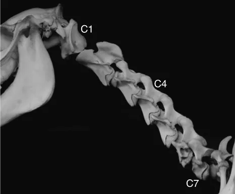
Almost all mammals, including the horse, possess seven cervical vertebrae, C1 to C7 (Figure 1.1). While C1 and C2 are extremely modified for their particular functions, C3 to C7 are more homogenous in structure. C3, C4 and C5 in particular are usually thought of as the ‘typical’ cervical vertebrae (Figure 1.2).
Figure 1.1 Lateral view of an articulated osteological preparation of the neck of a young horse.
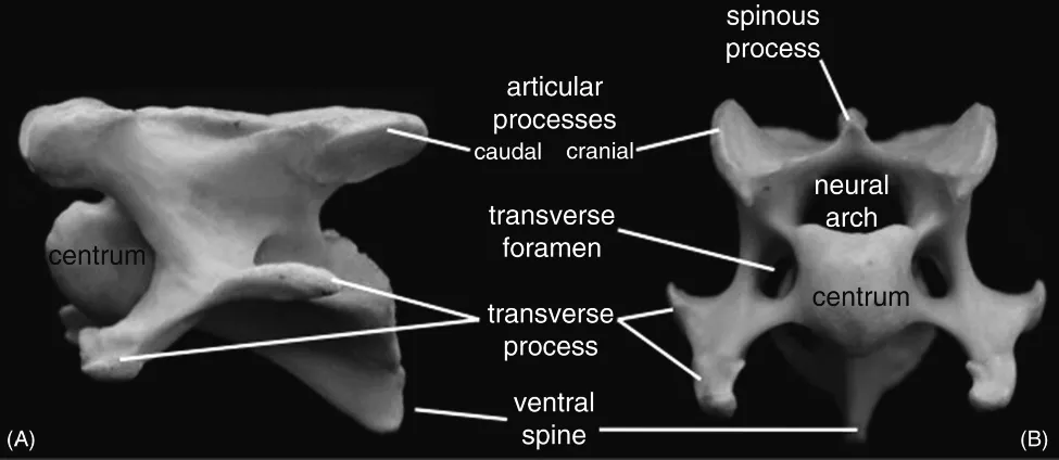
Figure 1.2 (A) Lateral view of equine C4 vertebra and (B) cranial view of C5.
Vertebrae C3–C7 consist of an approximately cylindrical body or centrum, a structure present in all jawed vertebrates to resist longitudinal compression of the spinal column. The centra of the equine neck are the longest in the body, but become progressively smaller caudally. Those of C3–C7 possess a distinctively convex cranial surface, the head, and a correspondingly concave caudal surface, the fossa. Thus the intervertebral joints, which are far more mobile than in the trunk, may be thought of functionally as ball-and-socket joints, although their constituent parts are very structurally different from those of synovial ball-and-socket joints.
Dorsal to the centrum is the neural arch, formed from bilateral bony laminae, which surrounds and protects the spinal cord and its associated structures. The vertebral canal formed by successive arches is relatively wide in the neck, especially cranially, to allow the spinal cord, which is wide in this region, to flex freely. The vertebrae C3–C7 each develop from three primary centres of ossification – one in the centrum and one in each of the two laminae. Formation of cervical neural arches, which are either statically or dynamically stenotic, is thought to be a cause of equine cervical ‘wobbler syndrome’ [3].
The centrum and arch are adorned with a variety of bony processes for the attachment of ligaments and muscles, and which often develop as secondary centres of ossification. These vertebral processes are a feature evolved by land vertebrates to permit complex movements in three dimensions and resist torsional forces.
The single dorsal midline spinous process is distinctively short in equine C3–C5.
In contrast, all equine cervical vertebrae bear a characteristically large ventral crest, often with a pronounced caudal tubercle.
The bilateral transverse processes are large but squat, and thought to incorporate vestigial ribs, sometimes yielding the name ‘costotransverse processes’. In C3–6, the processes are bifid and slanted, with a cranial ventral tubercle and caudal dorsal tubercle. The transverse processes of C1–6 are perforated by a large transverse foramen, which conveys the vertebral artery and vein.
Lateral to the neural arch lie the large, irregular articular processes, with their smooth ovoid articular surfaces. The caudal facets are directed ventrolaterally, and the complementary cranial facets dorsomedially. Ventral to the caudal process lies a notch for passage of the laterally coursing spinal nerve.
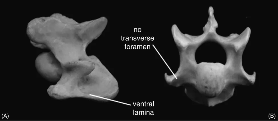
The sixth cervical vertebra (Figure 1.3) differs from its cranial neighbours in that it bears pronounced paired bony sheets, the ventral laminae, which act as a site of attachment and force redirection of muscles, especially longus colli. In the horse these laminae are elaborated into cranial and caudal tubercles. C6 also possesses a longer spinous process than C3, 4 and 5 – a reflection of a gradual transition to a more ‘thoracic’ morphology.
Figure 1.3 (A) Lateral view of equine C6 vertebra and (B) cranial view of C7.
This trend continues in C7 (Figure 1.3), which has an even longer spinous process, non-bifid transverse processes, and no transverse foramen – the vertebral arteries arise too far cranially to pass through C7. However, C7 does possess a caudal notch for the passage of a spinal nerve, but it should be emphasised that the nomenclature of the spinal nerves in inconsistent. Unlike the rest of the body, cervical spinal nerves emerge cranial to the vertebra of the same number, and the nerves emerging caudal to vertebra C7 are named C8, even though there is no corresponding C8 vertebra. Finally, the centra of C7 caudally bear unconvincing costal facets for the articulation of the cranial extremities of the capitula of the first ribs [4].
Atlas and Axis, C1 and C2
The anatomy of the caudal part of the axis, C2 (Figure 1.4), is similar to that of the more caudal cervical vertebrae – with centrum and neural arch formed from the same three centres of ossification, as well as the spinous process, ventral crest and tubercle, caudal articular facets and dorsal tubercle of the transverse process. However, the cranial part of the bone is markedly aberrant to allow the unique rotational, trochoid, ‘head-shaking’ movement of the atlanto-axial joint. Its unusual shape results from the incorporation of embryonic elements of C1.
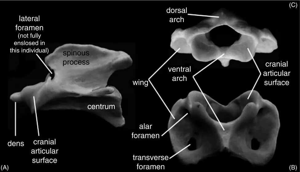
Figure 1.4 (A) Lateral view of equine C2 vertebra, and (B) ventral and (C) cranial views of C1.
A fourth primary centre of ossification, actually the annexed centrum of C1, forms the dens (‘tooth’) or odontoid process of C2. This cranially directed process is attached ventrally to the main centrum of C2 by a base formed from a further, secondary centre, which represents the cranial epiphysis of C2. The dens articulates closely with the ventral part of C1, and thus is smooth on its ventral surface, but is roughened dorsally with a midline gutter to allow attachment of stabilising ligaments. The smooth articular region of the dens is continuous with the large bilateral saddle-...
Table of contents
Cover
Title Page
Copyright
Dedication
List of Contributors
Chapter 1: The Normal Anatomy of the Neck
Chapter 2: The Normal Anatomy of the Osseous and Soft Tissue Structures of the Back and Pelvis
Chapter 3: The Normal Anatomy of the Nervous System
Chapter 4: Kinematics
Chapter 5: Neurological Examination of the Back and Pelvis
Chapter 6: Clinical Examination
Chapter 7: Radiography of the Cervical Spine
Chapter 8: Radiography of the Back
Chapter 9: Nuclear Scintigraphy and Computed Tomography of the Neck, Back and Pelvis
Chapter 10: Ultrasonography
Chapter 11: Thermography
Chapter 12: Neck Pathology
Chapter 13: Back Pathology
Chapter 14: Sacroiliac Dysfunction
Chapter 15: Muscular Disorders
Chapter 16: Integrative and Physical Therapies
Chapter 17: Rehabilitation
Index
End User License Agreement
Frequently asked questions
Yes, you can cancel anytime from the Subscription tab in your account settings on the Perlego website. Your subscription will stay active until the end of your current billing period. Learn how to cancel your subscription
No, books cannot be downloaded as external files, such as PDFs, for use outside of Perlego. However, you can download books within the Perlego app for offline reading on mobile or tablet. Learn how to download books offline
Perlego offers two plans: Essential and Complete
Essential is ideal for learners and professionals who enjoy exploring a wide range of subjects. Access the Essential Library with 800,000+ trusted titles and best-sellers across business, personal growth, and the humanities. Includes unlimited reading time and Standard Read Aloud voice.
Complete: Perfect for advanced learners and researchers needing full, unrestricted access. Unlock 1.5M+ books across hundreds of subjects, including academic and specialized titles. The Complete Plan also includes advanced features like Premium Read Aloud and Research Assistant.
Both plans are available with monthly, semester, or annual billing cycles.
We are an online textbook subscription service, where you can get access to an entire online library for less than the price of a single book per month. With over 1.5 million books across 990+ topics, we’ve got you covered! Learn about our mission
Look out for the read-aloud symbol on your next book to see if you can listen to it. The read-aloud tool reads text aloud for you, highlighting the text as it is being read. You can pause it, speed it up and slow it down. Learn more about Read Aloud
Yes! You can use the Perlego app on both iOS and Android devices to read anytime, anywhere — even offline. Perfect for commutes or when you’re on the go. Please note we cannot support devices running on iOS 13 and Android 7 or earlier. Learn more about using the app
Yes, you can access Equine Neck and Back Pathology by Frances M. D. Henson in PDF and/or ePUB format, as well as other popular books in Medicine & Equine Veterinary Science. We have over 1.5 million books available in our catalogue for you to explore.