The Pediatric Cardiac Anesthesia Handbook
eBook - ePub

The Pediatric Cardiac Anesthesia Handbook

  1. English
  2. ePUB (mobile friendly)
  3. Available on iOS & Android
eBook - ePub

The Pediatric Cardiac Anesthesia Handbook

Book details
Book preview
Table of contents
Citations

About This Book

Written by an expert author team at the renowned Boston Children's Hospital, USA, The Pediatric Cardiac Anesthesia Handbook provides a comprehensive yet concise overview of the anesthetic management of pediatric patients with congenital heart disease.

This book is divided into two parts. The first part provides an introduction to the basic assessment of patients, including cardiovascular physiology, pathophysiology and the underlying concepts in coronary heart disease, preoperative evaluation, intraoperative management, and interpretation of cardiac catheterization data. The second part of the book addresses disorders and diseases in a templated manner covering the anatomical and physiological features, surgical therapies, anesthetic approach and postoperative management. Chapters on the heart and heart and lung transplantation consider the additional complexities of those patients and anesthetic considerations for non-cardiac surgery after heart transplantation.

The anesthesiologist caring for patients with congenital heart disease faces a myriad of challenges in the perioperative management of these complex individuals. This book provides:

  • A concise and easily referable guide ideal for use during anesthesiology residency training and fellowships.
  • A templated chapter layout ideal for easy referral by wider members of the multidisciplinary team, such as cardiologists, cardiac intensivists, perfusionists, and surgeons.
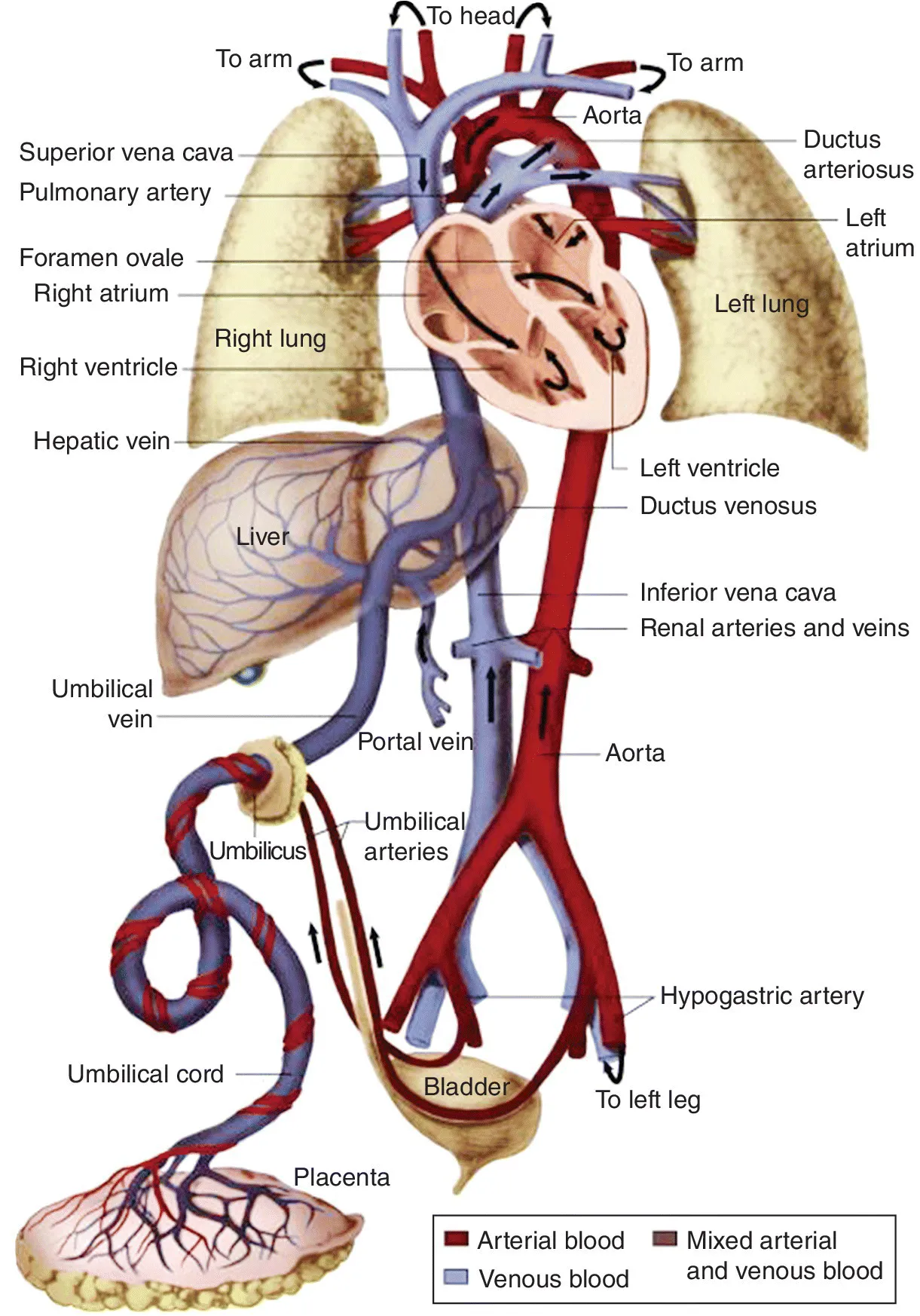
  • Helpful illustrations and a bulleted content for rapid reference.
  • Guidelines on specific lesions for the pediatric anesthesiologist caring for cardiac patients presenting for non-cardiac surgery.

This book is a valuable resource for all anesthesiology and cardiac critical care providers who manage patients with congenital heart disease, and an ideal study aid.

Frequently asked questions

Simply head over to the account section in settings and click on “Cancel Subscription” - it’s as simple as that. After you cancel, your membership will stay active for the remainder of the time you’ve paid for. Learn more here.
At the moment all of our mobile-responsive ePub books are available to download via the app. Most of our PDFs are also available to download and we're working on making the final remaining ones downloadable now. Learn more here.
Both plans give you full access to the library and all of Perlego’s features. The only differences are the price and subscription period: With the annual plan you’ll save around 30% compared to 12 months on the monthly plan.
We are an online textbook subscription service, where you can get access to an entire online library for less than the price of a single book per month. With over 1 million books across 1000+ topics, we’ve got you covered! Learn more here.
Look out for the read-aloud symbol on your next book to see if you can listen to it. The read-aloud tool reads text aloud for you, highlighting the text as it is being read. You can pause it, speed it up and slow it down. Learn more here.
Yes, you can access The Pediatric Cardiac Anesthesia Handbook by Viviane G. Nasr, James A. DiNardo in PDF and/or ePUB format, as well as other popular books in Medicine & Anesthesiology & Pain Management. We have over one million books available in our catalogue for you to explore.

Information

Year
2017
ISBN
9781119095545

Part I
The Basics

1
Cardiovascular Development

The incidence of congenital heart disease (CHD) is approximately 7 to 10 per 1000 live births. Most congenital heart defects are the result of an interaction of genetic predisposition and environmental factors. Environmental factors such as drugs, viral infection, maternal diabetes, or maternal alcohol abuse may account for specific lesions. Knowledge of cardiac development is a must to understand congenital heart lesions. This chapter reviews the embryology and cardiovascular physiology at birth.

Embryology

It is essential to understand the basic embryology and origin of the cardiac structures in order to appreciate the specific lesions described in the next section of the Handbook. It is beyond the scope of this Handbook to discuss the details of cardiac development, including : (i) cardiac sidedness or asymmetry; (ii) cardiac looping; (iii) formation of outflow tracts; and (iv) septation. The embryologic structures and their corresponding adult structures are listed in Table 1.1.
Table 1.1 Cardiovascular embryologic structure and the corresponding structures in adults.
Embryologic structureAdult structure
Truncus arteriosusAorta
Pulmonary trunk
Bulbus cordisSmooth part of right ventricle (conus arteriosus)
Smooth part of left ventricle (aortic vestibule)
Primitive ventricleTrabeculated part of right ventricle
Trabeculated part of left ventricle
Primitive atriumTrabeculated part of right atrium
Trabeculated part of left atrium
Sinus venosusSmooth part of right atrium (sinus venarum)
Coronary sinus
Oblique vein of left atrium
Aortic arches
1*
2*
3Common carotid arteries
Internal carotid arteries (proximal part)
4Right subclavian artery (proximal part)
Part of the aortic arch
5Regresses in the human
6Pulmonary arteries (proximal part)
Ductus arteriosus

Cardiovascular Physiology

Circulatory changes occur at birth and continue over the first few days and the first months of life, and are considerable. They need to be appreciated in order to understand their profound effects on neonatal cardiovascular physiology. It is not coincidental that 50% of the neonates born with CHD will become ill enough during the first days or weeks of life to require medical or surgical intervention. Optimal perioperative and anesthetic management of the neonate with CHD must be based on a firm understanding of these developmental changes.

Fetal Circulation

Fetal circulatory channels shunt blood away from the lung such that both ventricles, in parallel, contribute to systemic oxygen delivery by pumping blood to the systemic arterial system. This parallel circulation permits normal fetal growth and development even in fetuses with cardiac malformations.
Oxygenated blood from the placenta returns to the fetus via the umbilical vein, which enters the portal venous system. The ductus venosus connects the left portal vein to the left hepatic vein at its junction with the inferior vena cava (IVC). This allows approximately 50% of umbilical venous blood to bypass the hepatic sinuses. The remainder of the umbilical venous flow passes through the liver and enters the IVC via the hepatic veins. Fetal IVC blood is a combination of blood from the lower fetal body, umbilical vein, and hepatic veins. The stream of blood from the ductus venosus has a higher velocity in the IVC than the stream from the lower body and hepatic veins. This higher velocity facilitates delivery of this higher‐oxygen content blood across the foramen ovale (FO) into the left atrium (LA) (Figure 1.1).
image
Figure 1.1 Course of the fetal circulation in late gestation. Note the selective blood flow patterns across the foramen ovale and the ductus arteriosus.
Reproduced from Greeley, W.J., Berkowitz, D.H., Nathan, A.T. (2010) Anesthesia for pediatric cardiac surgery, in Anesthesia, 7th edition (ed. R.D. Miller), Churchill Livingstone, Philadelphia.
The IVC blood enters the right atrium (RA) and, due to the position of the Eustachian valve, Chiari network and FO, enters the LA during 80% of the cardiac cycle. During the other 20% (atrial systole), IVC blood crosses the tricuspid valve and enters the right ventricle (RV). The overwhelming majority of superior vena cava (SVC) blood crosses the tricuspid valve and also enters the RV. Blood from the RV is ejected into the pulmonary artery (PA). Approximately 10–15% of blood from the PA passes through the lungs to reach the LA, and the rest is shunted to the distal aorta via the ductus arteriosus (DA). As a result, two‐thirds of the total fetal cardiac output is provided by the RV, with the remaining one‐third provided via the LV.
The dynamics of shunting at the level of the ductus venosus, FO, and DA result in a preferential delivery of the most highly oxygenated blood to the coronary and cerebral circulations. Obviously, this preferential delivery of oxygenated blood may be compromised in utero by cardiac lesions that prevent or reduce left ventricular output. At birth, a series circulation is established in which each ventricle pumps into a specific vascular bed (RV to ...

Table of contents

  1. Cover
  2. Title Page
  3. Table of Contents
  4. Preface
  5. Part I: The Basics
  6. Part II: Specific Lesions
  7. Index
  8. End User License Agreement