Matrix Metalloproteinase Biology
eBook - ePub

Matrix Metalloproteinase Biology

  1. English
  2. ePUB (mobile friendly)
  3. Available on iOS & Android
eBook - ePub

Matrix Metalloproteinase Biology

Book details
Book preview
Table of contents
Citations

About This Book

Discussing recent advances in the field of matrix metalloproteinase (MMP) research from a multidisciplinary perspective, Matrix Metalloproteinase Biology is a collection of chapters written by leaders in the field of MMPs. The book focuses on the challenges of understanding the mechanisms substrate degradation by MMPs, as well as how these enzymes are able to degrade large, highly ordered substrates such as collagen. All topics addressed are considered in relation to disease progression including roles in cancer metastasis, rheumatoid arthritis and other inflammatory diseases. The text first provides an overview of MMPs, focusing on the history, the development and failures of small molecule inhibitors in clinical trials, and work with TIMPS, the endogenous inhibitors of MMPs. These introductory chapters establish the foundation for later discussion of the recent progress on the design of different types of inhibitors, including novel antibody based therapeutics. The following section emphasizes research using novel methods to further the study of the MMPs. The third and final section focuses on in vivo research, particularly with respect to cancer models, degradation of the extracellular matrix, and MMP involvement in other disease states.

Written and edited by leaders in the field, Matrix Metalloproteinase Biology addresses the rapidly growth in MMP research, and will be an invaluable resource to advanced students and researchers studying cell and molecular biology.

Frequently asked questions

Simply head over to the account section in settings and click on “Cancel Subscription” - it’s as simple as that. After you cancel, your membership will stay active for the remainder of the time you’ve paid for. Learn more here.
At the moment all of our mobile-responsive ePub books are available to download via the app. Most of our PDFs are also available to download and we're working on making the final remaining ones downloadable now. Learn more here.
Both plans give you full access to the library and all of Perlego’s features. The only differences are the price and subscription period: With the annual plan you’ll save around 30% compared to 12 months on the monthly plan.
We are an online textbook subscription service, where you can get access to an entire online library for less than the price of a single book per month. With over 1 million books across 1000+ topics, we’ve got you covered! Learn more here.
Look out for the read-aloud symbol on your next book to see if you can listen to it. The read-aloud tool reads text aloud for you, highlighting the text as it is being read. You can pause it, speed it up and slow it down. Learn more here.
Yes, you can access Matrix Metalloproteinase Biology by Irit Sagi, Jean Gaffney in PDF and/or ePUB format, as well as other popular books in Biological Sciences & Biology. We have over one million books available in our catalogue for you to explore.

Information

Year
2015
ISBN
9781118772386
Edition
1

Chapter 1
Matrix Metalloproteinases: From Structure to Function

Maciej J. Stawikowski1 and Gregg B. Fields2
Departments of Chemistry and Biology, Torrey Pines Institute for Molecular Studies, Port St. Lucie, USA

1.1 Introduction

Members of the matrix metalloproteinase (MMP) family are known to catalyze the hydrolysis of a great variety of biological macromolecules. Proteomic approaches have significantly expanded the number of known MMP substrates. However, the mechanisms by which macromolecular substrates are processed have often proved elusive. X-ray crystallography and NMR spectroscopy have yielded detailed information on structures of MMP domains and, in a few cases, full-length MMPs. As structures of MMPs and their substrates have been reported, examination of MMP•substrate complexes has provided insight into mechanisms of action. We examine the structures of MMPs and their substrates and consider how the various structural elements of MMPs contribute to the hydrolysis of biological macromolecules.

1.2 Structures of MMPs

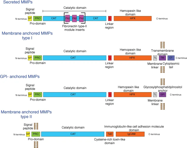
1.2.1 General MMP structure and domain organization

MMPs belong to the M10 zinc metalloproteinase family [1]. All MMPs have the characteristic zinc binding motif HExxHxxGxxH in their catalytic domain. MMPs possess similar domain organizations. Most MMPs consist of a signal peptide followed by four distinct domains, the N-terminal prodomain (propeptide), catalytic (CAT) domain, linker (hinge) region, and C-terminal hemopexin-like (HPX) domain (Fig. 1.1). The membrane-type (MT) MMPs contain an additional transmembrane (TM) domain that anchors them to the cell membrane. Following the TM domain is a small cytoplasmic “tail”.
c01f001
Figure 1.1 General domain organization of MMPs.
There are several exceptions to this general domain organization. MMP-7 and MMP-26 (matrilysins) lack the linker region and HPX domain and thus are referred to as “minimal MMPs”. MMP-2 and MMP-9 possess three repeats of fibronectin type II-like motifs within the CAT domain. MMP-17 and MMP-25 are type I TM enzymes anchored to membranes through a C-terminal glycosylphosphatidylinositol (GPI) residue [2]. The N-terminal MMP-23 pro-domain contains a type II TM domain that anchors the protein to the plasma membrane. Instead of the C-terminal HPX domain common to other MMPs, MMP-23 contains a small toxin-like domain (TxD) and an immunoglobulin-like cell adhesion molecule (IgCAM) domain.

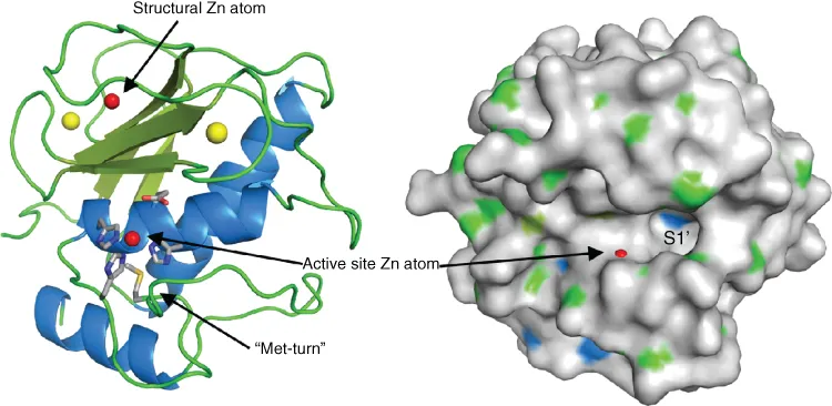
1.2.2 Catalytic domain

The topology of the CAT domain is similar among all MMPs. The CAT domain is composed of a five- stranded β-sheet which is interrupted by three α-helices (Fig. 1.2). Four of the five β-strands are aligned in a parallel fashion, while only the smallest “edge” strand runs in the opposite direction. Between strands III and IV there is an S-loop fixed by a structural Zn atom. The center of the catalytic site is located at helix B and the loop connecting it with helix C. This center helix provides the first and second His residues of the Zn-binding motif along with “catalytic” Glu residue. The loop behind this helix provides the third zinc binding His residue. Further down along this loop there is a 1,4 β-turn forming Met residue. This residue is highly conserved among metzincins and is believed essential for the structural integrity of the zinc-binding site. However, MMP-2 mutants where the conserved Met was replaced with Leu or Ser were able to cleave gelatin, type I collagen, and chemokine monocyte chemoattractant protein-3 with similar efficiency as wild-type MMP-2 [3].
c01f002
Figure 1.2 Typical structure of the CAT domain of MMPs. Characteristic structural elements are highlighted with arrows. Figure generated using MMP-8 structure (PDB 2OY2) [4].

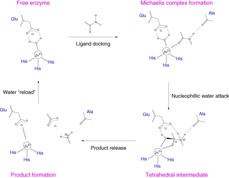
1.2.3 Catalytic mechanism

On the basis of early structural information, a catalytic mechanism for MMPs was proposed (Fig. 1.3) [5, 6]. The carbonyl group of the scissile bond coordinates to the active site zinc (II) ion. A water molecule is hydrogen bonded to a conserved Glu residue and coordinated to the zinc (II) ion. The water molecule donates a proton to the Glu residue, allowing the generated hydroxide ion to attack the carbonyl at the scissile bond. This attack results in a tetrahedral intermediate, which is stabilized by the zinc (II) ion. The Glu residue transfers a proton to the nitrogen of the scissile amide, the tetrahedral intermediate rearranges, and amide bond hydrolysis occurs. During this catalytic process, the carbonyl from a conserved Ala residue helps to stabilize the positive charge at the nitrogen of the scissile amide.
c01f003
Figure 1.3 Mechanism of proteolysis catalyzed by MMPs. (Figure prepared based on mechanism proposed by Lovejoy et al. [5]).

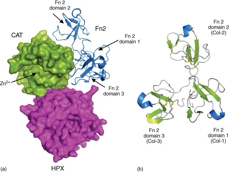
1.2.4 Fibronectin type II-like inserts

Gelatinases (MMP-2 and MMP-9) bind to gelatin and collagen with significant contribution from their three fibronectin type II-like (FN2) repeats. MMP-2 and MMP-9 are unique among the MMPs in that the three FN2 modules (Col-1, Col-2, and Col-3) are inserted in their CAT domain in the vicinity of the active site [7]. More specifically, the FN2 modules of MMP-2 and MMP-9 are inserted between the fifth β-strand and helix B in the CAT domain (according to active enzyme domain organization). The basic fold of the FN2 module comprises a pair of β-sheets, each made from two antiparallel strands, connected by a short ι-helix (Fig. 1.4). The two β-sheets form a hydrophobic pocket that is part of a hairpin turn, which orients the surrounding aromatic side chains into the hydrophobic pocket. These pockets are the structural hallmark of the FN2 modules and contribute to substrate binding (see below) [8].
c01f004
Figure 1.4 Fibronectin type II-like module structure and organization. (a) General orientation of FN2 modules of MMP-2. (b) Top view of FN2 modules. Figure prepared using MMP-2 structure (PDB 1CK7) [8].

1.2.5 Linker region

The CAT domain is connected to the HPX domain via a linker (hinge) region. The length of this linker varies from 8 to 72 amino acids, depending on the enzyme (Fig. 1.5). The linker regions may be posttranslationally modified with sugar moieties. The conformational flexibility of the linker region contributes to MMP function. For example, in the case of MMP-9, it has been suggested that the long (72 residue), glycosylated, and flexible linker region mediates protein-substrate interactions by allowing the independent movement of the enzyme CAT and HPX domains [9]. Independent domain movements were also proposed to mediate enzyme translocation on collagen fibrils [10–12]. Domain flexibility may contribute to MMP activation via promoting long-range conformational transitions induced by the binding of activator proteins or ligand [13–15]. Finally, the linker region may help to re-orient the CAT domain with respect to the HPX domain during catalysis of collagen [16]. Domain flexibility may be rationalized for most MMPs by considering the amino acid composition (i.e., Gly and Pro residues) and the various lengths of linker regions (Fig. 1.5). The linker region and HPX domain of MT1-MMP and MMP-9 are proposed to offer allosteric control of enzyme dimer forma...

Table of contents

  1. Cover
  2. Title Page
  3. Copyright
  4. Table of Contents
  5. List of Contributors
  6. Chapter 1: Matrix Metalloproteinases: From Structure to Function
  7. Chapter 2: Dynamics and Mechanism of Substrate Recognition by Matrix Metalloproteases
  8. Chapter 3: Matrix Metalloproteinases: From Structure to Function
  9. Chapter 4: Metzincin Modulators
  10. Chapter 5: Therapeutics Targeting Matrix Metalloproteinases
  11. Chapter 6: Matrix Metalloproteinase Modification of Extracellular Matrix-Mediated Signaling
  12. Chapter 7: Meprin and ADAM Metalloproteases: Two Sides of the Same Coin?
  13. Chapter 8: Subtracting Matrix out of the Equation: New Key Roles of Matrix Metalloproteinases in Innate Immunity and Disease
  14. Chapter 9: MMPs: From Genomics to Degradomics
  15. Chapter 10: MMPs in Biology and Medicine
  16. Index
  17. End User License Agreement