Cleft Lip and Palate Management: A Comprehensive Atlas —with more than 400 photographs and illustrations—provides the latest concepts about the surgical/orthodontic interrelation in cleft lip and palate treatment. Dr. Bennun and his team detail the diagnostic techniques to determine the best treatment protocols for optimal results and decreased chance of retreatment. The first part explains the principles of cleft and palate treatment, including the role of tissue engineering in craniofacial surgery. Part 2 details the aspects of primary surgical reconstruction, Part 3 discusses orthodontic treatments of cleft lip and palate, including a chapter on adult treatment, and Part 4 covers how to improve results in interdisciplinary treatment. Case presentations include results of treatment after 20-year follow up visits. Ideal for oral and maxillofacial surgeons, pediatric plastic surgeons, orthodontists, pediatric dentists, and residents in these specialties.
Frequently asked questions
Simply head over to the account section in settings and click on “Cancel Subscription” - it’s as simple as that. After you cancel, your membership will stay active for the remainder of the time you’ve paid for. Learn more here.
At the moment all of our mobile-responsive ePub books are available to download via the app. Most of our PDFs are also available to download and we're working on making the final remaining ones downloadable now. Learn more here.
Both plans give you full access to the library and all of Perlego’s features. The only differences are the price and subscription period: With the annual plan you’ll save around 30% compared to 12 months on the monthly plan.
We are an online textbook subscription service, where you can get access to an entire online library for less than the price of a single book per month. With over 1 million books across 1000+ topics, we’ve got you covered! Learn more here.
Look out for the read-aloud symbol on your next book to see if you can listen to it. The read-aloud tool reads text aloud for you, highlighting the text as it is being read. You can pause it, speed it up and slow it down. Learn more here.
Yes, you can access Cleft Lip and Palate Management by George K. B. Sándor, David Genecov, Ricardo D. Bennun, Julia F. Harfin in PDF and/or ePUB format, as well as other popular books in Medicine & Surgery & Surgical Medicine. We have over one million books available in our catalogue for you to explore.
Chapter 1 Mechanisms of cleft palate: developmental field analysis
Michael H. Carstens
Saint Louis University and Universidad Nacional Autonoma de Nicaragua, Nicaragua
The purpose of this chapter is to present concepts of cleft palate repair based on a single unifying concept: the embryology of the oronasopharynx. We shall begin with an in-depth discussion of how the bone and soft tissue structures are assembled, based upon the developmental field model. Next, we shall consider how this normal process is altered when a disruption of the neurovascular pedicle to an individual field results in a deficiency state such that the affected field is unable to fuse with its partner fields. Attention will also be give to the effect that such a deficiency state has on the subsequent development of the partner fields. Surgical procedures based on the embryologic model are designed to restore functional tissue relationships.
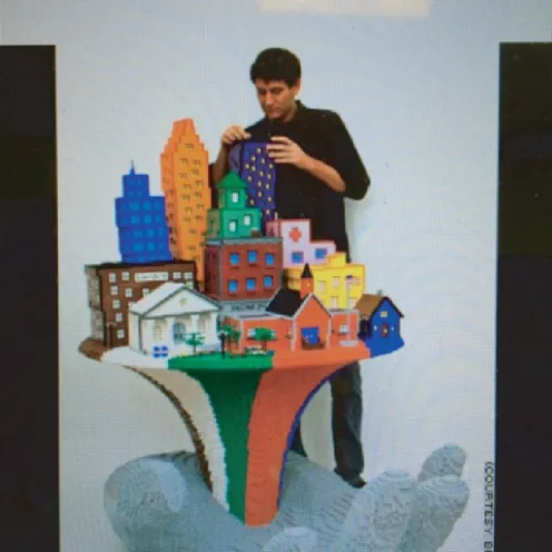
Craniofacial development: the Lego® model
The anatomic structures of the head and neck are assembled from tissue units known as developmental fields, each of which has a distinct neurovascular pedicle providing sensory and/or autonomic control and blood supply. Fields are often composite structures containing mesenchymal elements such as cartilage, bone, fascia, muscle and so on. They may have an associated epithelium such as skin or mucosa. Adjacent fields interact. Muscles with a primary attachment to bone or cartilage within one field may have a secondary attachment site in an adjacent field.
Fields develop in a strict spatio-temporal sequence. Congenital conditions that reduce the size or content of a field will affect subsequent growth. In the Pierre Robin sequence, the relative decrease in volume of the mandibular ramus leads to a posterior position of the chin and subsequent relationships of the infrahyoid musculature. The reduction of the frontal process of the premaxilla seen in the typical orofacial cleft causes a relative narrowing of the nasal fossa, malposition of the internal nasal valve, and respiratory dysfunction (Figure 1.1).
Figure 1.1 Craniofacial fields are composite blocks of tissue supplied by a specific neurovascular pedicle. Fields grow in relation to each other over time, each with a different volume and rate of growth. Deficiency or absence of a field results in collapse of adjacent partner fields. The leaning tower of Pisa is a classic example of what happens when a supporting field is absent – the entire complex is displaced and, if the upper stories of the tower were made of soft plastic, would become distorted as well. A “cleft” is really a condition of excess or deficiency in a given field that results in displacement and/or distortion of adjacent fields.
The anatomic defects seen in clefts of the hard and soft palate present as a spectrum involving several fields. Many cases involve deficiency states of the piriform fossa and/or premaxilla and soft tissues of the lip and nose. In other, rarer conditions, such as the Tessier 3 cleft, a cleft palate defect coincides with defects in seemingly unrelated anatomic zones, such as the inferior turbinate and medial maxillary wall. For this reason, it is necessary to have a comprehensive picture of the neurovascular anatomy of the oronasopharynx.
The zones of anatomic interest are all supplied by arterial axes running in parallel with the various sensory branches of V1 and V2. Development of the pedicles is a reciprocal process. Neuronal growth cones secrete vascular endothelial growth factor (VEGF) while the arterial growth cone secretes nerve growth factor (NGF). Like all cranial nerves, the trigeminal complex is constructed from neural crest, whereas the histologic composition of the arteries consists of a tubular conduit of endothelial cells made from paraxial mesoderm embraced by pericytes. These latter cells are contractile and control capillary permeability. Pericytes are ubiquitous throughout the human body (Figure 1.2). They are the precursor for mesenchymal stem cells. They also elaborate paracrine factors that are essential for survival of the vascular growth cone. Thus, we come to a very simple and powerful idea: dysfunction of a vascular growth cone will result in either a reduction of volume of mesenchymal structures within the target field, or the outright loss of the field itself. In the first case, the physical effect of the small field is to constrain subsequent growth of surrounding fields. If a frank tissue defect exists (i.e., a cleft) adjacent fields actually collapse into the site.
Figure 1.2 (a, b) Pericytes are ubiquitous throughout the body. They surround all vessels, especially capillaries, providing control of diameter and permeability. Pericytes have contractile fibers. They are interconnected, including between adjacent vessels. Pericytes may have a connection with neural crest, can detach under conditions of inflammation, are the source of white fat, and also give rise to all mesenchymal stem cells of the body. (c) Demonstrated are the multiple physiologic functions of pericytes.
The reader will note here terminology that may be unfamiliar: it harkens back to those embryology lectures that we endured … an endless list of structures that morphed into a final result via mechanisms that were unknown. The molecular revolution transformed the science into developmental biology with a tight connection to genetics (these fields formerly co-existed in virtual isolation from each other). In the following section we shall consider the tissue composition of developmental fields, how they are arranged in the intermediate state as pharyngeal arches, and how, with growth-driven folding of the embryo, these fields become physically repositioned and interactive (Carlson, 2013; Gilbert, 2013).
The embryonic period lasts 8 weeks and is divided into 23 anatomic stages (see Figure 1.3). In the first three stages, the embryo is a rapidly dividing ball of cells. Stages 4–5 are all about survival as the embryo implants itself into the uterine wall and begins the process by which blood supply will come from the mother. The stage 4 embryo secretes fluid into its center, becoming a hollow blastocyst with a single l...
Table of contents
Cover
Title Page
Copyright
Table of Contents
List of contributors
Preface
Acknowledgments
Part 1: Principles
Part 2: Present primary surgical reconstruction
Part 3: Orthodontic treatment
Part 4: Improving results in cleft lip and palate interdisciplinary management