This is a test
- English
- ePUB (mobile friendly)
- Available on iOS & Android
eBook - ePub
Human Neuroanatomy
Book details
Book preview
Table of contents
Citations
About This Book
Human Neuroanatomy, 2nd Edition is a comprehensive overview of the anatomy of the human brain and spinal cord. The book is written at a level to be of use as a text for advanced students and a foundational reference for researchers, clinicians in the field. Building on the foundations of first edition, this revision looks to increase user-friendliness and clinical applicability through improved figures and the addition of illustrative case studies.
Written by James R. Augustine, with decades of experience teaching and researching in the field, Human Neuroanatomy, authoritatively covers this fundamental area of study within the neurosciences.
Frequently asked questions
At the moment all of our mobile-responsive ePub books are available to download via the app. Most of our PDFs are also available to download and we're working on making the final remaining ones downloadable now. Learn more here.
Both plans give you full access to the library and all of Perlego’s features. The only differences are the price and subscription period: With the annual plan you’ll save around 30% compared to 12 months on the monthly plan.
We are an online textbook subscription service, where you can get access to an entire online library for less than the price of a single book per month. With over 1 million books across 1000+ topics, we’ve got you covered! Learn more here.
Look out for the read-aloud symbol on your next book to see if you can listen to it. The read-aloud tool reads text aloud for you, highlighting the text as it is being read. You can pause it, speed it up and slow it down. Learn more here.
Yes, you can access Human Neuroanatomy by James R. Augustine in PDF and/or ePUB format, as well as other popular books in Biological Sciences & Human Anatomy & Physiology. We have over one million books available in our catalogue for you to explore.
Information
CHAPTER 1
Introduction to the Nervous System
The human nervous system is a specialized complex of excitable cells, called neurons. There are many functions associated with neurons, including (1) reception of stimuli, (2) transformation of these stimuli into nerve impulses, (3) conduction of nerve impulses, (4) neuron to neuron communication at points of functional contact between neurons called synapses, and (5) the integration, association, correlation, and interpretation of impulses such that the nervous system may act on, or respond to, these impulses. The nervous system resembles a well‐organized and extremely complex communicational system designed to receive information from the external and internal environment, and assimilate, record, and use such information as a basis for immediate and intended behavior. The ability of neurons to communicate with one another is one way in which neurons differ from other cells in the body. Such communication between neurons often involves chemical messengers called neurotransmitters.
The human nervous system consists of the central nervous system (CNS) and the peripheral nervous system (PNS). The CNS, surrounded and protected by bones of the skull and vertebral column, consists of the brain and spinal cord. The term “brain” refers to the following structures: brain stem, cerebellum, diencephalon, and the cerebral hemispheres. The PNS includes all cranial, spinal, and autonomic nerves and also their ganglia, and associated sensory and motor endings.
1.1 NEURONS
The structural unit of the nervous system is the neuron with its neuronal cell body (or soma) and numerous, elaborate neuronal processes. There are many contacts between neurons through these processes. The volume of cytoplasm in the processes of a neuron greatly exceeds that found in its cell body. A collection of neuronal cell bodies in the PNS is a ganglion; a population of neuronal cell bodies in the CNS is a nucleus. An example of the former is a spinal ganglion and of the latter is the dorsal vagal nucleus – a collection of neuronal cell bodies in the brain stem whose processes contribute to the formation of the vagal nerve [X].
1.1.1 Neuronal cell body (soma)
The central part of a neuron without its many processes is the neuronal cell body (Fig. 1.1). It has a prominent, central nucleus (with a large nucleolus), various organelles, and inclusions such as the chromatophil (Nissl) substance, neurofibrils (aggregates of neurofilaments), microtubules, and actin filaments (microfilaments). The neuronal cell body contains the complex machinery needed for continuous protein synthesis – a characteristic feature of neurons. It also has an area devoid of chromatophil substance that corresponds to the point of origin of the axon called the axon hillock (Fig. 1.1). With proper staining and then examined microscopically, the chromatophil substance appears as intensely basophil aggregates of rough endoplasmic reticulum. There is an age‐related increase of the endogenous pigment lipofuscin, a marker of cellular aging often termed “age pigment,” in lysosomes of postmitotic neurons and in some glial cells of the human brain. Lipofuscin consists of a pigment matrix in association with varying amounts of lipid droplets. Another age pigment, neuromelanin makes its appearance by 11–12 months of life in the human locus coeruleus and by about 3 years of life in the human substantia nigra. This brownish to black pigment undergoes age‐related reduction in both these nuclear groups and is marker for catecholaminergic neurons.
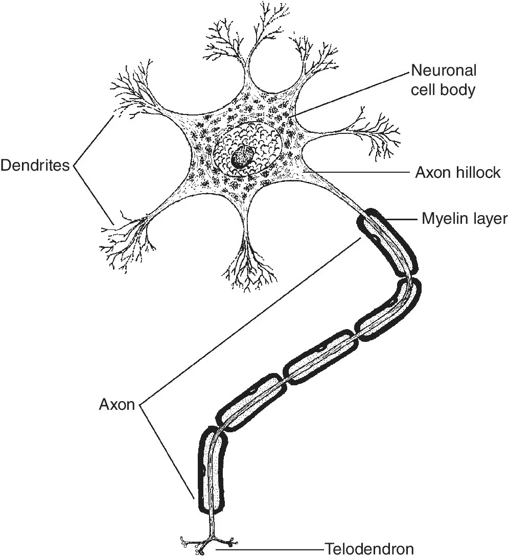
Figure 1.1 Component parts of a neuron.
Neuronal cytoskeleton
Neurofibrils, microtubules, and actin filaments in the neuronal cell body make up the neuronal cytoskeleton that supports and organizes organelles and inclusions, determines cell shape, and generates mechanical forces in the cytoplasm. Injury to the neuronal cell body or its processes due to genetic causes, mechanical damage, or exposure to toxic substances will disrupt the neuronal cytoskeleton. Neurofibrils, identifiable with a light microscope as linear fibrillary structures, are aggregates of neurofilaments when viewed with the electron microscope. Neurofilaments are slender, tubular structures 8–14 nm in diameter occurring only in neurons. Neurofilaments help maintain the radius of larger axons. Microtubules are longer, with a hollow‐core, and have an outside diameter of about 22–25 nm. Their protein subunit is composed of α‐and β‐tubulin. They form paths or “streets” through the center of the axoplasm that are traveled by substances transported from the neuronal cell body and destined for the axon terminal. In the terminal, such substances may participate in the renewal of axonal membranes and for making synaptic vesicles. Actin filaments (microfilaments, F‐actin) are in the neuronal cell body where they measure about 7 nm in diameter. The protein actin is the subunit of these neuronal actin filaments.
Neurofibrillary degenerations
Neurofilaments increase in number, thicken, or become tangled during normal aging and in certain diseases such as Alzheimer disease and Down syndrome. These diseases are termed neurofibrillary degenerations because of the involvement of neurofilaments. Alzheimer disease is the sixth leading cause of death in the United States and the fifth leading cause of death for those aged 65 years and older. Approximately 5.2 million Americans have Alzheimer disease. By 2050, the number of people living with Alzheimer disease in the United States is likely to reach about 13.8 million. This is an irreversible degenerative disease with an insidious onset, inexorable progression, and fatal outcome. Alzheimer disease involves loss of memory and independent living skills, confusion, disorientation, language disturbances, and a generalized intellectual deficit involving personality changes that ultimately result in the loss of identity (“Mr. Jones is no longer the same person”). Progression of symptoms occurs over an average of 5–15 years. Eventually, patients with Alzheimer disease become confused and disoriented, lose control of voluntary motor activity, become bedridden and incontinent, and cannot feed themselves.
Neuritic plaques, neurofibrillary tangles, and neuropil threads
Small numbers of plaques and tangles characterize the brain of normal individuals 65 years of age and over. Neuritic plaques, neurofibrillary tangles, and neuropil threads, however, are structural changes characteristic of the brains of patients with Alzheimer disease. These structural changes may occur in neuronal populations in various parts of the human brain. Other elements such as 10 and 15 nm straight neurofilaments, various‐sized dense granules, and microtubule‐associated proteins, especially the tau protein, also occur in this disease. Neurofibrillary tangles occur in the neuronal cytoplasm and have a paired helical structure that consists of pairs of 14–18 nm neurofilaments linked by thin cross‐bridging filaments that coil around each other at regular 70–90 nm intervals. These paired helical filaments, unlike any neuronal organelle and unique to the human brain, are formed by one or more modified polypeptides that have unusual solubility properties but originate from neurofilament or other normal cytoskeletal proteins. Antibodies raised against the microtubule‐associated protein, tau, are a useful marker that recognizes the presence of this protein in these neurofibrillary tangles. The tau protein helps organize and stabilize the neuronal cytoskeleton. Proponents of the “tau theory” of Alzheimer disease suggest that the phosphorylated form of this protein is a central mediator of the disease as it loses its ability to maintain the neuronal cytoskeleton, eventually aggregating into neurofibrillary tangles. Neuropil threads (curly fibers) are fine, extensively altered neurites in the cerebral cortex consisting of paired helical filaments or nonhelical straight filaments with no neurofilaments. They occur primarily in dendrites.
Degenerating neuronal processes along with an extracellular glycoprotein called amyloid precursor protein or β‐amyloid protein (β‐AP) form neuritic plaques. These plaques are of three types: primitive plaques composed of distorted neuronal processes with a few reactive cells, classical plaques of neuritic processes around an amyloid core, and end‐stage plaques with a central amyloid core surrounded by few or no processes. Proponents of the “amyloid hypothesis” of Alzheimer disease regard the production and accumulation of β‐amyloid protein in the brain and its consequent neuronal toxicity as a key event in this disease. In addition to the amyloid hypothesis and the “tau theory,” other possible cau...
Table of contents
- Cover
- Title Page
- Table of Contents
- Preface
- About the companion website
- CHAPTER 1: Introduction to the Nervous System
- CHAPTER 2: Development of the Nervous System
- CHAPTER 3: The Spinal Cord
- CHAPTER 4: The Brain Stem
- CHAPTER 5: The Forebrain
- CHAPTER 6: Introduction to Ascending Sensory Paths
- CHAPTER 7: Paths for Pain and Temperature
- CHAPTER 8: Paths for Touch, Pressure, Proprioception, and Vibration
- CHAPTER 9: The Reticular Formation
- CHAPTER 10: The Auditory System
- CHAPTER 11: The Vestibular System
- CHAPTER 12: The Visual System
- CHAPTER 13: Ocular Movements and Visual Reflexes
- CHAPTER 14: The Thalamus
- CHAPTER 15: Lower Motor Neurons and the Pyramidal System
- CHAPTER 16: The Extrapyramidal System and Cerebellum
- CHAPTER 17: The Olfactory and Gustatory Systems
- CHAPTER 18: The Limbic System
- CHAPTER 19: The Hypothalamus
- CHAPTER 20: The Autonomic Nervous System
- CHAPTER 21: The Cerebral Hemispheres
- CHAPTER 22: Blood Supply to the Central Nervous System
- CHAPTER 23: The Meninges, Ventricular System, and Cerebrospinal Fluid
- Figure and Table References
- Index
- End User License Agreement