A guide to the applications of holographic techniques for microwave and millimeter wave imaging
Real-Time Three-Dimensional Imaging of Dielectric Bodies Using Microwave/Millimeter Wave Holography offers an authoritative guide to the field of microwave holography for the specific application of imaging dielectric bodies. The authors—noted experts on the topic—review the early works in the area of optical and microwave holographic imaging and explore recent advances of the microwave and millimeter wave imaging techniques. These techniques are based on the measurement of both magnitude and phase over an aperture and then implementing digital image reconstruction.
The book presents developments in the microwave holographic techniques for near-field imaging applications such as biomedical imaging and non-destructive testing of materials. The authors also examine novel holographic techniques to gain super-resolution or quantitative images. The book also includes a discussion of the capabilities and limitations of holographic reconstruction techniques and provides recommendations for overcoming many of the limitations. This important book:
• Describes the evolution of wide-band microwave holography techniques from synthetic aperture radar principles
• Explores two major approaches to near-field microwave holography: Using the incident field and Green's function information and using point-spread function of the imaging system
• Introduces the "diffraction limit" in the resolution for techniques that are based on the Born approximation, and provides techniques to overcome this limit
Written for students and research associates in microwave and millimeter wave engineering, Real-Time Three-Dimensional Imaging of Dielectric Bodies Using Microwave/Millimeter Wave Holography reviews microwave and millimeter-wave imaging techniques based on the holographic principles and provides information on the most current developments.
Trusted by 375,005 students
Access to over 1.5 million titles for a fair monthly price.
Microwave (300 MHz to 30 GHz) and millimeter‐wave (30–300 GHz) imaging (MMI) technologies exploit electromagnetic (EM) waves with much lower frequencies than the visible light. This allows for sufficient penetration into various dielectric materials for inspection purposes and the ability to “see” through media which cannot be inspected via optical or acoustic means. These technologies have underwent rapid growth during the last few decades due to a steady demand for new applications and services along with remarkable miniaturization and diversification of the high‐frequency electronics. This has resonated well with the significant developments in computing technologies and processing algorithms leading to a wide range of applications such as imaging various composites, cement, soil, wood, ceramics, plastics, clothing, living tissues, etc.
Another major motivation to develop these technologies is that the microwave and millimeter‐wave radiation is nonionizing and is not considered hazardous at moderate power levels. This makes such technologies favorable compared to the competing X‐ray imaging technology which is ionizing, i.e. it has enough energy to potentially cause cellular and DNA damage or to elevate the risk of developing cancer in living tissues [1].
Moreover, the possibility of measuring wideband data as well as acquiring both phase and magnitude (which is typically challenging at frequencies higher than microwave and millimeter‐wave) makes these technologies favorable for three‐dimensional (3D) imaging applications, e.g. see [2]. All the aforementioned advantages have motivated the researchers in academic and industrial institutions to pursue development of the microwave technologies toward various imaging and sensing applications.
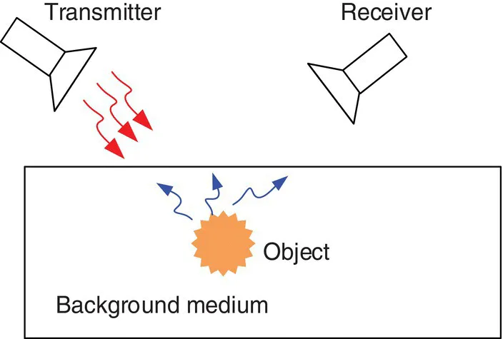
Figure 1.1 illustrates a typical MMI system consisting of a transmitting antenna illuminating the inspected medium. The inspected medium includes the background along with any objects that are electrically sufficiently different from the background to cause scattering of the EM waves. The scattered waves due to the presence of the object along with the scattered waves due to the background medium are acquired by a receiving antenna. The acquired data are then processed to reconstruct two‐dimensional (2D) or 3D images of the object.
Figure 1.1 Illustration of a MMI setup. The object inside the background medium scatters the EM waves emitted by the transmitter provided its electrical properties are different from those of the background. Some of the scattered waves are picked up by the receiving antenna and are processed to detect the object.
In various MMI systems, the transmission and reception can be implemented by the same antenna (monostatic system), or one antenna can transmit while another receives (bistatic system), or an array of antennas (multistatic) can receive the back‐scattered waves. Besides, the data acquisition can be implemented while the antennas are stationary or the antennas may perform scanning of the back‐scattered waves over an aperture to construct a synthetic aperture. An alternative solution to avoid mechanical scanning of the antennas (that may be time‐consuming and may suffer from positioning errors) is the use of stationary antenna arrays that can be switched electronically to capture the distribution of the back‐scattered waves over an aperture.
1.1 Some Emerging Applications of MMI
Some of the applications of microwave imaging technology, such as synthetic aperture radar (SAR) [3] and ground penetrating radar (GPR) [4], have been well‐established, commercialized, and employed for several decades. These technologies provide high‐resolution qualitative images of the earth surface, landscape, weather condition, underground objects, hydrocarbon reservoirs, etc. These have been typically long‐range applications of MMI since the distance between the imaged objects and the antennas (sensors) is typically much larger than the operation wavelength.
Recently, researchers have been pursuing the capabilities of MMI in some new applications, demonstrating promising imaging results. These new applications, such as concealed weapon detection [5], nondestructive testing (NDT) [6], through‐the‐wall imaging [7], and biomedical imaging [8], are under rapid development and commercialization. Their success depends on how they can overcome challenges encountered by the competing technologies of ultrasound imaging, X‐ray imaging, magnetic resonance imaging (MRI), etc. in each particular application domain.
The conventional MMI techniques such as SAR and GPR suffer from the well‐known “diffraction limit” in the resolution. The diffraction limit states that the spatial resolution of the far‐field techniques is proportional to the operation wavelength, i.e. the shorter the wavelength, the smaller the size of the shapes that can be resolved in the image. However, near‐field MMI techniques may offer better spatial resolution which is typically proportional to the sensor's sensing dimensions (e.g. see [9]). This is due to the use of evanescent waves, which contain information about finer details of the object but decay exponentially versus the distance between the object and the antenna. Thus, while using the evanescent‐wave information is possible in the near‐field MMI applications (leading to better spatial resolution), in the far‐field, receiving the evanescent waves is practically impossible leading to degradation of the spatial resolution down to the diffraction limit. Later in this book, we discuss the resolution of MMI systems in more detail.
The case of interest in the recent applications of MMI is that of an object in the near field of the antenna. This necessitates the development of new algorithms and techniques for image‐reconstruction purposes. In the following, we briefly describe the recent progress in some of the emerging and most promising applications of MMI technologies which are mostly mid‐field to near‐field applications.
One revolutionizing application of the MMI is in security screening in public places such as airports. This technology allows for penetration of the EM waves through clothes and for the formation of a whole‐body image of the scanned person in order to seek for prohibited concealed items [5, 10–13]. So far, several countries have chosen scanners based on this technology for security screening in the airports [14] over the competing technology, which is back‐scattered X‐ray. These millimeter‐wave scanners can be divided into two categories: active and passive. Active scanners illuminate the inspected domain with millimeter‐wave energy and then measure and process the reflected energy for image reconstruction [5, 11–13]. Passive systems create images using only the millimeter‐wave spectrum of the ambient thermal radiation and that emitted from the inspected body [15–17].
Figure 1.2 shows a millimeter‐wave imaging setup used by transportation security administration (TSA) in the US airports for whole‐body scanning. This figure also shows some sample reconstructed images by this system. It is worth noting that the resolution of the produced images using millimeter‐wave technology in the airport security screening is so high that it has raised privacy concerns. This has led to the prohibition of displaying detailed images by the US congress and applying automatic target recognition software (which provides a generic body outline with largest contrasts due to the concealed prohibited objects). This is to emphasize that very high‐resolution, robust, and accurate images can be captured using this technology.
Figure 1.2 (a) Millimeter‐wave whole body scanning setup based on the technology developed for airport security screening. (b) Sample images of screened persons.
Source: Reprinted with permission from [14].
Another application of the MMI, which is under active research in the microwave society, is in biomedical imaging [18]. This includes imaging of isolated organs or in situ imaging such as imaging of the brain for stroke detection [19–21], cerebral edema [22], breast cancer [8, 23–25], bone imaging [26, 27], heart imaging [28–33], and joint‐tissue imaging [34]. In these applications, the tissue is illuminated with low‐power microwaves which are safe and allow for frequent examinations. The scattered waves are then measured and processed to provide images of the interior of the tissue. The images show various tissues such as muscle, fibro‐glandular and fatty tissues, which exhibit various contrasts in the dielectric properties. This provides a basis for distinguishing the normal tissue from the malignant tissue (tumor). The malignant tissue typically has larger dielectric properties compared to normal tissue due to the higher concentration of blood vessels and water content. Figure 1.3 shows the measurement setup for brain imaging using a physical head phantom and the Vivaldi antenna proposed in [35]. This figure also shows the reconstructed images using the confocal method showing damage due to stroke at two different locations.
Figure 1.3 (a) Measurement setup for brain imaging using a physical head phantom and the Vivaldi antenna. (b) Reconstructed images using the confocal method showing position of stroke at two different loca...
Table of contents
Cover
Table of Contents
Preface
Acknowledgments
1 Introduction
2 Microwave/Millimeter Wave Holography Based on the Concepts of Optical Holography
3 Direct and Quasi‐Microwave/Millimeter‐Wave Holography for Far‐Field Imaging Applications
4 Microwave/Millimeter‐Wave Holography for Near‐Field Imaging Applications
5 Increasing the Resolution and Accuracy of Microwave/Millimeter‐Wave Holography
6 Conclusion
Appendix: Diffraction Limit for the Spatial Resolution in Far‐Field Imaging
References
Index
IEEE Press Series on RF and Microwave Technology
End User License Agreement
Frequently asked questions
Yes, you can cancel anytime from the Subscription tab in your account settings on the Perlego website. Your subscription will stay active until the end of your current billing period. Learn how to cancel your subscription
No, books cannot be downloaded as external files, such as PDFs, for use outside of Perlego. However, you can download books within the Perlego app for offline reading on mobile or tablet. Learn how to download books offline
Perlego offers two plans: Essential and Complete
Essential is ideal for learners and professionals who enjoy exploring a wide range of subjects. Access the Essential Library with 800,000+ trusted titles and best-sellers across business, personal growth, and the humanities. Includes unlimited reading time and Standard Read Aloud voice.
Complete: Perfect for advanced learners and researchers needing full, unrestricted access. Unlock 1.5M+ books across hundreds of subjects, including academic and specialized titles. The Complete Plan also includes advanced features like Premium Read Aloud and Research Assistant.
Both plans are available with monthly, semester, or annual billing cycles.
We are an online textbook subscription service, where you can get access to an entire online library for less than the price of a single book per month. With over 1.5 million books across 990+ topics, we’ve got you covered! Learn about our mission
Look out for the read-aloud symbol on your next book to see if you can listen to it. The read-aloud tool reads text aloud for you, highlighting the text as it is being read. You can pause it, speed it up and slow it down. Learn more about Read Aloud
Yes! You can use the Perlego app on both iOS and Android devices to read anytime, anywhere — even offline. Perfect for commutes or when you’re on the go. Please note we cannot support devices running on iOS 13 and Android 7 or earlier. Learn more about using the app
Yes, you can access Real-Time Three-Dimensional Imaging of Dielectric Bodies Using Microwave/Millimeter Wave Holography by Reza K. Amineh,Natalia K. Nikolova,Maryam Ravan in PDF and/or ePUB format, as well as other popular books in Tecnología e ingeniería & Microondas. We have over 1.5 million books available in our catalogue for you to explore.