Atlas of Pediatric Oral and Dental Developmental Anomalies
eBook - ePub

Atlas of Pediatric Oral and Dental Developmental Anomalies

  1. English
  2. ePUB (mobile friendly)
  3. Available on iOS & Android
eBook - ePub

Atlas of Pediatric Oral and Dental Developmental Anomalies

Book details
Book preview
Table of contents
Citations

About This Book

A quick reference diagnostic guide for students and clinicians, covering a wide range of oral and dental developmental anomalies in children and adolescents

Written by world-renowned pediatric dentists, this easily accessible, well-illustrated reference covers a wide range of oral and dental developmental anomalies in children and adolescents, and includes rare as well as more common conditions.

Divided into two parts, the first part is dedicated to normal tissue initiation, formation, and development in the orodental region. The second part offers comprehensive pictorial descriptions of each condition and discussions of the treatment options available.

  • A useful, quick reference atlas helping students and clinicians diagnose a wide range of oral and dental developmental anomalies in children and adolescents
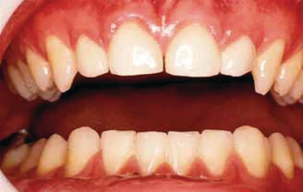
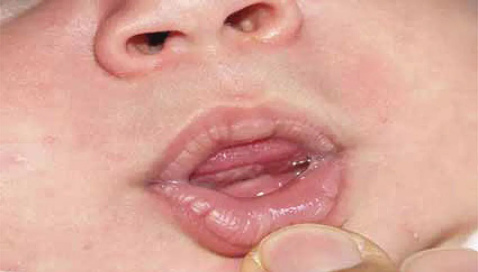
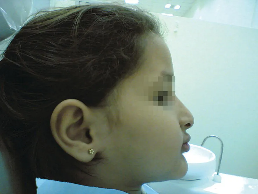
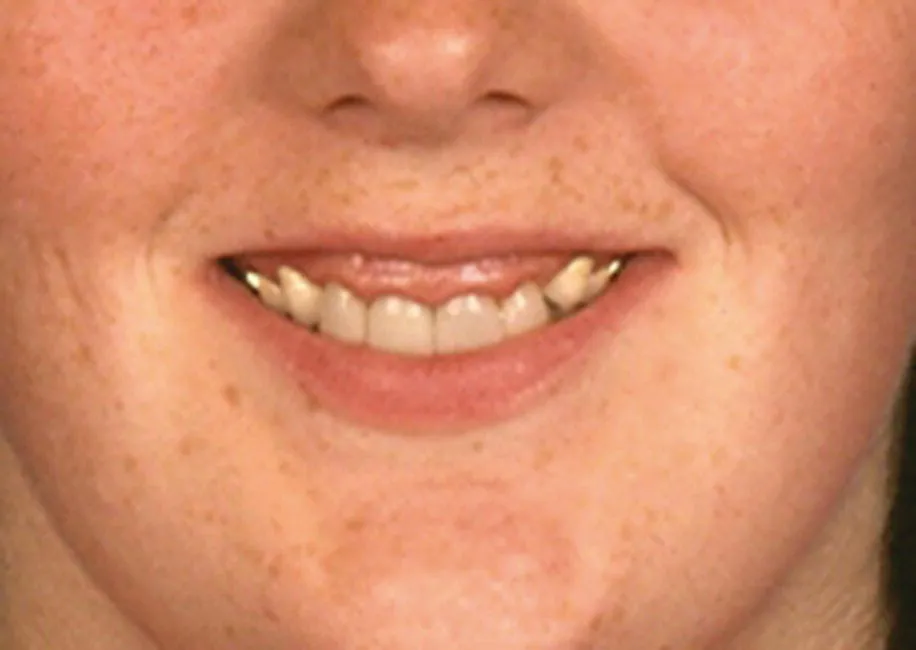
  • Highly illustrated with clinical photographs
  • Describes both common and rare conditions, and explores treatment options

Atlas of Pediatric Oral and Dental Developmental Anomalies is an excellent resource for undergraduate dentistry students, postgraduate pediatric dentistry students, and pediatric dental practitioners.

Frequently asked questions

Simply head over to the account section in settings and click on “Cancel Subscription” - it’s as simple as that. After you cancel, your membership will stay active for the remainder of the time you’ve paid for. Learn more here.
At the moment all of our mobile-responsive ePub books are available to download via the app. Most of our PDFs are also available to download and we're working on making the final remaining ones downloadable now. Learn more here.
Both plans give you full access to the library and all of Perlego’s features. The only differences are the price and subscription period: With the annual plan you’ll save around 30% compared to 12 months on the monthly plan.
We are an online textbook subscription service, where you can get access to an entire online library for less than the price of a single book per month. With over 1 million books across 1000+ topics, we’ve got you covered! Learn more here.
Look out for the read-aloud symbol on your next book to see if you can listen to it. The read-aloud tool reads text aloud for you, highlighting the text as it is being read. You can pause it, speed it up and slow it down. Learn more here.
Yes, you can access Atlas of Pediatric Oral and Dental Developmental Anomalies by Ghassem Ansari, Mojtaba Vahid Golpayegani, Richard Welbury in PDF and/or ePUB format, as well as other popular books in Medicine & Dentistry. We have over one million books available in our catalogue for you to explore.

Information

Year
2018
ISBN
9781119380917
Edition
1
Subtopic
Dentistry

1
Oral and Dental Anatomy

The oral cavity consists of soft and hard tissues. The lips, cheeks, tongue, gingivae, palate, and tonsils are the former, while the teeth are the latter. The oral cavity is bounded by the lips anteriorly, the nasopharynx posteriorly, the cheeks laterally, the tongue and sublingual tissues inferiorly, and the soft and hard palate superiorly. Various muscles, nerves, and vascular systems contribute to these surrounding structures. The muscles of the oral cavity include mylohyoid, geniohyoid, stylohyoid, hyoglossus, glossopharyngeal, thyroglossus, buccinator, masseter, medial and lateral pterygoid, orbicularis oris, and temporalis. These muscles, together with their tendons, nerves, and blood vessels, keep the oral cavity functional.

1.1 The Lips: Macro Anatomy

The lips are composed of the muscular layer of orbicularis oris, connective tissue, dermis, and mucosa (Figure 1.1). The red vermilion border and its junction with the skin and mucosa at its outer and inner borders may vary in width between races and genders. Lips may have a different posture at rest, including: (i) sealed or competent, and (ii) not sealed or incompetent. Lip position may affect the alignment and profile view of teeth and occlusion (Figures 1.2–1.4). In certain circumstances, the lips appear shorter than normal, or the jaws are not in normal skeletal relationship, with a large part of the maxillary labial gingiva being visible during speech and smile. This condition is often referred to as “gummy” smile (Figure 1.5). Alternatively, there are cases where a longer‐than‐normal lip length or lost vertical height is noted due to lost or missing teeth – for example, ectodermal dysplasia. This in turn could cause the lips to overlap heavily, producing the appearance of an edentulous individual (Figure 1.4).
Frontal view of normal intraoral tissue and teeth.
Figure 1.1 Normal intraoral tissue and teeth.
Image described by caption.
Figure 1.2 Lips of a newborn.
Image described by caption.
Figure 1.3 Lips of an 8‐year‐old.
Right lateral of the head of the patient displaying lateral lip pattern in ectodermal dysplasia.
Figure 1.4 Lateral lip pattern in ectodermal dysplasia.
Image described by caption.
Figure 1.5 High upper lip resulting in a “gummy” smile.

1.2 The Palate

The palate is divided into two major parts – soft and hard palate, with each of them having specific characteristics related directly to the role they play in different oral functions. The hard palate is supported by a hard, bony structure in the roof of the mouth, while the soft palate is mainly supported by fibrous tissue. The hard palate is covered with keratinized membrane, with a prominent eminence at the anterior mid‐line located on top of the incisive foramen of maxillary bone, called the “incisive papilla.” The nasopalatine nerve and blood supply pass through this foramen. “Rugae” are the anterior rough mucosal folds of the palate located on either s...

Table of contents

  1. Cover
  2. Table of Contents
  3. Preface
  4. 1 Oral and Dental Anatomy
  5. 2 Histology and Embryology of the Teeth and Periodontium
  6. 3 Epidemiology and Diagnosis of Teeth Developmental Disturbances
  7. 4 Etiology and Pathology of Teeth Disturbances
  8. 5 Eruption Disturbances of Teeth – Etiology and Diagnosis
  9. 6 Self‐evaluation Section
  10. Bibliography
  11. Self‐evaluation Answer
  12. Index
  13. End User License Agreement