eBook - ePub
Principles of Stem Cell Biology and Cancer
Future Applications and Therapeutics
- English
- ePUB (mobile friendly)
- Available on iOS & Android
eBook - ePub
Principles of Stem Cell Biology and Cancer
Future Applications and Therapeutics
About this book
Principles of Stem Cell Biology and Cancer: Future Applications and Therapeutics Tarik Regad, The John van Geest Cancer Research Centre, Nottingham Trent University, UK, Thomas J. Sayers, Centre for Cancer Research, National Cancer Institute, Frederick, USA and Robert Rees The John van Geest Cancer Research Centre, Nottingham Trent University, UK The field of cancer stem cells is expanding rapidly, with many groups focusing on isolating and identifying cancer stem cell populations. Although some progress has been made developing efficient cancer therapies, targeting cancer stem cells remains one of the important challenges facing the growing stem cell research community. Principles of Stem Cell Biology and Cancer brings together original contributions from international experts in the field to present the very latest information linking stem cell biology and cancer. Divided into two parts, the book begins with a detailed introduction to stem cell biology with a focus on the characterization of these cells, progress that has been made in their identification, as well as future therapeutic applications of stem cells. The second part focuses on cancer stem cells and their role in cancer development, progression and chemo-resistance. This section of the book includes an overview of recent progress concerning therapies targeting cancer stem cells. Features: An authoritative introduction to the link between stem cell biology and cancer. Includes contributions from leading international experts in the field. Well-illustrated with full colour figures throughout. This book will prove an invaluable resource for basic and applied researchers and clinicians working on the development of new cancer treatments and therapies, providing a timely publication of high quality reviews outlining the current progress and exciting future possibilities for stem cell research.
Trusted by 375,005 students
Access to over 1.5 million titles for a fair monthly price.
Study more efficiently using our study tools.
Information
Part I
Stem Cells
Chapter 1
Isolation and Characterization of Human Embryonic Stem Cells and Future Applications in Tissue Engineering Therapies
Christian Unger, James Hackland, David Preskey and Harry Moore
Centre for Stem Cell Biology, Department of Biomedical Sciences, University of Sheffield, Sheffield, UK
1.1 Derivation of human embryonic stem cells from the ICM
1.1.1 Early development of the ICM: the cells of origin for hESCs
The mammalian zygote (fertilized ovum) is defined as being totipotent, as it is capable of developing into a new offspring and the placenta required for full gestation. The zygote initially undergoes cleavage-stage cell division, forming cells (early blastomeres) that remain totipotent. With further development to the preimplantation blastocyst stage, a primary cell differentiation results in outside trophectoderm cells (TE) and an inside aggregate of inner cell mass (ICM) cells. The TE forms placental tissue and membranes, while the ICM forms the foetus and extra-embryonic membranes. Therefore, ICM cells are defined as being pluripotent, forming all cells of the developing offspring other than the complete placenta (unless genetically manipulated). Embryonic stem cells (ESCs) are derived in vitro from ICM cells, which adapt to specific conducive conditions that enable indefinite cell proliferation (self-renewal) without further differentiation and thereby confer a pluripotent capacity. This in vitro pluripotent state is due principally to the induction and maintenance of expression of key ‘gate-keeper’ genes, including Oct4, Nanog and Sox2, which then regulate one another (Silva & Smith, 2008). The capacity for self-renewal is sustained by high telomerase activity, which protects chromosome telomeres from degradation during mitosis (Blasco, 2007).
Mammalian ESCs were first derived in the mouse (mESC) (Evans and Kaufman, 1981; Martin, 1981). When mESCs are integrated into an embryo and returned to a recipient, they can contribute to all cell lineages, including germ cells. Their utility soon became invaluable for many transgenic procedures. Successful derivation of human (hESC) lines was reported by Thomson et al. (1998), who essentially followed the same procedure as used for the mouse. ICMs isolated from preimplantation human blastocysts were plated on to mitotically inactivated mouse embryonic feeders in culture medium with basic fibroblast growth factor (bFGF) and foetal calf serum (FCS). This culture medium was also supplemented with leukaemia inhibitory factor (LIF), a cytokine necessary to maintain mESCs (Smith et al., 1988), although (as is now known) not necessary for standard hESC derivation. Human ESCs display (or lose on differentiation) plasma membrane expression of stage-specific embryonic antigens (SSEAs) that correlate with the preimplantation morphological development of human embryos (Henderson et al., 2002) and form teratomas (benign tumours) in immune-deficient mice that can contain cell phenotypes from the three major cell lineages (endoderm, mesoderm and ectoderm), as well as trophoblast. The differentiation of trophoblast cells indicates that hESCs are not entirely equivalent to mESCs, as usually defined, but align with slightly later LIF-independent mouse epiblast pluripotent stem cells, which have the propensity to differentiate to trophoblast in vitro (Brons et al., 2007).
1.1.2 Derivation of hESCs
Success in the derivation of hESCs depends in part on the quality of the human embryos used (usually blastocysts from days 5 to 8), although cell lines have been generated from morphologically poor embryos. Numerous hESC lines have been derived (Figure 1.1) from normal, aneuploid and mutant embryos from patients undergoing treatment for assisted conception (IVF, ICSI) or preimplantation genetic diagnosis (PGD) who consent to donate them for stem cell research. Some of these cell lines have been extensively characterized and compared, enabling international standards to be established (Adewumi et al., 2007).
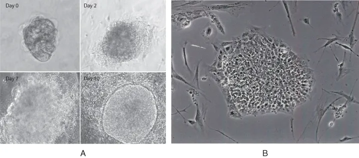
Figure 1.1 (A) Outgrowth of hESCs over 10 days of culture from ICM. In this instance, a clearly defined colony was observed by 10 days, which was mechanically passaged. (B) hESC line Shef1 plated on ECM.
1.1.2.1 Evolution to a more efficient and better-defined derivation method: drivers and technologies
Over the last 15 years, continuous improvements have been made in the process of deriving and maintaining hESC lines. The emphasis initially was on improving efficiency and consistency in the stem cell laboratory. But as hESC lines have become readily available for research in many countries, the focus has changed to devising methods for deriving clinical-grade cell lines that comply with health care regulatory authorities (e.g. Federal Drug Administration, FDA; European Medicines Agency, EMA), which can be used as starting materials for potential cell-therapy trials. Xeno-free methods (free of nonhuman animal components) are preferable as they minimize the risk of cross-species contamination with adventitious agents. An important early improvement was the replacement of FCS with a serum extract (knockout serum replacement, KOSR) to reduce hESC differentiation. This modification also minimized batch variation (inherent in FCS) between culture media, and allowed consistency in the proliferation of the cells after passaging (transfer of cells to a new culture vessel). Subsequently, more defined culture media (xeno-free) have been devised, which, in combination with a variety of extracellular matrix (ECM) compositions, facilitate the proliferation and passage of pluripotent hESCs in the absence of feeder cells (mouse or human), which otherwise remain an ill-defined and inconsistent component of the cell culture. Manipulation of the embryo has also changed over time. Initially, the ICM was isolated according to mouse protocols using enzymatic (protease) removal of the zona pellucida (ECM surrounding blastocyst) and immunosurgical lysis of TE with antitrophoblast antibody to prevent TE culture outgrowth from inhibiting early ESC proliferation. However, xeno-free methods using laser-assisted removal of the zona and plating of the intact blastocyst or the ICM on to a defined matrix (e.g. laminin 521) with a defined culture medium is the method of choice, leading to successful feeder/xeno-free cell line production in ∼20–40% of attempts with good-quality human embryos (Hasegawa et al., 2010). With further improvements to the cell adhesion matrix and cell medium, the efficiency of hESC line derivation is likely to increase further, although the quality of the embryo used to develop ICM cells remains a crucial factor.
Another important consideration is the genetic character and stability of the hESC line. Generally, most hESC outgrowths and initial cell lines derived from unselected embryos (i.e. not PGD selected) are determined to be karyotypically normal within the precision of the chromosomal analysis. However, hESCs acquire genetic mutations in culture, which may endow them with a selective cell culture advantage, so that mutated cells predominate (Baker et al., 2007). Since derivation and ESC passage represent key stress events for ESC cultures, minimization of selective pressure on cells at these stages may help to maintain their normal karyotype. For example, the proliferation of cells by mechanical division of hESC colonies into smaller aggregates may be preferable to enzymatic disaggregation to single cells, which will initiate apoptotic stress pathways unless inhibited from doing so by a chemical inhibitor (i.e. ROCK inhibitor).
1.1.3 Regulation of ...
Table of contents
- Cover
- Title Page
- Copyright
- Table of Contents
- List of Contributors
- Preface
- Part I: Stem Cells
- Part II: Cancer Stem Cells
- Index
- End User License Agreement
Frequently asked questions
Yes, you can cancel anytime from the Subscription tab in your account settings on the Perlego website. Your subscription will stay active until the end of your current billing period. Learn how to cancel your subscription
No, books cannot be downloaded as external files, such as PDFs, for use outside of Perlego. However, you can download books within the Perlego app for offline reading on mobile or tablet. Learn how to download books offline
Perlego offers two plans: Essential and Complete
- Essential is ideal for learners and professionals who enjoy exploring a wide range of subjects. Access the Essential Library with 800,000+ trusted titles and best-sellers across business, personal growth, and the humanities. Includes unlimited reading time and Standard Read Aloud voice.
- Complete: Perfect for advanced learners and researchers needing full, unrestricted access. Unlock 1.5M+ books across hundreds of subjects, including academic and specialized titles. The Complete Plan also includes advanced features like Premium Read Aloud and Research Assistant.
We are an online textbook subscription service, where you can get access to an entire online library for less than the price of a single book per month. With over 1.5 million books across 990+ topics, we’ve got you covered! Learn about our mission
Look out for the read-aloud symbol on your next book to see if you can listen to it. The read-aloud tool reads text aloud for you, highlighting the text as it is being read. You can pause it, speed it up and slow it down. Learn more about Read Aloud
Yes! You can use the Perlego app on both iOS and Android devices to read anytime, anywhere — even offline. Perfect for commutes or when you’re on the go.
Please note we cannot support devices running on iOS 13 and Android 7 or earlier. Learn more about using the app
Please note we cannot support devices running on iOS 13 and Android 7 or earlier. Learn more about using the app
Yes, you can access Principles of Stem Cell Biology and Cancer by Tarik Regad,Thomas Sayers,Robert Rees in PDF and/or ePUB format, as well as other popular books in Biological Sciences & Cell Biology. We have over 1.5 million books available in our catalogue for you to explore.