A PRACTICAL GUIDE TO VULVAL DISEASE DIAGNOSIS AND MANAGEMENT
A PRACTICAL GUIDE TO VULVAL DISEASE DIAGNOSIS AND MANAGEMENT
Patients with vulval disease frequently experience delays in diagnosis due to a lack of training for physicians. A Practical Guide to Vulval Disease: Diagnosis and Management offers practical, up-to-date and expert guidance on the diagnosis and management of vulval disorders. It provides the knowledge required for diagnosis and treatment of these conditions at both trainee and specialist level. Key information about diagnosis, investigation and basic management is included, with a section on signs and symptoms to direct the reader to the appropriate chapter for the particular disease. Current classification and terminology of vulval disease is featured, along with guidance on when a patient should be referred to a specialist.
Well illustrated, with 185 high quality photographs, this user-friendly clinical guidebook integrates clinical and histological features of vulval disorders, so the reader can understand the disease from a microscopic to macroscopic level.
Written by an experienced author team, A Practical Guide to Vulval Disease: Diagnosis and Management is essential reading for gynaecologists, dermatologists, genito-urinary physicians, general practitioners and nurses, both in practice and in training.
Trusted by 375,005 students
Access to over 1.5 million titles for a fair monthly price.
The vulva is a complex organ, due to its embryologic derivation from the three germ layers belonging to the embryonic disc:
ectoderm (squamous epithelium);
mesoderm (connective epithelium);
endoderm (vulval vestibule).
This embryological derivation is responsible for the different variants in morphology that occur during the development of the vulva.
A correct and thorough knowledge of the ‘normal’ vulva is vital for several reasons. Firstly, it is important in order to recognize some of the normal anatomical variants in order to differentiate them from pathological features. This will prevent unnecessary excision and treatment of normal areas. Secondly, it leads to a more specific and logical approach in treating vulval disorders. In some conditions, the normal anatomy of the vulva is altered and this can give diagnostic clues. It is important to note that the ‘normal’ vulva modifies itself during a woman’s lifetime, depending on age, obstetrical and gynaecological history.
Normal vulval anatomy
The vulva may be considered as the combination of the mucosal, cutaneous, muscular and connective tissue structures that compose the lower part of the female genital tract. The peculiarity of this localization means that the vulva is in close association with urological structures (urethra and bladder), gynaecological structures (vagina), and intestinal structures (rectum and anus).
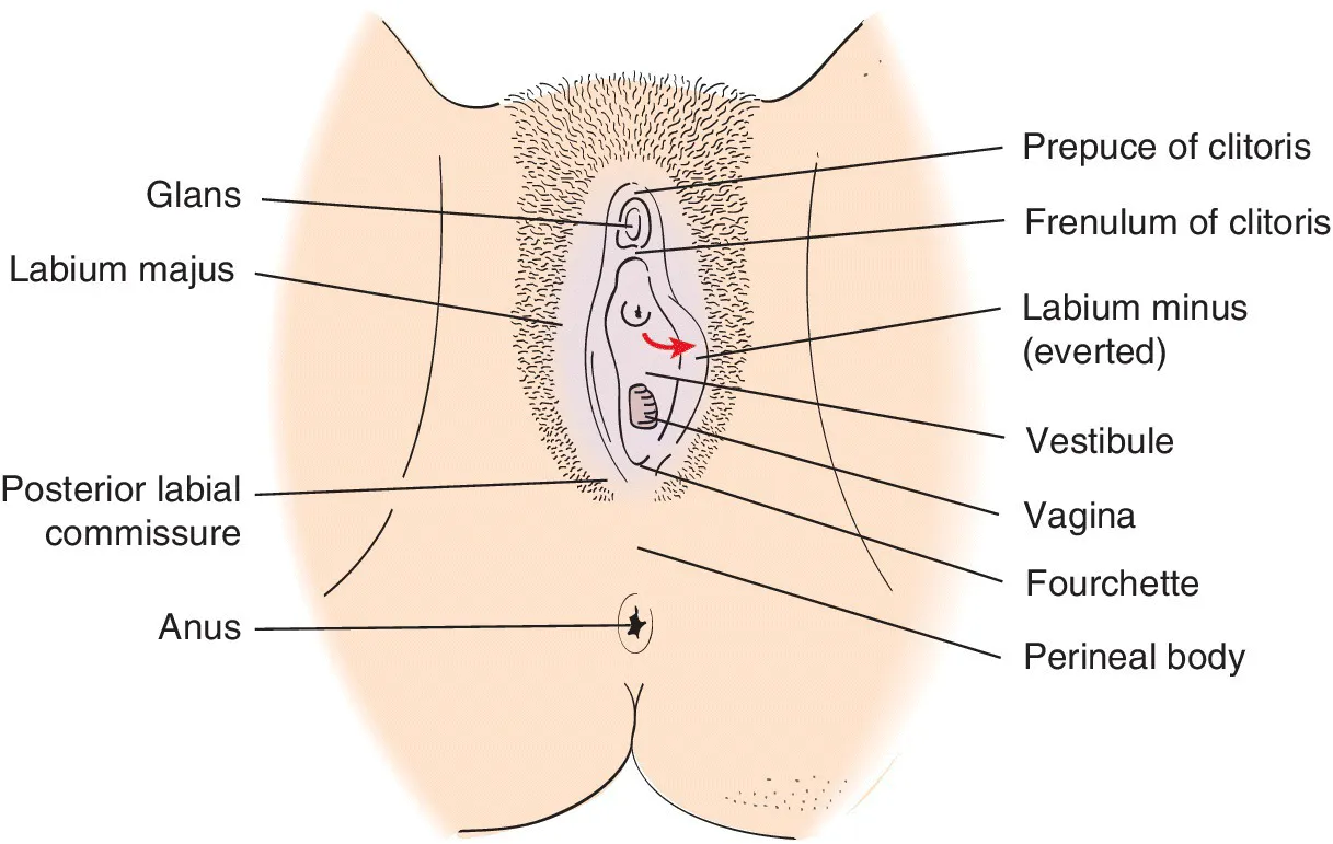
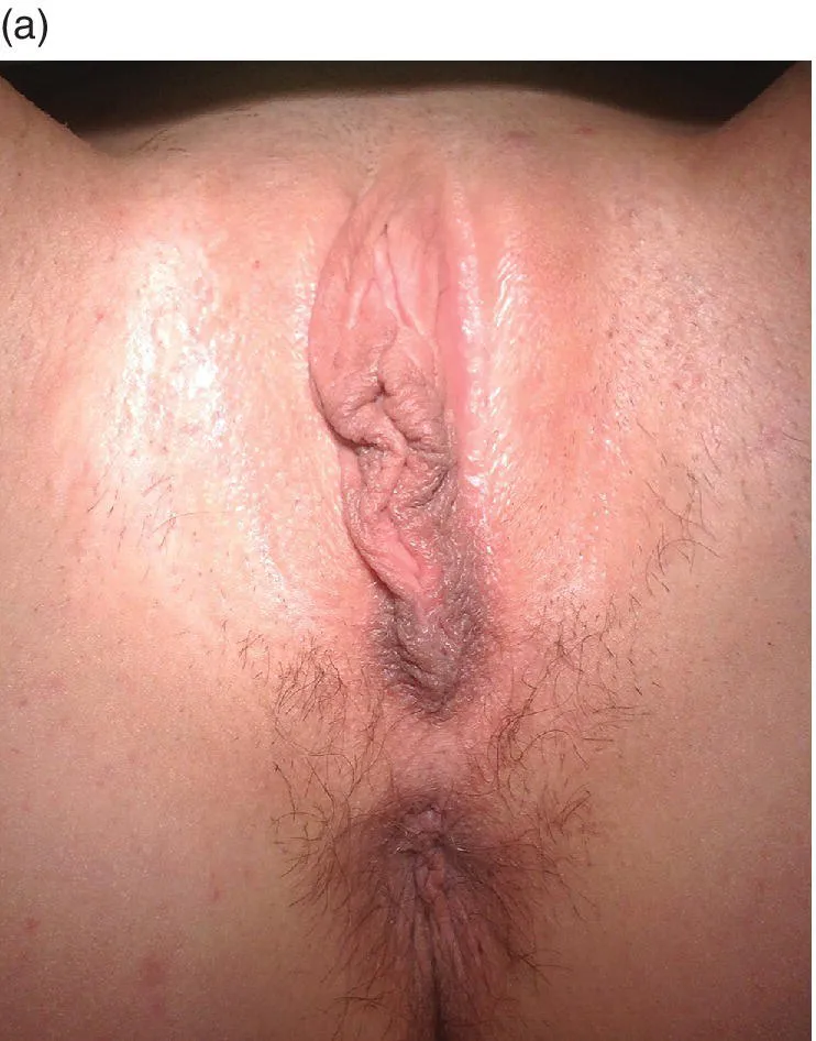
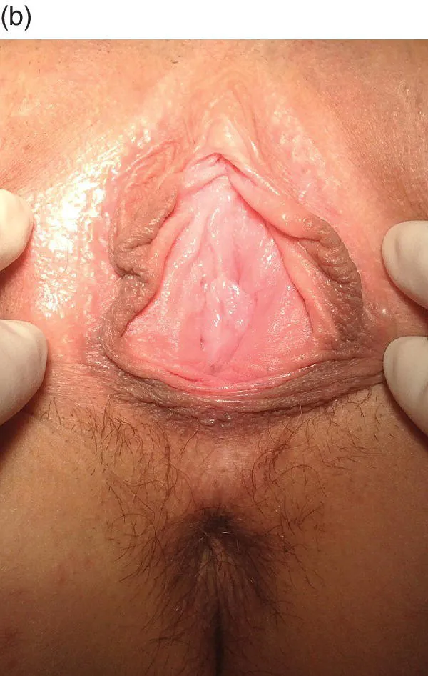
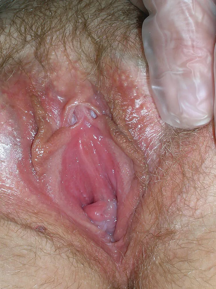
The borders of the vulva are: mons pubis anteriorly, perineal body posteriorly, genital crural folds laterally and hymen medially (Figure 1.1). In this triangular‐shaped region, with naked‐eye examination, five distinct structures clearly appear: the labia majora, the clitoris, the vestibule, the labia minora and the hymen (Figure 1.2 a, b).
Figure 1.1 The vulva.
Figure 1.2 Normal vulva (a) – outer and (b) – inner vulva.
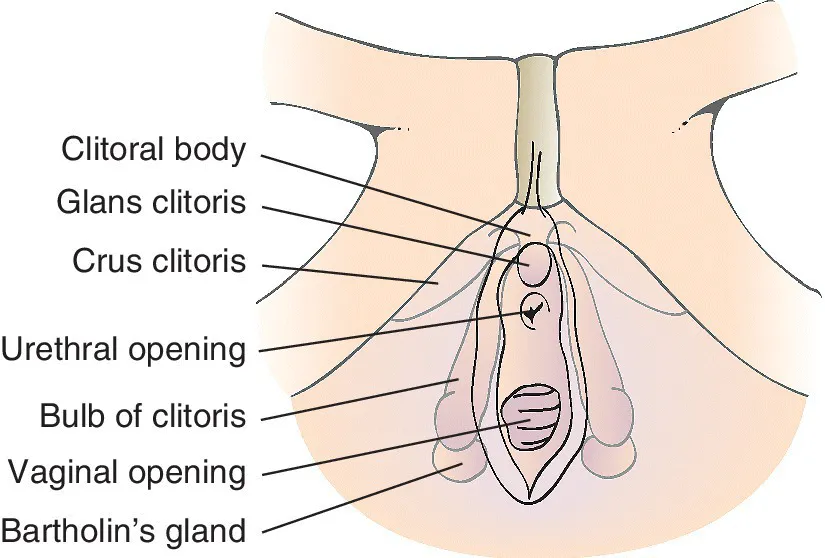
There is usually a limited description of the internal structures of the vulva in gynaecological and dermatological textbooks. These structures reach the plane of the perineal fascia (or urogenital membrane) under the skin. A knowledge of the anatomy of these structures and planes then encompasses the clitoral body, the minor vestibular bulbs and glands, the urethral opening and the paraurethral glands, which are all part of the vulva. A good understanding of the anatomy, together with its embryological development, allows a comprehensive approach to vulval morphology and correct surgical dissection if required.
The labia majora are two cutaneous folds, even and symmetrical, arising from the lateral portions of the mons pubis and extending to the posterior triangle of the perineum. Laterally they terminate on the genito‐crural fold, and medially continue to the external aspects of the labia minora, forming the interlabial sulci. On the outer surface, they are covered by hair‐bearing skin. The hair follicles are lost on the inner surface but many sebaceous glands remain.
The labia minora are two thin structures that are connected anteriorly to form the clitoral hood and, below the clitoral body, form the frenulum. Posteriorly the labia minora unite to define the fourchette The epithelium starting from the internal side of the fourchette to the hymen is called the navicular fossa. The labia minora do not have hair follicles but they are covered by numerous sebaceous glands and sweat glands.
The clitoris develops from an outgrowth in the embryo called the genital tubercle. It contains trabeculated erectile tissue, similar to the male penis, and is composed of the body (the shaft and the glans) and the crura. The glans is covered by the clitoral hood, formed by the fusion of the anterior portions of the labia minora. The body of the clitoris continues in each crus (singular form of ‘crura’), attached to the corresponding ischial ramus, beneath the descending pubic rami. Hence only about 30% of the clitoris is visible (Figure 1.3).
Figure 1.3 The clitoris.
The vestibule is the space between the hymenal ring and the internal aspect of labia minora. Its boundaries are the clitoris anteriorly, the fourchette posteriorly and the ‘Hart’s line’ laterally, which runs down the internal side of the labia minora. It represents the junction between the mucosal epithelium and the keratinized skin of the vestibule (Figure 1.4). Some authors define the lateral extension of the vestibule as the free edge of labia minora, therefore including the two types of epithelium (mucosa and skin).
Figure 1.4 Hart’s line.
Several structures open into the vestibule. The urethral open...
Table of contents
Cover
Title Page
Table of Contents
Acknowledgements
1 The Normal Vulva
2 Taking a History and Examination
3 How to Take a Vulval Biopsy and the Importance of Clinico-Pathological Correlation
4 Basic Histology of the Vulva
5 Investigations in Vulval Disease
6 Topical Treatment in Vulval Disease
7 Symptoms in Vulval Disease
8 Signs in Vulval Disease
9 Eczema, Allergy and the Vulva
10 Psoriasis
11 Lichen Simplex
12 Lichen Sclerosus
13 Lichen Planus
14 Hidradenitis Suppurativa and Crohn's Disease
15 Disorders of Pigmentation on the Vulva
16 Other Dermatoses
17 Vulval Infection – Sexually Transmitted
18 Vulval Infection – Nonsexually Transmitted
19 Vulval Intraepithelial Neoplasia
20 Extramammary Paget’s Disease
21 Vulval Squamous Cell Carcinoma
22 Other Vulval Cancers
23 Vulvodynia
24 Psychosexual Aspects of Vulval Disease
25 Benign Lesions
Index
End User License Agreement
Frequently asked questions
Yes, you can cancel anytime from the Subscription tab in your account settings on the Perlego website. Your subscription will stay active until the end of your current billing period. Learn how to cancel your subscription
No, books cannot be downloaded as external files, such as PDFs, for use outside of Perlego. However, you can download books within the Perlego app for offline reading on mobile or tablet. Learn how to download books offline
Perlego offers two plans: Essential and Complete
Essential is ideal for learners and professionals who enjoy exploring a wide range of subjects. Access the Essential Library with 800,000+ trusted titles and best-sellers across business, personal growth, and the humanities. Includes unlimited reading time and Standard Read Aloud voice.
Complete: Perfect for advanced learners and researchers needing full, unrestricted access. Unlock 1.5M+ books across hundreds of subjects, including academic and specialized titles. The Complete Plan also includes advanced features like Premium Read Aloud and Research Assistant.
Both plans are available with monthly, semester, or annual billing cycles.
We are an online textbook subscription service, where you can get access to an entire online library for less than the price of a single book per month. With over 1.5 million books across 990+ topics, we’ve got you covered! Learn about our mission
Look out for the read-aloud symbol on your next book to see if you can listen to it. The read-aloud tool reads text aloud for you, highlighting the text as it is being read. You can pause it, speed it up and slow it down. Learn more about Read Aloud
Yes! You can use the Perlego app on both iOS and Android devices to read anytime, anywhere — even offline. Perfect for commutes or when you’re on the go. Please note we cannot support devices running on iOS 13 and Android 7 or earlier. Learn more about using the app
Yes, you can access A Practical Guide to Vulval Disease by Fiona M. Lewis,Fabrizio Bogliatto,Marc van Beurden in PDF and/or ePUB format, as well as other popular books in Medicine & Gynecology, Obstetrics & Midwifery. We have over 1.5 million books available in our catalogue for you to explore.