Anatomy and Physiology of Domestic Animals
eBook - ePub

Anatomy and Physiology of Domestic Animals

  1. English
  2. ePUB (mobile friendly)
  3. Available on iOS & Android
eBook - ePub

Anatomy and Physiology of Domestic Animals

Book details
Book preview
Table of contents
Citations

About This Book

Anatomy and Physiology of Domestic Animals, Second Edition offers a detailed introduction to the foundations of anatomy and physiology in a wide range of domestic species. Well illustrated throughout, the book provides in-depth information on the guiding principles of this key area of study for animal science students, fostering a thorough understanding of the complex make-up of domestic animals. This Second Edition includes access to supplementary material online, including images and tables available for download in PowerPoint, a test bank of questions for instructors, and self-study questions for students at www.wiley.com/go/akers/anatomy.

Taking a logical systems-based approach, this new edition is fully updated and now provides more practical information, with descriptions of anatomic or physiological events in pets or domestic animals to demonstrate everyday applications. Offering greater depth of information than other books in this area, Anatomy and Physiology of Domestic Animals is an invaluable textbook for animal science students and professionals in this area.

Frequently asked questions

Simply head over to the account section in settings and click on “Cancel Subscription” - it’s as simple as that. After you cancel, your membership will stay active for the remainder of the time you’ve paid for. Learn more here.
At the moment all of our mobile-responsive ePub books are available to download via the app. Most of our PDFs are also available to download and we're working on making the final remaining ones downloadable now. Learn more here.
Both plans give you full access to the library and all of Perlego’s features. The only differences are the price and subscription period: With the annual plan you’ll save around 30% compared to 12 months on the monthly plan.
We are an online textbook subscription service, where you can get access to an entire online library for less than the price of a single book per month. With over 1 million books across 1000+ topics, we’ve got you covered! Learn more here.
Look out for the read-aloud symbol on your next book to see if you can listen to it. The read-aloud tool reads text aloud for you, highlighting the text as it is being read. You can pause it, speed it up and slow it down. Learn more here.
Yes, you can access Anatomy and Physiology of Domestic Animals by R. Michael Akers, D. Michael Denbow in PDF and/or ePUB format, as well as other popular books in Medicine & Veterinary Medicine. We have over one million books available in our catalogue for you to explore.

Information

Year
2013
ISBN
9781118688601
Edition
2
1
Introduction to anatomy and physiology
Contents
Anatomy and Physiology
Levels of Organization
Homeostasis
Homeostatic Regulatory Mechanisms
Feedback Systems
Negative Feedback System
Positive Feedback System
Anatomical Nomenclature
Directional and Positional Terms
Body Planes
Body Cavities and Membranes
Although there are many good anatomy and physiology texts that focus on humans, there is a paucity of such options for animals. Since animals have distinct physiological and anatomical differences relative to humans, a human-focused text does not do the study of animals justice. Animals walk on four legs, whereas humans walk on two. Ruminant animals have adaptations to their digestive system that make them unique from humans. The respiratory system of birds differs from that of humans, thus making birds able to fly at high altitudes. The focus of this text will be to emphasize the anatomy and physiology of animals to appreciate their unique physiological systems.

Anatomy and physiology

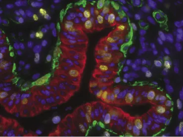
Anatomy (derived from the Greek words meaning “to cut open”) is the study of the morphology, or structure, of organisms. Thus, strictly speaking, anatomy deals with form rather than function. It can be divided into macroscopic (gross) or microscopic anatomy. Macroscopic anatomy deals with structure that can be seen with the naked eye, whereas microscopic anatomy deals with structure that can only be seen with the aid of a microscope. It is also important to appreciate that it is also much more than simply looking at smears of cells or stained tissue sections. Use of immunocytochemistry, fluorescence-labeled markers, multispectral cameras and sophisticated imaging software, and so on is revolutionizing our understanding of cell and tissue biology. Figure 1.1 provides an example of the ability to localize specific proteins within various cells of the mammary gland.
Fig. 1.1. Photomicrograph of a developing mammary duct. Taken from a Holstein calf, this tissue section was stained with specific antibodies and fluorescent tags to detect cell nuclei (blue), cytokeratin 18 (red, a marker specific for epithelial cells), CD10 (green, a marker of myoepithelial cells), and Ki67 (yellow, a protein produced in nuclei of cells that are about to divide). The tissue section is from a study to evaluate the effects of the ovary on ontogeny of myoepithelial cells in the bovine mammary gland. Image is courtesy of Dr. Steve Ellis, Clemson University.
c1-fig-0001
Macroscopic anatomy can be approached in different ways. Regional anatomy, as the name implies, deals with all the structures, such as nerves, bones, muscles, and blood vessels, in a defined region such as the head or hip. Systemic anatomy entails the study of a given organ system such as the muscular or skeletal system. It also involves the study of organ systems that are groups of organs that work together for a specific function, such as the digestive or urinary system. Surface anatomy considers markings that are visible from the outside. These may include knowledge of the muscles, such as sternocleidomastoid muscle, as a landmark to find another structure, such as the carotid artery.
Microscopic anatomy includes cytology and histology. Cytology is the study of the structure of individual cells that constitute the smallest units of life, at least in the sense of animal physiology. Histology is the study of tissues. Tissues are a collection of specialized cells and their products that perform a specific function. Tissues combine to form organs such as the heart, liver, and brain, and will be explained in greater detail in Chapter 4.
Developmental anatomy is the study of the changes in structure that occur throughout life. Embryology is a subdivision of developmental anatomy that traces the developmental changes prior to birth. Many sys­tems of the body are not completely developed at birth, hence the need to continue to follow their development after parturition. Specific to farm mammals, understanding and management of the postnatal devel­opment of the mammary gland and reproductive system are essential for the success of dairies, cow/calf operations, flocks of sheep and goats, piggeries, and so on.
Physiology is the study of the function of living systems. While various systems will be presented separately throughout this book, it must be recognized that all systems work together to maintain the normal functioning of an animal. Therefore, the cardiovascular system does not work in isolation from the respiratory or nervous system, but instead they work in unison to coordinate the distribution of oxygen and removal of carbon dioxide throughout the body. As in anatomy, there are levels of complexity.
Cellular physiology is the study of how cells work. This includes the study of events at the chemical, molecular, and genetic levels. Organ physiology includes the study of specific organs, that is, cardiac or ovarian. Systems physiology includes the study of the functions of specific systems such as the cardiovascular, respiratory, or reproductive systems.
As you study anatomy and physiology, it will become apparent that structure and function have evolved to complement each other. The complementarity of structure and function is an essential concept. At multiple levels, a return to this fundamental idea will hasten your grasp of what sometimes seems to be an overwhelming amount of information and detail. But ultimately, the point is for you to understand how an animal works and to understand limitations. This relationship between form and function is evident beginning at the cellular level. For example, the epithelial cells that line the internal surface of the small intestine have so-called tight junctions that act to restrict the movement of materials into the body from the gastrointestinal tract, whereas the epithelial linings (endothelial cells) of capillaries have modified junctions. The linings of capillaries must be sufficiently porous to allow solutes to move readily in either direction across the capillary wall to nourish the tissue and remove waste products.
As another example, there are structural differences between birds and mammals that allow flight. Birds contain pneumatic bones, that is, bones that are hollow, which are connected to the respiratory system. These bones include the skull, humerus, clavicle, keel, sacrum, and lumbar vertebrae. In addition, the lumbar and sacral vertebrae are fused as an adaptation for flight. This provides yet another example of complementary structure and function.

Levels of organization

The animal body has a complex organization ex­tending from the most microscopic levels up to the macroscopic (Fig. 1.2). Beginning with the smallest microscopic units of stability, the levels of organization are as follows:
  • Chemical level. Atoms are the smallest units of matter that have properties of an element. They combine with covalent bonds to form molecules such as molecular oxygen (O2), glucose (C6H12O6), or methane (CH4). The properties of various chemicals have a major influence on physiology. For example, at a low pH, a chemical may not be ionized and can thus cross a cellular membrane whereas above a certain pH, the same molecule may become ionized and thus unable to cross a lipid bilayer.
  • Cellular level. As the smallest unit of life, cells have various sizes, shapes, and properties that allow them to carry out specialized functions. Some cells have cilium that allow them to move materials across their surfaces (i.e., the epithelial lining the bronchioles or cells lining the oviduct), whereas other cells are adapted to store lipids, produce collagen, or contract when stimulated.
  • Tissue level. A tissue is a group of cells having a common structure and function. The four types of tissue include muscle, epithelia, nervous, and connective tissue.
  • Organ level. Two or more tissues working for a given function form an organ. All four tissue types combine to form skin, the largest organ of the body, or the cochlea in the ear, the smallest organ of the body.
  • Organ system level. Organs can work together for a common function. For example, the alimentary canal works with the liver, gallbladder, and pancreas to form part of the digestive system. The pancreas also functions as part of the endocrine system because of the pancreatic islets that produce insulin and glucagon. The organ systems include the integumentary, skeletal, muscular, nervous, endocrine, respiratory, digestive, lymphatic, urinary, and reproductive systems (Fig. 1.3).
  • Organismal level. The organismal level, or the whole animal, includes all of the organ syste...

Table of contents

  1. Cover
  2. Title page
  3. Copyright page
  4. Preface
  5. Preface to the First Edition
  6. About the Companion Website
  7. 1: Introduction to anatomy and physiology
  8. 2: The cell: The common physiological denominator
  9. 3: Fundamental biochemical pathways and processes in cellular physiology
  10. 4: Tissue structure and organization
  11. 5: Integumentary system
  12. 6: Bones and skeletal system
  13. 7: Muscular tissue
  14. 8: Introduction to the nervous system
  15. 9: Central nervous system
  16. 10: Peripheral and autonomic nervous system
  17. 11: Special senses
  18. 12: Endocrine system
  19. 13: Cardiovascular system
  20. 14: Respiratory system
  21. 15: Immunity
  22. 16: Urinary system
  23. 17: Digestive system
  24. 18: Lactation
  25. 19: Reproduction
  26. Glossary
  27. Index