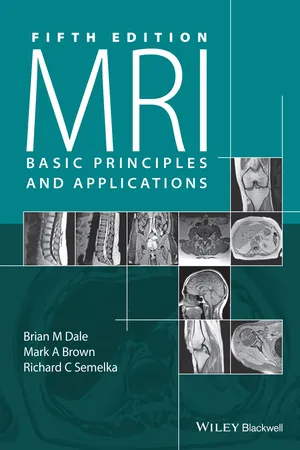
- English
- ePUB (mobile friendly)
- Available on iOS & Android
eBook - ePub
About this book
This fifth edition of the most accessible introduction to MRI principles and applications from renowned teachers in the field provides an understandable yet comprehensive update.
- Accessible introductory guide from renowned teachers in the field
- Provides a concise yet thorough introduction for MRI focusing on fundamental physics, pulse sequences, and clinical applications without presenting advanced math
- Takes a practical approach, including up-to-date protocols, and supports technical concepts with thorough explanations and illustrations
- Highlights sections that are directly relevant to radiology board exams
- Presents new information on the latest scan techniques and applications including 3 Tesla whole body scanners, safety issues, and the nephrotoxic effects of gadolinium-based contrast media
Tools to learn more effectively

Saving Books

Keyword Search

Annotating Text

Listen to it instead
Information
Chapter 1
Production of net magnetization
Magnetic resonance (MR) is a measurement technique used to examine atoms and molecules. It is based upon the interaction between an applied magnetic field and a particle that possesses spin and charge. While electrons and other subatomic particles possess spin (or more precisely, spin angular momentum) and can be examined using MR techniques, this book focuses on nuclei and the use of MR techniques for their study, formally known as Nuclear Magnetic Resonance, or NMR. Nuclear spin, or more precisely nuclear spin angular momentum, is one of several intrinsic properties of an atom and its value depends on the precise atomic composition. Every element in the Periodic Table except argon and cerium has at least one naturally occurring isotope that possesses nuclear spin. Thus, in principle, nearly every element can be examined using MR, and the basic ideas of resonance absorption and relaxation are common for all of these elements. The precise details will vary from nucleus to nucleus and from system to system.
1.1 Magnetic fields

Magnetic fields often vary over time and/or space, and will be coupled to the electric field, producing electromagnetic waves. Magnetic fields, particularly those in electromagnetic waves, are characterized by their frequency (the time between two consecutive “peaks” in the field). In MR, there are magnetic fields, which are constant in time, which vary at acoustic frequencies (a few kilohertz), and which vary at radio frequencies (RF) (several megahertz).
1.2 Nuclear spin
The structure of an atom is an essential component of the MR experiment. Atoms consist of three fundamental particles: protons, which possess a positive charge; neutrons, which have no charge; and electrons, which have a negative charge. The protons and neutrons are located in the nucleus or core of an atom; thus all nuclei are positively charged. The electrons are located in shells or orbitals surrounding the nucleus. The characteristic chemical reactions of elements depend upon the particular number of each of these particles. The properties most commonly used to categorize elements are the atomic number and the atomic weight. The atomic number is the number of protons in the nucleus and is the primary index used to differentiate atoms. All atoms of an element have the same atomic n...
Table of contents
- Cover
- Title Page
- Copyright
- Table of Contents
- Preface
- ABR study guide topics
- Chapter 1: Production of net magnetization
- Chapter 2: Concepts of magnetic resonance
- Chapter 3: Relaxation
- Chapter 4: Principles of magnetic resonance imaging – 1
- Chapter 5: Principles of magnetic resonance imaging – 2
- Chapter 6: Pulse sequences
- Chapter 7: Measurement parameters and image contrast
- Chapter 8: Signal suppression techniques
- Chapter 9: Artifacts
- Chapter 10: Motion artifact reduction techniques
- Chapter 11: Magnetic resonance angiography
- Chapter 12: Advanced imaging applications
- Chapter 13: Magnetic resonance spectroscopy
- Chapter 14: Instrumentation
- Chapter 15: Contrast agents
- Chapter 16: Safety
- Chapter 17: Clinical applications
- References and suggested readings
- Index
- End User License Agreement
Frequently asked questions
Yes, you can cancel anytime from the Subscription tab in your account settings on the Perlego website. Your subscription will stay active until the end of your current billing period. Learn how to cancel your subscription
No, books cannot be downloaded as external files, such as PDFs, for use outside of Perlego. However, you can download books within the Perlego app for offline reading on mobile or tablet. Learn how to download books offline
Perlego offers two plans: Essential and Complete
- Essential is ideal for learners and professionals who enjoy exploring a wide range of subjects. Access the Essential Library with 800,000+ trusted titles and best-sellers across business, personal growth, and the humanities. Includes unlimited reading time and Standard Read Aloud voice.
- Complete: Perfect for advanced learners and researchers needing full, unrestricted access. Unlock 1.4M+ books across hundreds of subjects, including academic and specialized titles. The Complete Plan also includes advanced features like Premium Read Aloud and Research Assistant.
We are an online textbook subscription service, where you can get access to an entire online library for less than the price of a single book per month. With over 1 million books across 990+ topics, we’ve got you covered! Learn about our mission
Look out for the read-aloud symbol on your next book to see if you can listen to it. The read-aloud tool reads text aloud for you, highlighting the text as it is being read. You can pause it, speed it up and slow it down. Learn more about Read Aloud
Yes! You can use the Perlego app on both iOS and Android devices to read anytime, anywhere — even offline. Perfect for commutes or when you’re on the go.
Please note we cannot support devices running on iOS 13 and Android 7 or earlier. Learn more about using the app
Please note we cannot support devices running on iOS 13 and Android 7 or earlier. Learn more about using the app
Yes, you can access MRI by Brian M. Dale,Mark A. Brown,Richard C. Semelka in PDF and/or ePUB format, as well as other popular books in Medicine & Radiology, Radiotherapy & Nuclear Medicine. We have over one million books available in our catalogue for you to explore.