Step by step course companion for dental nurses studying for the Certificate in Oral Health Education. Topics covered include dental structures, anatomy and physiology, oral diseases and prevention, the principles of education, oral health and society, promoting oral health in the 21st century, patient communication, project planning and workplace assignments. This second edition has been thoroughly updated in line with the substantial changes to the role of the dental nurse since the 1st edition was published. To address this, a brand new section has been added on education and research.
eBook - ePub
Basic Guide to Oral Health Education and Promotion
- English
- ePUB (mobile friendly)
- Available on iOS & Android
eBook - ePub
Basic Guide to Oral Health Education and Promotion
SECTION 1
STRUCTURE AND FUNCTIONS OF THE ORAL CAVITY
INTRODUCTION
This section looks at the structure and functions of the oral cavity in some detail. It includes the development of the oral cavity in utero, the structure of the tooth and its supporting tissues, plus eruption dates for primary and secondary dentitions.
It also includes the functions of the tongue in maintaining oral health and common conditions associated with it, plus the composition and role of saliva in keeping the mouth healthy.
Chapter 1
The oral cavity in health
LEARNING OUTCOMES
By the end of this chapter you should be able to:
1. Describe how the oral cavity, jaws and face develop in utero.
2. Explain the structure and function of the tissues and fluid of the oral cavity, including teeth, supporting structures, the tongue and saliva.
3. List primary and secondary dentition eruption dates.
INTRODUCTION
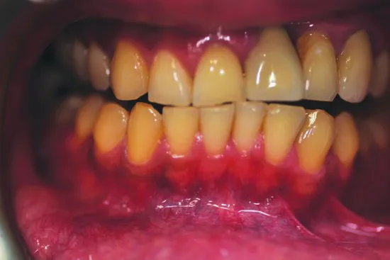
Before oral health educators (OHEs) can deliver dental health messages to patients, and confidently discuss oral care and disease with them, they will need a basic understanding of how the mouth develops in utero, the anatomy of the oral cavity (Figures 1.1, 1.2, 1.3 and 1.4) and how the following structures function within it:
Figure 1.1 Structure of the oral cavity (© Elsevier 2002. Reproduced with permission from Reference 1)
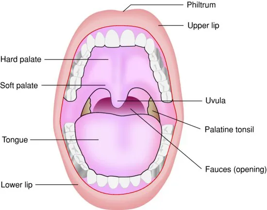
Figure 1.2 A healthy mouth, white person (© John Wiley & Sons, Ltd 2003. Reproduced with permission from Reference 2)
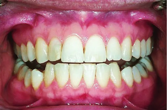
Figure 1.3 A healthy mouth, black person (source: Alison Chapman)
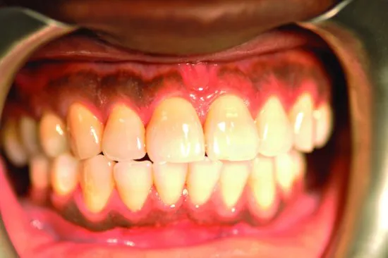
Figure 1.4 A healthy mouth, Asian person (source: Alison Chapman)
- Teeth (including dentition).
- Periodontium (the supporting structure of the tooth).
- Tongue.
- Salivary glands (and saliva).
ORAL EMBRYOLOGY
A basic understanding of the development of the face, oral cavity and jaws in the embryo and developing foetus will enable the OHE to discuss with patients certain oral manifestations of conditions that stem from in utero development (notably cleft lip and palate – Figure 1.5). (An embryo describes the growing organism up to 8 weeks in utero; a foetus describes the growing organism from 8 weeks in utero.)
Figure 1.5 Cleft lip (© iStockphoto.com/April Anderton)

Development of the face
At approximately week 4 in utero (Figure 1.6), the embryo begins to develop five facial processes (projections), which eventually form the face, oral cavity, palate and jaws by week 8 [3]:
Figure 1.6 Facial development at 4 weeks in utero (© John Wiley & Sons, Ltd. Reproduced with permission from Reference 3)
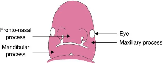
- Frontonasal process – forms the forehead, nose and philtrum (groove in upper lip).
- Maxillary process (two projections) – forms the middle face and upper lip.
- Mandibular process (two projections) – forms the mandible and lower lip.
Development of the palate and nasal cavities
Week 5
The frontonasal and maxillary processes begin to form the nose and maxilla. However, if the nasal and maxillary processes fail to fuse a cleft lip and palate will result [3]. A cleft lip can be anything from a small notch in the lip (incomplete cleft) to a wide gap that runs up to the nostril (complete cleft lip). One type of cleft palate (submucous) can be hidden.
There are two types of cleft lip:
- Unilateral – appears on one side of the lip at the philtrum.
- Bilateral – occurs on both sides of the lip, both sides of the philtrum.
Week 6
By week 6, the primary palate and nasal septum have developed. The septum divides the nasal cavity into two.
Week 8
By week 8, the palate is divided into oral and nasal cavities.
Development of the jaws (mandible and maxilla)
Week 6
By week 6, a band of dense fibrous tissue (Meckel's cartilage) forms and provides the structure around which the mandible forms.
Week 7
By week 7, bone develops, outlining the body of the mandible, and as the bone grows backwards two secondary cartilages develop; these eventually become the condyle and coronoid processes. As the bone grows forward, the two sides are separated by a cartilage called the mandibular symphysis. The two sides will finally fuse into one bone approximately 2 years after birth. Upward growth of bone begins along the mandibular arch forming the alveolar process, which will go on to surround the developing tooth germs.
Week 8
By week 8, ossification (bone development) of the maxilla begins.
Tooth germ development in the foetus
Tooth germ (tissue mass) develops in three stages known as bud, cap and bell. The developing tooth germ can be affected by the mother's health (see Chapter 20).
1. Bud – at 8 weeks, clumps of cells form swellings known as enamel organs. Each enamel organ is responsible for the development of a tooth.
2. Cap – the enamel organ continues to grow and by 12 weeks (the late cap stage), cells have formed the inner enamel epithelium and the outer enamel epithelium. Beneath the inner enamel epithelium, the concentration of cells will eventually become the pulp. The enamel organ is surrounded by a fibrous capsule (the dental follicle), which will eventually form t...
Table of contents
- Cover
- Endorsements
- Title Page
- Copyright
- Foreword
- Preface
- Acknowledgements
- Section 1: Structure and Functions of the Oral Cavity
- Section 2: Diseases and Conditions of the Oral Cavity
- Section 3: Oral Disease Prevention
- Section 4: Delivering Oral Health Messages
- Section 5: Oral Health Target Groups and Case Studies
- Section 6: Oral Health and Society
- Index
Frequently asked questions
Yes, you can cancel anytime from the Subscription tab in your account settings on the Perlego website. Your subscription will stay active until the end of your current billing period. Learn how to cancel your subscription
No, books cannot be downloaded as external files, such as PDFs, for use outside of Perlego. However, you can download books within the Perlego app for offline reading on mobile or tablet. Learn how to download books offline
Perlego offers two plans: Essential and Complete
- Essential is ideal for learners and professionals who enjoy exploring a wide range of subjects. Access the Essential Library with 800,000+ trusted titles and best-sellers across business, personal growth, and the humanities. Includes unlimited reading time and Standard Read Aloud voice.
- Complete: Perfect for advanced learners and researchers needing full, unrestricted access. Unlock 1.5M+ books across hundreds of subjects, including academic and specialized titles. The Complete Plan also includes advanced features like Premium Read Aloud and Research Assistant.
We are an online textbook subscription service, where you can get access to an entire online library for less than the price of a single book per month. With over 1.5 million books across 990+ topics, we’ve got you covered! Learn about our mission
Look out for the read-aloud symbol on your next book to see if you can listen to it. The read-aloud tool reads text aloud for you, highlighting the text as it is being read. You can pause it, speed it up and slow it down. Learn more about Read Aloud
Yes! You can use the Perlego app on both iOS and Android devices to read anytime, anywhere — even offline. Perfect for commutes or when you’re on the go.
Please note we cannot support devices running on iOS 13 and Android 7 or earlier. Learn more about using the app
Please note we cannot support devices running on iOS 13 and Android 7 or earlier. Learn more about using the app
Yes, you can access Basic Guide to Oral Health Education and Promotion by Simon Felton,Alison Chapman,Simon H. Felton in PDF and/or ePUB format, as well as other popular books in Medicine & Dentistry. We have over 1.5 million books available in our catalogue for you to explore.