eBook - ePub
Liver Imaging
MRI with CT Correlation
- English
- ePUB (mobile friendly)
- Available on iOS & Android
eBook - ePub
Liver Imaging
MRI with CT Correlation
About this book
- The first single source work to deal with the two primary radiologic modalities in diagnosing and treating benign and malignant diseases of the liver, presented with clearly laid out MRI and CT correlations. Developed by an editor team led by one of the world's leading authorities in abdominal imaging, Richard C. Semelka MD.
- User-friendly, atlas-style presentation, with over 1500 MRI and CT images in over 320 figures featuring state-of-the-art MR and CT imaging sequences, multidetector row CT images, 3D reformatted images, breath-hold MRI sequences, and cutting-edge MR 3T images
- Highly practical approach for imaging of focal and diffuse liver lesions, complete relevant and systematic (differential) diagnostic information, the latest references to primary literature and clinical evidence, and patient management possibilities
- Reflects a pattern-recognition approach to MRI and CT imaging, assisting with efficient scanning of images and assessment and diagnosis of disorders
Tools to learn more effectively

Saving Books

Keyword Search

Annotating Text

Listen to it instead
Information
Chapter 1
The cross-sectional anatomy of the liver and normal variations
Ersan Altun1, Mohamed El-Azzazi1,2,3,4, and Richard C. Semelka1
1The University of North Carolina at Chapel Hill, Department of Radiology, Chapel Hill, NC, USA
2University of Dammam, Department of Radiology, Dammam, Saudi Arabia
3King Fahd Hospital of the University, Department of Radiology, Khobar, Saudi Arabia
4University of Al Azhar, Department of Radiology, Cairo, Egypt
Knowledge of cross-sectional anatomy of the liver is essential for the determination of localization of disease processes and for their management. To have a good knowledge and sense of the cross-sectional anatomy of the liver on computerized tomography (CT) and magnetic resonance imaging (MRI) studies, the segmental anatomy of the hepatic parenchyma and the anatomy of hepatic fissures, hepatic vessels, and bile ducts should be understood.
Liver anatomy can be described using two different approaches, including morphological anatomy and functional anatomy (1).
Morphological anatomy of the liver describes the liver anatomy depending on external appearance of the liver (1). Four lobes of the liver including the right, left, caudate and quadrate can be identified on the basis of the fissures of the liver surface (1). Morphological anatomy is not sufficient for the needs of modern radiology, hepatology, and hepatobiliary surgery.
Functional anatomy of the liver describes the functional segments of the liver on the basis of the anatomy of hepatic vessels and bile ducts (1). Functional anatomy is necessary to meet the needs of modern radiology, hepatology, and hepatobiliary surgery. Functional anatomy of the liver has been described by a number of different nomenclature systems for the determination of anatomic segments of the liver. A single, universally accepted classification system for the functional segmental anatomy of the liver does not exist. The Goldsmith and Woodburne system (1957), the Couinaud system (1957) and the Bismuth system (1982) are the most commonly used nomenclaturel systems (1).
Functional anatomy
The segments of the liver
The Bismuth system, which is a modified version of the Couinaud system, is the most commonly used anatomic nomenclature system, particularly in the United States. This hepatic segmental nomenclature system meets the needs of modern surgical techniques (Table 1.1) (1–5) and allows hepatobiliary surgeons, hepatologists, and radiologists to use a common nomenclature that meets their needs and enables them to understand each other.
Table 1.1 Description of the liver segments according to the three most commonly used nomenclature systems.
| Part | Nomenclature system | |||||
| N. Goldsmith and R. Woodburne (1957) | C. Couinaud (1957) | H. Bismuth (1982) | ||||
| Segment | Subsegment | Sector | Segment | Sector | Segment | |
| Dorsal | Caudate L. | Caudate L. | I | Caudate L. | I | |
| Left | Lateral | Superior | Lateral | II | Posterior | II |
| Inferior | Paramedian | III | Anterior | III | ||
| Left | Medial | Superior | IV | IVa | ||
| Inferior | IVb | |||||
| Right | Anterior | Inferior | Paramedian | V | Anteromedial | V |
| Superior | VIII | VIII | ||||
| Right | Posterior | Inferior | Lateral | VI | Posterolateral | VI |
| Superior | VII | VII | ||||
The three vertical planes (scissurae) hosting the hepatic veins, and a transverse plane passing through the right and left portal vein branches are used to describe the segments of the liver (1, 5).
The three vertical scissurae hosting the hepatic veins divide the liver into four sectors and a transverse plane passing through the right and left portal vein branches divides these sectors into the eight segments, which are numbered clockwise on the frontal view. These segments can be described in a straightforward approach by combining the definitions of two systems including the Bismuth, and Goldsmith and Woodburne systems (Table 1.1). These liver segments, including the caudate lobe, can be described on the basis of this approach as follows: caudate lobe (I), left lateral superior (II), left lateral inferior (III), left medial superior (IVa), left medial inferior (IVb), right anterior inferior (V), right posterior inferior (VI), right posterior superior (VII), and right anterior superior (VIII) (Figures 1.1 and 1.2).
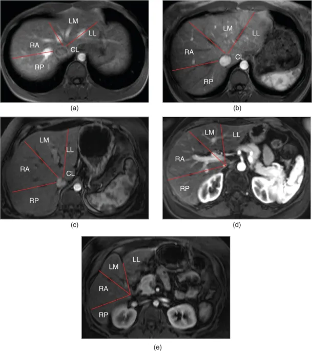
Figure 1.1 Segments of the Liver. T1-weighted axial hepatic venous (a) and hepatic arterial dominant (b–e) phase 3D-GE images acquired at different levels demonstrate the segments of liver, which are determined based on the distribution of diagonal planes (lines) hosting hepatic veins according to Goldsmith and Woodburne classification. RP, Right lobe posterior segment; RA, Right lobe anterior segment; LM, Left lobe medial segment; LL, Left lobe lateral segment; CL, Caudate lobe.
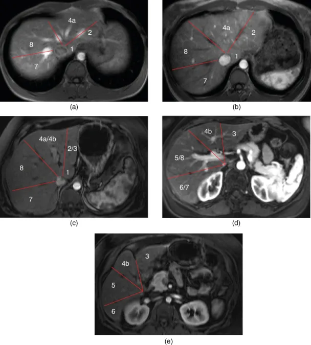
Figure 1.2 Segments of the Liver. T1-weighted axial hepatic venous (a) and hepatic arterial dominant (b–e) phase 3D-GE images acquired at different levels demonstrate the segments of liver, which are determined based on the distribution of diagonal planes hosting hepatic veins (lines) and transverse planes hosting portal veins according to Bismuth classification. 1: Caudate lobe. 2: Left Lateral inferior segment. 3: Left lateral superior segment. 4a: Left medial superior segment. 4b: Left medial inferior segment. 5: Right anterior inferior segment. 6: Right posterior inferior segment. 7: Right posterior superior segment. 8: Right anterior superior segment.
In the Bismuth system, each segment has an independent vascular supply, including arterial, portal, and venous supplies, as well as independent lymphatic and biliary drainage (1–5).
The caudate lobe has been described as a separate sector in the Bismuth system (1, 5). The caudate lob ...
Table of contents
- Cover
- Title Page
- Series Page
- Copyright
- Table of Contents
- List of Contributors
- Preface
- Chapter 1: The cross-sectional anatomy of the liver and normal variations
- Chapter 2: The cross-sectional imaging techniques and diagnostic approach
- Chapter 3: Safety of MRI and CT
- Chapter 4: Cystic diseases of the liver
- Chapter 5: Benign solid liver lesions
- Chapter 6: Liver metastases
- Chapter 7: Hepatocellular carcinoma
- Chapter 8: Rare primary and secondary tumors of the liver
- Chapter 9: Cholangiocarcinoma
- Chapter 10: Infectious diseases of the liver
- Chapter 11: Chronic hepatitis and liver cirrhosis
- Chapter 12: Hepatic fat and iron deposition
- Chapter 13: Inflammatory liver diseases
- Chapter 14: Vascular disorders of the liver
- Chapter 15: Post-treatment changes in the liver
- Chapter 16: Liver trauma
- Index
- End User License Agreement
Frequently asked questions
Yes, you can cancel anytime from the Subscription tab in your account settings on the Perlego website. Your subscription will stay active until the end of your current billing period. Learn how to cancel your subscription
No, books cannot be downloaded as external files, such as PDFs, for use outside of Perlego. However, you can download books within the Perlego app for offline reading on mobile or tablet. Learn how to download books offline
Perlego offers two plans: Essential and Complete
- Essential is ideal for learners and professionals who enjoy exploring a wide range of subjects. Access the Essential Library with 800,000+ trusted titles and best-sellers across business, personal growth, and the humanities. Includes unlimited reading time and Standard Read Aloud voice.
- Complete: Perfect for advanced learners and researchers needing full, unrestricted access. Unlock 1.4M+ books across hundreds of subjects, including academic and specialized titles. The Complete Plan also includes advanced features like Premium Read Aloud and Research Assistant.
We are an online textbook subscription service, where you can get access to an entire online library for less than the price of a single book per month. With over 1 million books across 990+ topics, we’ve got you covered! Learn about our mission
Look out for the read-aloud symbol on your next book to see if you can listen to it. The read-aloud tool reads text aloud for you, highlighting the text as it is being read. You can pause it, speed it up and slow it down. Learn more about Read Aloud
Yes! You can use the Perlego app on both iOS and Android devices to read anytime, anywhere — even offline. Perfect for commutes or when you’re on the go.
Please note we cannot support devices running on iOS 13 and Android 7 or earlier. Learn more about using the app
Please note we cannot support devices running on iOS 13 and Android 7 or earlier. Learn more about using the app
Yes, you can access Liver Imaging by Ersan Altun, Mohamed El-Azzazi, Richard C. Semelka, Ersan Altun,Mohamed El-Azzazi,Richard C. Semelka in PDF and/or ePUB format, as well as other popular books in Medicine & Radiology, Radiotherapy & Nuclear Medicine. We have over one million books available in our catalogue for you to explore.