![]() PART 1
PART 1
Dermatologic Surgery![]()
CHAPTER 1
Cutaneous Anatomy in Dermatologic Surgery
Diana Bolotin1, Lucille White2, and Murad Alam3
1Section of Dermatology, University of Chicago, Chicago, IL, USA
2Pearland Dermatology, Pearland, TX, USA
3Department of Dermatology, Northwestern University, Chicago, IL, USA
Introduction
Knowledge of anatomy is essential to every surgeon. The subtleties of surface anatomy require a special, fine-tuned understanding and are meaningful for a successful dermatologic procedure in both cutaneous oncology and cosmetic surgery. Understanding the cutaneous anatomy of the head and neck is essential in directing appropriate anesthesia, reducing postoperative complications, and providing for an acceptable cosmetic outcome. The anatomy of the face is often regarded in terms of cosmetic subunits and will be discussed as such in this chapter.
Scalp and Forehead
The frontal hairline separates the forehead from the scalp superiorly and laterally, and the temporal region is separated from the scalp by the temporal hairline.1 The forehead ends at the zygomatic arch inferiorly, while the inferior scalp is separated from the neck by the nuchal line inferiorly. The anatomy of the layers of the scalp can be recalled by the mnemonic S-C-A-L-P, which refers to the skin, connective tissue, aponeurotic galeal layer, loose connective tissue, and periosteum. In its most anterior segment, the skin of the scalp measures about 3–4 mm in thickness and reaches up to 8 mm in the more posterior segments. Blood vessels, lymphatics, and cutaneous adnexa reside within the connective tissue layers, while the underlying musculature connects to the galea aponeurotica. Muscles of the forehead and scalp include frontalis, temporalis, procerus, corrugator supercilii, and superior fibers of the orbicularis oculi (Figure 1.1a). Deep to the aponeurotic layer is the loose connective tissue that is largely avascular but does contain perforating emissary veins. Lastly, the periosteum envelops the bony skull and contains another layer of vasculature.
Vasculature
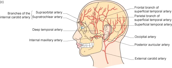
The blood supply to the forehead and scalp subunits is provided by branches of both the internal and the external carotid arteries. The supratrochlear and supraorbital arteries supply the central forehead and anterior scalp and originate from the ophthalmic artery branch of the internal carotid (Figure 1.1b). The lateral forehead and scalp are supplied by the superficial temporal and posterior auricular branches of the external carotid artery (Figure 1.1c). The posterior scalp is supplied by the occipital artery, another branch of the external carotid (Figure 1.1c).
Nerves
The motor and sensory anatomy of the forehead and scalp are a crucial part of surgery on these subunits. Motor innervation of the forehead is provided by the temporal branch of the facial nerve (CN VII). This branch runs along the temple and the zygomatic arch but courses more superficially superiorly to innervate the frontalis muscle at its deep surface. It is therefore most susceptible to injury at its superficial course, which may result in ipsilateral brow ptosis and pronounced asymmetry of the face (Figure 1.1a).
The sensory innervation of the forehead and scalp is provided by branches of all three divisions of the trigeminal nerve (CN V). The supraorbital and supratrochlear nerves branch off from the ophthalmic nerve (CNV1) to supply the scalp up to the vertex (Figure 1.1a). The zygomaticotemporal nerve, arising from the maxillary division of the trigeminal nerve (CN V2), supplies sensory innervations to the anterior temple. The auriculotemporal nerve, a branch of the mandibular division (CN V3), supplies the rest of the temporal area. Branches of the cervical spinal nerves (C2, C3) innervate the posterior scalp.
Lymphatic Drainage
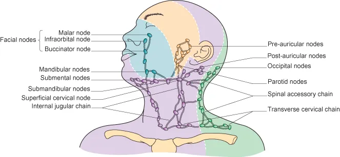
The lymphatic drainage of the scalp is collected by the occipital and posterior auricular lymph nodes (Figure 1.2). The basin responsible for lymph drainage of the forehead subunit is located within the parotid glands bilaterally.
Mid-Face
Nasal Subunit
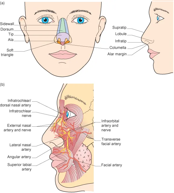
The nasal subunit is generally further subdivided into a number of cosmetic subunits.2 These consist of the nasal sidewall, nasal dorsum, tip, alae, soft triangles, and the columella (Figure 1.3a). To achieve the best cosmetic outcome, it is recommended that incision lines during nasal reconstruction are placed at the borders of these cosmetic units. The nasal cartilaginous structures, consisting of the lateral nasal and the lower lateral cartilage, are essential to the integrity of the nose (Figure 1.3a). Certain infiltrative tumors may infiltrate the lower lateral nasal cartilage, requiring its excision. Failure to properly repair the cartilage in this situation may result in the loss of alar support leading to collapse of the nasal ala and inhibition of air flow into the nose.
Vasculature
Similar to the forehead and scalp region, the vasculature of the nose is derived from both the internal and external carotid arteries.3 In fact, arteries supplying the nasal area make up one of the essential anastomosing sites between the internal and external carotid arteries. The dorsal nasal and external nasal branches of the ophthalmic artery, which branches off from the external carotid, supply the dorsal nose (Figure 1.3b). Vascular supply to the nasal sidewalls, columella, and nasal alae is provided by branches of the angular artery, a branch of the facial artery that originates off from the external carotid (Figure 1.3b).
Nerves
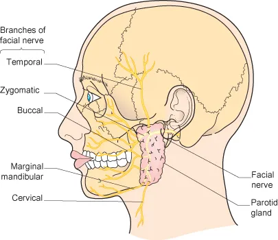
Motor innervations to the procerus muscle at the nasal root, depressor septi nasi and nasalis muscles are provided by the zygomatic and buccal branches of the facial nerve (CN VII) (Figure 1.4). The ophthalmic and maxillary divisions of the trigeminal nerve provide sensory innervation to the nose. The infratrochlear and external nasal branches of the ophthalmic division (CN V1) and the infraorbital branch of the maxillary division (CN V2) are the primary sources of sensory innervation for this subunit.
Lymphatic Drainage
Lymphatic drainage from the nose is collected primarily by the submandibular lymph nodes (Figure 1.2).
Perioral
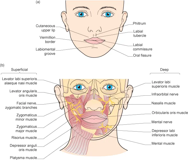
The surface anatomy of the lip is divided into the lateral wings of the cutaneous upper lip, philtrum, lower lip and the vermillion border which demarcates the red and white portion of the lips (Figure 1.5a).4 The underlying musculature includes the orbicularis oris, zygomaticus major and minor, levator anguli oris, depressor anguli oris, levator labii superioris, depressor labii inferioris, risorius, mentalis, and the buccinators (Figure 1.5b). The nasolabial crease is formed by the cutaneous insertion of lip elevator musculature.
Vasculature
Vascular supply to the lips is provided by the labial arteries that branch off the facial artery. Labial arteries are frequently resected during biopsies and lip surgery. Intraoperative ligation or electrosurgery is usually sufficient to prevent excessive bleeding.
Nerves
Perioral musculature is innervated by the zygomatic, buccal, marginal mandibular, and cervical branches of the facial nerve (CN VII) (Figure 1.4). The maxillary division (CN V2) provides sensory innervations to the upper perioral region via the infraorbital nerve. The mandibular division (CN V3) contributes to the sensory innervation of the lower lip via the mental nerve.
Lympha...