Color Atlas of Small Animal Anatomy
The Essentials
Thomas O. McCracken, Robert A. Kainer, David Carlson
- English
- ePUB (handyfreundlich)
- Über iOS und Android verfügbar
Color Atlas of Small Animal Anatomy
The Essentials
Thomas O. McCracken, Robert A. Kainer, David Carlson
Über dieses Buch
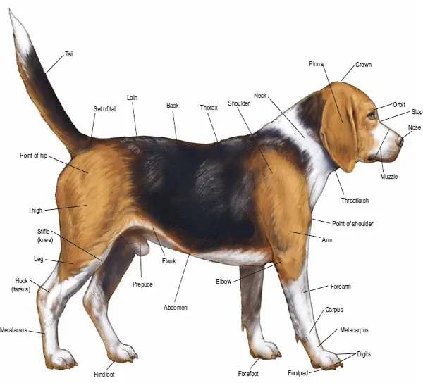
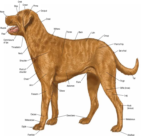
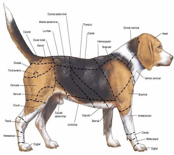
This new resource provides a basic foundation in small animal anatomy for students of veterinary medicine, animal science, and veterinary technology. Extraordinary accuracy and beautiful original artwork make this a truly unique learning tool that includes the anatomy of all organ systems in the dog, cat, rabbit, rat, and guinea pig - all described in a consistent manner.
Learning features include: carefully selected labeling helps students learn and remember structures and relationships; male and female of species are depicted on facing pages so topographic anatomy can be compared; structures common to various animals are labeled several times, whereas unique structures are labeled on one or two species so students can make rapid distinctions of the structures peculiar to certain animals; and an introduction that provides readers with a background in nomenclature and anatomic orientation so they can benefit from the atlas even if they lack training in anatomy. The Atlas depicts topographic relationships of major organs in a simple, yet technically accurate presentation that's free from extraneous material so that those using the atlas can concentrate on the essential aspects of anatomy. It will be an invaluable resource for veterinary students, teachers and practitioners alike.
Häufig gestellte Fragen
Information
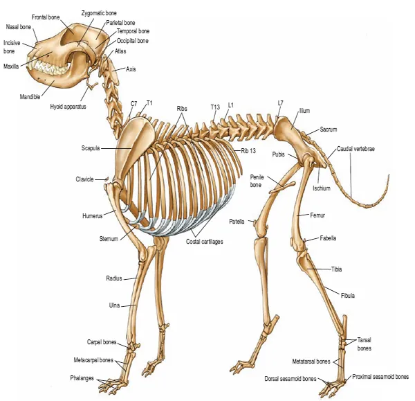
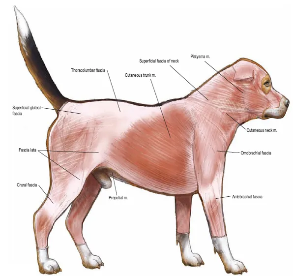
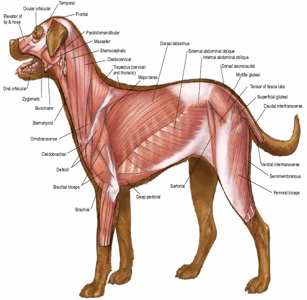
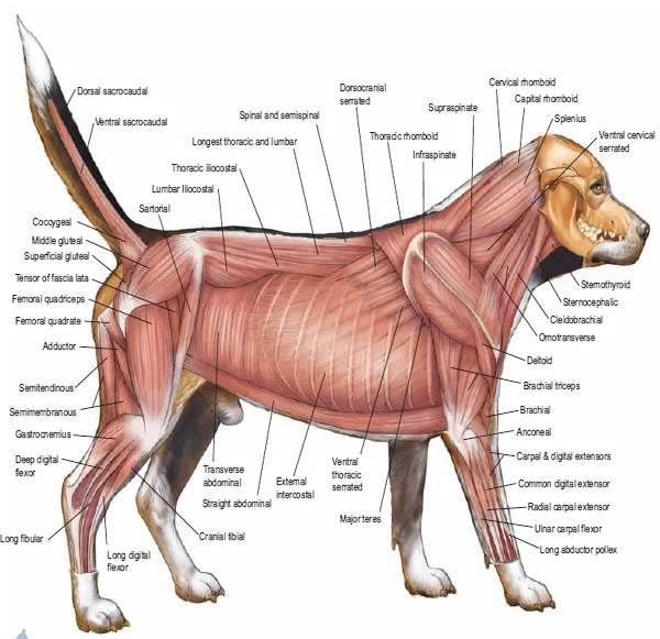
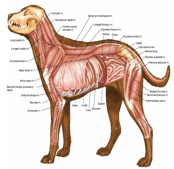
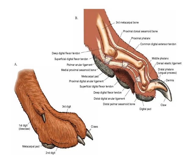
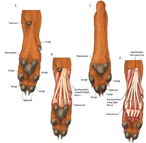
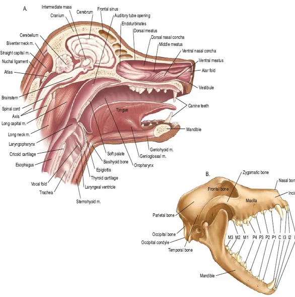
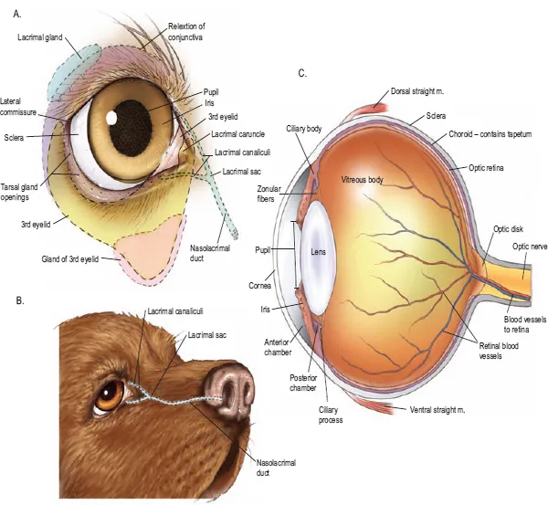
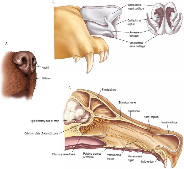
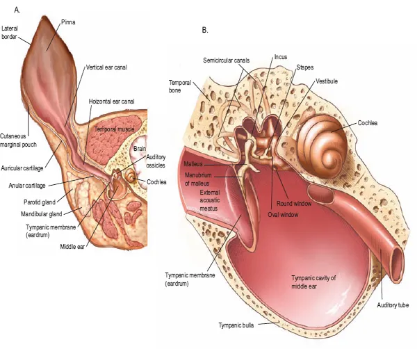
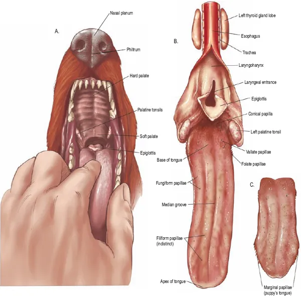
SECTION 1 THE DOG
Inhaltsverzeichnis
- COVER
- CONTENTS
- TITLE PAGE
- COPYRIGHT
- INTRODUCTION
- NOMENCLATURE AND ANATOMIC ORIENTATION
- SECTION 1 THE DOG
- SECTION 2 THE CAT
- SECTION 3 THE RABBIT
- SECTION 4 THE RAT
- SECTION 5 THE GUINEA PIG
- INDEX