Color Atlas of Small Animal Anatomy
eBook - ePub

Color Atlas of Small Animal Anatomy

The Essentials

Thomas O. McCracken, Robert A. Kainer, David Carlson

  1. English
  2. ePUB (apto para móviles)
  3. Disponible en iOS y Android
eBook - ePub

Color Atlas of Small Animal Anatomy

The Essentials

Thomas O. McCracken, Robert A. Kainer, David Carlson

Detalles del libro
Vista previa del libro
Índice
Citas

Información del libro

This new resource provides a basic foundation in small animal anatomy for students of veterinary medicine, animal science, and veterinary technology. Extraordinary accuracy and beautiful original artwork make this a truly unique learning tool that includes the anatomy of all organ systems in the dog, cat, rabbit, rat, and guinea pig - all described in a consistent manner.

Learning features include: carefully selected labeling helps students learn and remember structures and relationships; male and female of species are depicted on facing pages so topographic anatomy can be compared; structures common to various animals are labeled several times, whereas unique structures are labeled on one or two species so students can make rapid distinctions of the structures peculiar to certain animals; and an introduction that provides readers with a background in nomenclature and anatomic orientation so they can benefit from the atlas even if they lack training in anatomy. The Atlas depicts topographic relationships of major organs in a simple, yet technically accurate presentation that's free from extraneous material so that those using the atlas can concentrate on the essential aspects of anatomy. It will be an invaluable resource for veterinary students, teachers and practitioners alike.

Preguntas frecuentes

¿Cómo cancelo mi suscripción?
Simplemente, dirígete a la sección ajustes de la cuenta y haz clic en «Cancelar suscripción». Así de sencillo. Después de cancelar tu suscripción, esta permanecerá activa el tiempo restante que hayas pagado. Obtén más información aquí.
¿Cómo descargo los libros?
Por el momento, todos nuestros libros ePub adaptables a dispositivos móviles se pueden descargar a través de la aplicación. La mayor parte de nuestros PDF también se puede descargar y ya estamos trabajando para que el resto también sea descargable. Obtén más información aquí.
¿En qué se diferencian los planes de precios?
Ambos planes te permiten acceder por completo a la biblioteca y a todas las funciones de Perlego. Las únicas diferencias son el precio y el período de suscripción: con el plan anual ahorrarás en torno a un 30 % en comparación con 12 meses de un plan mensual.
¿Qué es Perlego?
Somos un servicio de suscripción de libros de texto en línea que te permite acceder a toda una biblioteca en línea por menos de lo que cuesta un libro al mes. Con más de un millón de libros sobre más de 1000 categorías, ¡tenemos todo lo que necesitas! Obtén más información aquí.
¿Perlego ofrece la función de texto a voz?
Busca el símbolo de lectura en voz alta en tu próximo libro para ver si puedes escucharlo. La herramienta de lectura en voz alta lee el texto en voz alta por ti, resaltando el texto a medida que se lee. Puedes pausarla, acelerarla y ralentizarla. Obtén más información aquí.
¿Es Color Atlas of Small Animal Anatomy un PDF/ePUB en línea?
Sí, puedes acceder a Color Atlas of Small Animal Anatomy de Thomas O. McCracken, Robert A. Kainer, David Carlson en formato PDF o ePUB, así como a otros libros populares de Medicina y Medicina veterinaria. Tenemos más de un millón de libros disponibles en nuestro catálogo para que explores.

Información

Año
2013
ISBN
9781118692110
Edición
1
Categoría
Medicina

SECTION 1 THE DOG


PLATES
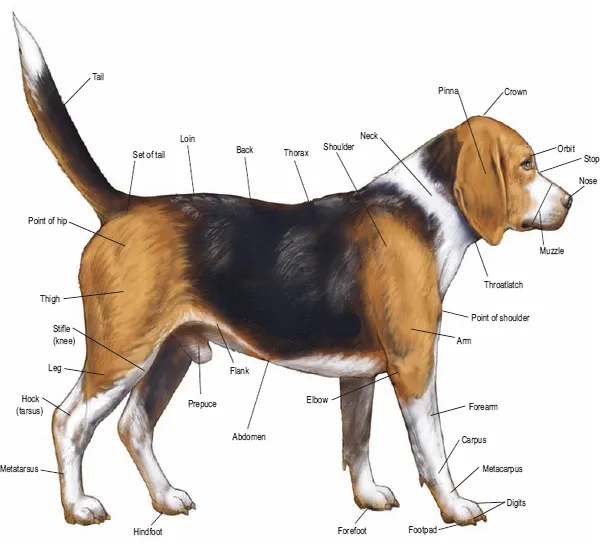
1.1 Surface view of the dog (Beagle)
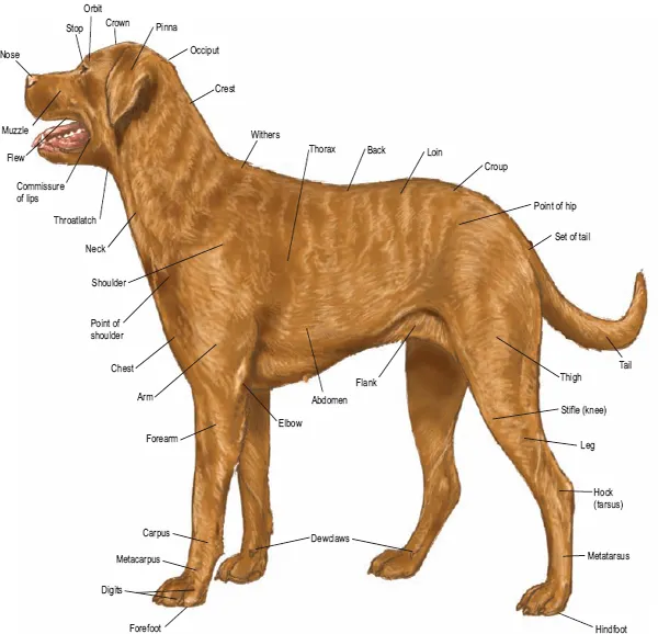
1.2 Surface view of the bitch (Chesapeake Bay Retriever)
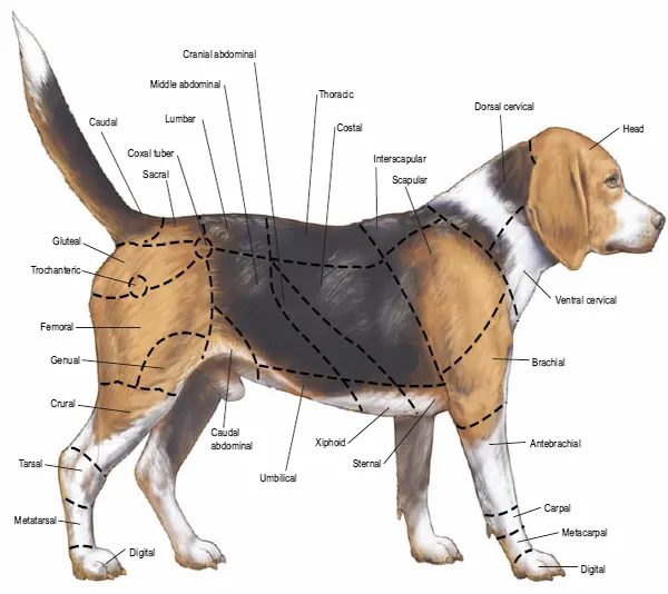
1.3 Body regions
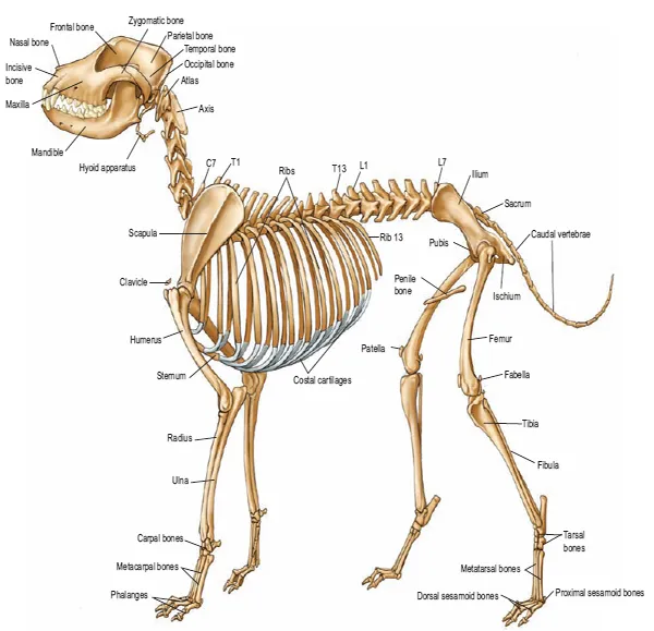
1.4 Skeleton
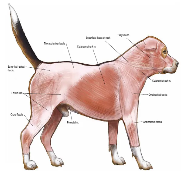
1.5 Cutaneous muscles and major fasciae of the dog
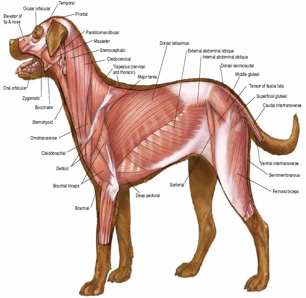
1.6 Superficial muscles of the bitch
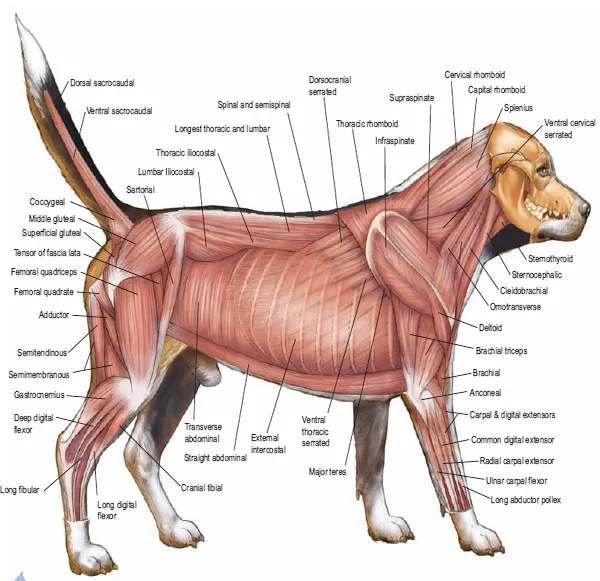
1.7 Deep muscles of the dog
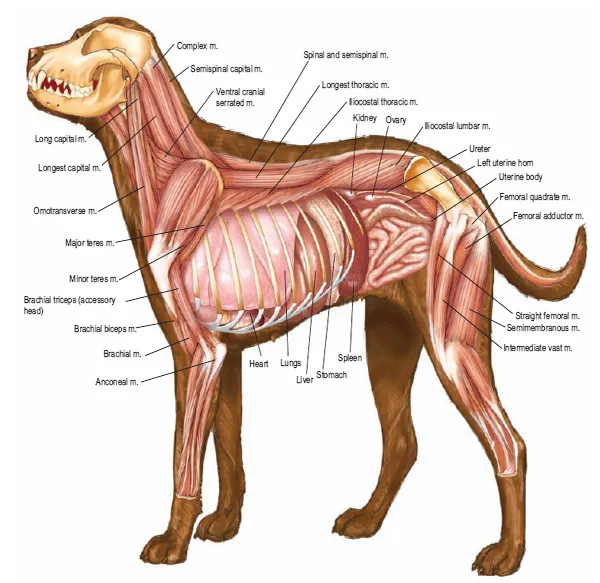
1.8 Deep cervical muscles, major joints, and in situ viscera of the bitch
1.9 Paraxial and medial view of the third digit
1.10 Palmar views of the major structures of the forepaw; plantar view of the major structures of the hindpaw
1.11 Median section of the head and dentition
1.12 Eye and adnexal ocular structures
1.13 Nasal fossae
1.14 External, middle and inner ear
1.15 Oral cavity, tongue, and esophagus
1.16 Ventral view of abdominal structures
1.17 Large intestine, anus and anal sacs
1.18 Body cavities and serous membranes
1.19 Thoracic, abdominal and pelvic viscera related to the skeleton of the dog
1.20 Thoracic, abdominal and pelvic viscera related to the skeleton of the bitch
1.21 Hip joint
1.22 Location of major endocrine organs
1.23 Relations of the reproductive organs of the dog
1.24 Relations of the reproductive organs of the bitch
1.25 Major veins
1.26 Major arteries
1.27 Lymph nodes and vessels
1.28 Central nervous system and spinal nerves
1.29 Autonomic nervous system
1.30 Brain

PLATE 1.1 Right lateral view of a male Beagle dog.
img_002_001.gif
PLATE 1.2 Left lateral view of a female Chesapeake Bay Retriever.
img_003_001.gif
PLATE 1.3 Body regions.
img_004_001.gif
PLATE 1.4 Skeleton; C = Cervical vertebrae, T = Thoracic vertebrae, L = Lumbar vertebrae.
img_005_001.gif
PLATE 1.5 Cutaneous muscles and major fasciae of the dog.
img_006_001.gif
PLATE 1.6 Superficial muscles of the bitch.
img_007_001.gif
PLATE 1.7 Deep muscles of the dog.
img_008_001.gif
PLATE 1.8 Deep cervical and back muscles (m.), major joints, and in situ viscera of the bitch.
img_009_001.gif
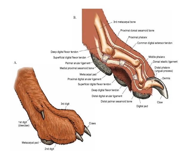
PLATE 1.9 Forepaw: A. Medial superficial view, B. Medial deep view.
img_010_001.gif
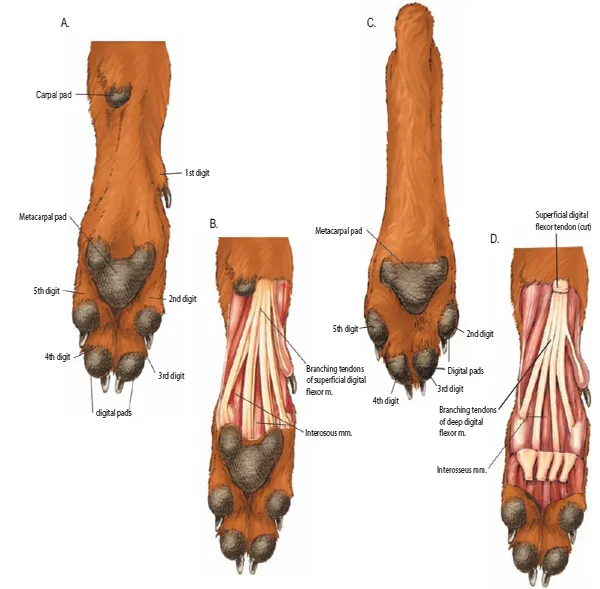
PLATE 1.10 A. Palmar view of the forepaw; B. Palmar view of superficial dissection of forepaw; C. Plantar view of hindpaw; D. Plantar view of superficial dissection of hindpaw.
img_011_001.gif
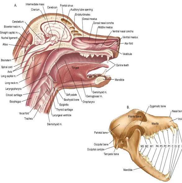
PLATE 1.11 A. Medial sagittal section of head. B. Skull and dentition (M- molar, I- incisor, C - canine, P - premolar).
img_012_001.gif
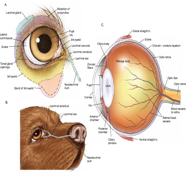
PLATE 1.12 A. The eye and adnexal ocular structures. B. Nasolacrimal duct. C. Median section of eye.
img_013_001.gif
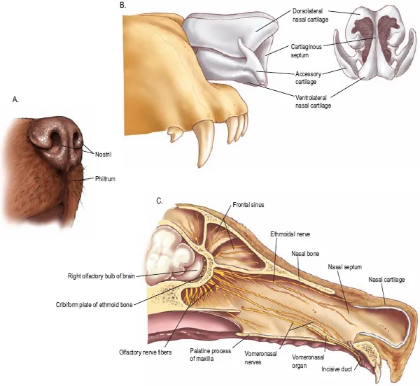
PLATE 1.13 Rt. nasal fossa. A. External nares. B. Nasal cartilages. C. Medial sagittal section of the muzzle.
img_014_001.gif
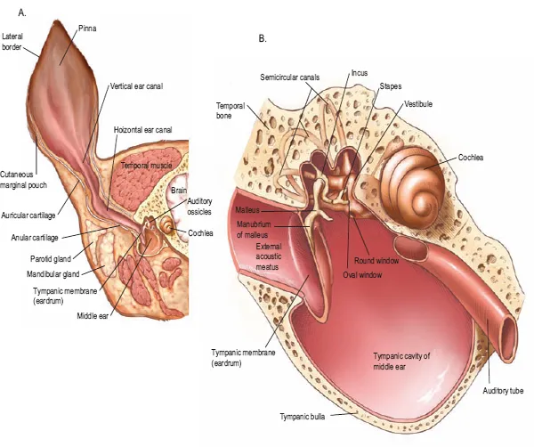
PLATE 1.14 The ear. A. Coronal section of the head and external ear. B. Middle and inner ear.
img_015_001.gif
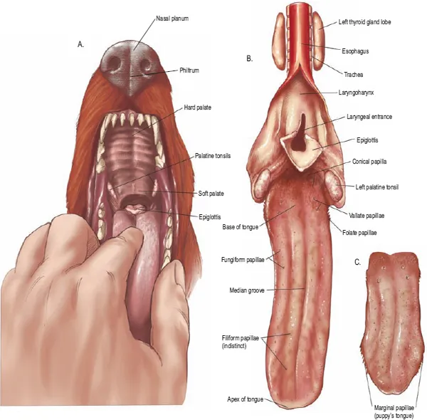
PLATE 1.15 Oral cavity, tongue, and esophagus
img_016_001.gif
P...

Índice

  1. COVER
  2. CONTENTS
  3. TITLE PAGE
  4. COPYRIGHT
  5. INTRODUCTION
  6. NOMENCLATURE AND ANATOMIC ORIENTATION
  7. SECTION 1 THE DOG
  8. SECTION 2 THE CAT
  9. SECTION 3 THE RABBIT
  10. SECTION 4 THE RAT
  11. SECTION 5 THE GUINEA PIG
  12. INDEX
Estilos de citas para Color Atlas of Small Animal Anatomy

APA 6 Citation

McCracken, T., Kainer, R., & Carlson, D. (2013). Color Atlas of Small Animal Anatomy (1st ed.). Wiley. Retrieved from https://www.perlego.com/book/1008555/color-atlas-of-small-animal-anatomy-the-essentials-pdf (Original work published 2013)

Chicago Citation

McCracken, Thomas, Robert Kainer, and David Carlson. (2013) 2013. Color Atlas of Small Animal Anatomy. 1st ed. Wiley. https://www.perlego.com/book/1008555/color-atlas-of-small-animal-anatomy-the-essentials-pdf.

Harvard Citation

McCracken, T., Kainer, R. and Carlson, D. (2013) Color Atlas of Small Animal Anatomy. 1st edn. Wiley. Available at: https://www.perlego.com/book/1008555/color-atlas-of-small-animal-anatomy-the-essentials-pdf (Accessed: 14 October 2022).

MLA 7 Citation

McCracken, Thomas, Robert Kainer, and David Carlson. Color Atlas of Small Animal Anatomy. 1st ed. Wiley, 2013. Web. 14 Oct. 2022.