CT at a Glance gets readers quickly up to speed with the core knowledge and competencies required for computed tomography (CT) scanning, as established by the major radiography organizations around the world, including the ASRT and the CAMRT. This brand new title describes the basic science behind CT with an emphasis on the theory that is essential for practice. Featuring an abundance of illustrations, succinct, straightforward explanations and clear, step-by-step guidance, it includes the fundamental physics, technical principles, and imaging strategies and procedures involved in CT scanning.
Over the course of twenty four, concise modular chapters, CT at a Glance covers all the bases for entry-to-practice students, including:
The basic physics underlying CT scanning
State-of-the-art multi-slice technologies
Data acquisition strategies
Equipment components—their functions and applications
Image reconstruction and image quality control
CT dose and dose optimization procedures
Quality control fundamentals
CT at a Glance is an indispensable learning resource for students in medical imaging technology courses, including those covering radiography, nuclear medicine, and radiation therapy, as well as for biomedical engineering technology students.
Häufig gestellte Fragen
Wie kann ich mein Abo kündigen?
Gehe einfach zum Kontobereich in den Einstellungen und klicke auf „Abo kündigen“ – ganz einfach. Nachdem du gekündigt hast, bleibt deine Mitgliedschaft für den verbleibenden Abozeitraum, den du bereits bezahlt hast, aktiv. Mehr Informationen hier.
(Wie) Kann ich Bücher herunterladen?
Derzeit stehen all unsere auf Mobilgeräte reagierenden ePub-Bücher zum Download über die App zur Verfügung. Die meisten unserer PDFs stehen ebenfalls zum Download bereit; wir arbeiten daran, auch die übrigen PDFs zum Download anzubieten, bei denen dies aktuell noch nicht möglich ist. Weitere Informationen hier.
Welcher Unterschied besteht bei den Preisen zwischen den Aboplänen?
Mit beiden Aboplänen erhältst du vollen Zugang zur Bibliothek und allen Funktionen von Perlego. Die einzigen Unterschiede bestehen im Preis und dem Abozeitraum: Mit dem Jahresabo sparst du auf 12 Monate gerechnet im Vergleich zum Monatsabo rund 30 %.
Was ist Perlego?
Wir sind ein Online-Abodienst für Lehrbücher, bei dem du für weniger als den Preis eines einzelnen Buches pro Monat Zugang zu einer ganzen Online-Bibliothek erhältst. Mit über 1 Million Büchern zu über 1.000 verschiedenen Themen haben wir bestimmt alles, was du brauchst! Weitere Informationen hier.
Unterstützt Perlego Text-zu-Sprache?
Achte auf das Symbol zum Vorlesen in deinem nächsten Buch, um zu sehen, ob du es dir auch anhören kannst. Bei diesem Tool wird dir Text laut vorgelesen, wobei der Text beim Vorlesen auch grafisch hervorgehoben wird. Du kannst das Vorlesen jederzeit anhalten, beschleunigen und verlangsamen. Weitere Informationen hier.
Ist CT at a Glance als Online-PDF/ePub verfügbar?
Ja, du hast Zugang zu CT at a Glance von Euclid Seeram im PDF- und/oder ePub-Format sowie zu anderen beliebten Büchern aus Medicine & Medical Technology & Supplies. Aus unserem Katalog stehen dir über 1 Million Bücher zur Verfügung.
A significant and important technological innovation that has now become a popular tool for diagnostic imaging of patients is computed tomography (CT), an imaging technique that was first investigated as early as 1967. Later, in 1971, a prototype CT scanner for imaging the brain was developed by EMI Limited (Electric and Musical Industries [a manufacturer of records and electronics; the Beatles recorded under the EMI label], now Thorn EMI) in Middlesex, UK. This prototype resulted in the first patient being scanned in 1971, and this development earned two pioneers of CT, Godfrey Hounsfield and Allan Cormack, the Nobel Prize in Medicine in 1979.
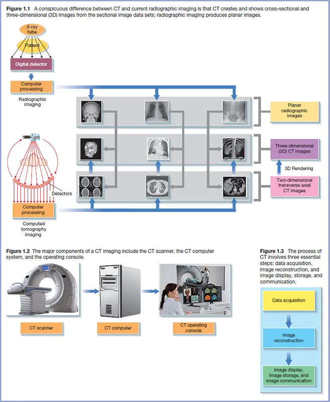
A striking fundamental difference between CT and current radiographic imaging is clearly illustrated in Figure 1.1 While CT creates and shows cross-sectional and three-dimensional (3D) images from the sectional image data sets, radiographic imaging produces planar images. There are other differences between these two imaging modalities, which will be described in later chapters. One such example is that CT uses much more sensitive electronic detectors, which can show very small differences in tissue attenuation compared to radiographic detectors. This characteristic results in CT providing much better tissue image contrast than radiography, and therefore the observer can see soft tissues much better than with radiography.
Radiographic imaging
The major components of radiographic imaging includes an X-ray tube and generator that provide the appropriate X-rays to image the patient, a detector that captures X-rays transmitted through the patient, a computer processing system, and an image display workstation (Figure 1.1). X-rays transmitted through the patient are converted into digital data for processing by the computer. The image output from the computer is subsequently displayed for viewing and interpretation by an observer. These radiographic images are usually referred to as planar images. The problems with these images are (i) superimposition of all structures on the detector (which makes it difficult and sometimes impossible to distinguish a particular detail) and (ii) the qualitative nature of radiographic imaging. The latter simply means that it is difficult to distinguish between a homogeneous object (one tissue type) of non-uniform thickness and a heterogeneous object (bone, soft tissue, and air) of uniform thickness. Finally, the beam used in radiography is an open beam (wide beam) and this creates more scattered rays, which get to the image and essentially destroy the image contrast.
CT imaging
CT overcomes these limitations by removing the superimposition of structures, improving image contrast, and imaging very small differences in tissue contrast.
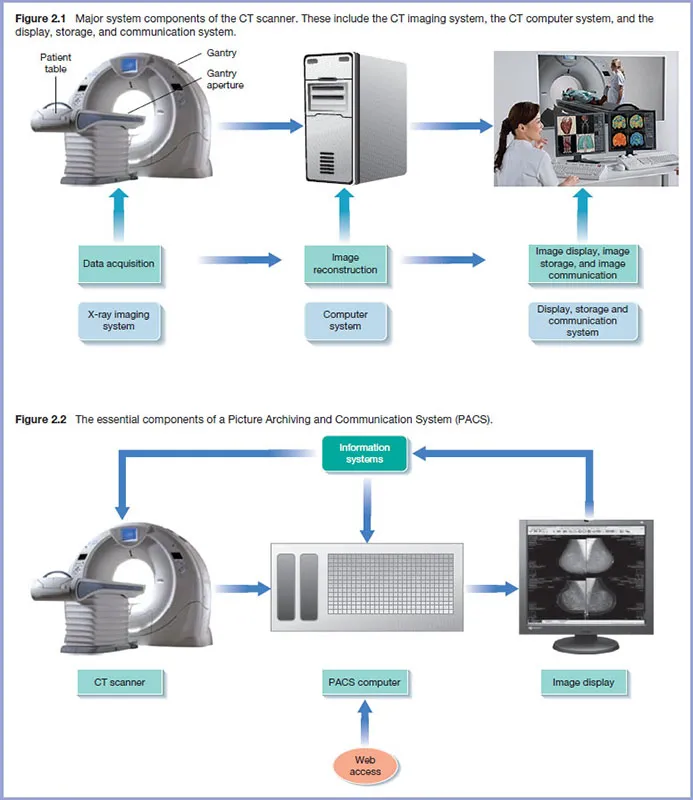
The major components of a CT imaging are shown in Figure 1.2 and include the CT scanner, the CT computer, and the CT operating console. Furthermore, the process of acquiring images of the patient involves three steps shown in Figure 1.3, data acquisition, image reconstruction, and image display, storage, and communication.
The CT scanner contains the X-ray tube and detectors, which rotate around the patient to collect attenuation data. These data are subsequently sent to the CT computer, which produces images using image reconstruction algorithms (computer programs that build up the image using the attenuation data). Furthermore, CT imaging now creates several 3D image types (Figure 1.1) using what is referred to as 3D rendering algorithms. These image types are intended to enhance diagnostic interpretation. Finally images are displayed for viewing and interpretation, after which they are stored for retrospective analysis, and sent to another location using computer network communications technology. One such popular technology is a Picture Archiving and Communication System (PACS).
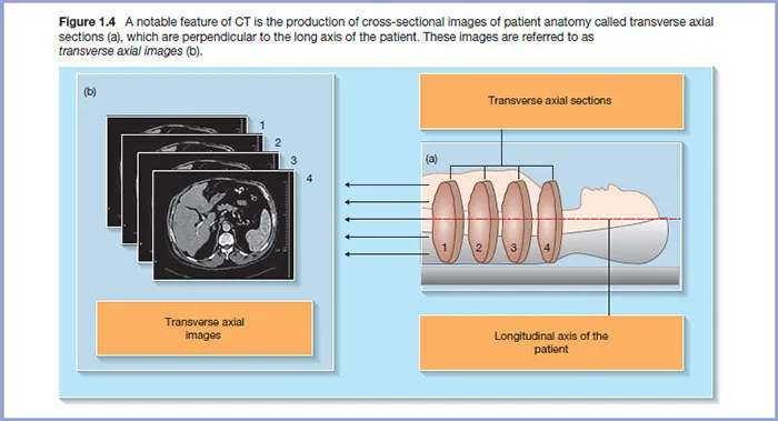
As noted earlier, CT produces cross-sectional images of patient anatomy, which are transverse axial sections (Figure 1.4a). These images are referred to as transverse axial images (Figure 1.4b).These sections are perpendicular to the long axis of the patient as illustrated in Figure 1.4a.
Nobel prize for the invention of the CT scanner
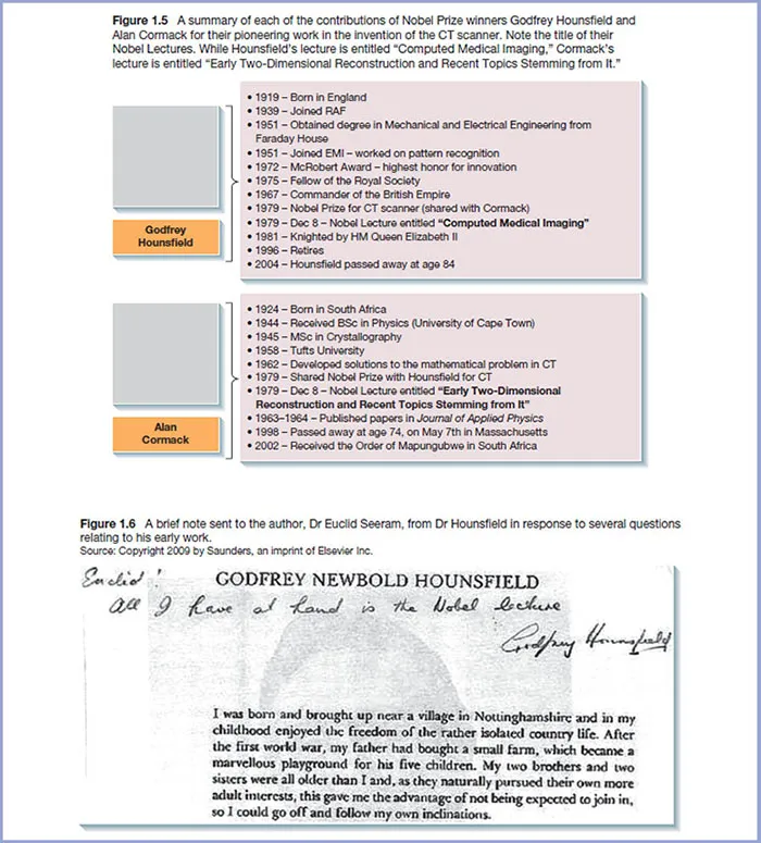
The invention of the CT scanner is credited to two individuals (Godfrey Hounsfield and Alan Cormack) working in two separate countries and who shared the Nobel Prize in Medicine in 1979 for their contributions to the development of the scanner. Photographs and detailed notes of their work are published by the Nobel Foundation (http://www.nobelprize.org). A summary of each of their contributions is shown in Figure 1.5. It is interesting to note the title of their Nobel Lectures. While Hounsfield’s lecture is entitled “Computed Medical Imaging,” Cormack’s lecture is entitled “Early Two-Dimensional Reconstruction and Recent Topics Stemming from It.” Both of these pioneers worked out the mathematical solutions to the problem in CT, but Hounsfield developed the first useful clinical CT scanner. Figure 1.6 shows a note sent to the author, Dr Euclid Seeram, from Dr Hounsfield in response to several questions relating to his early work.
The technical evolution of CT
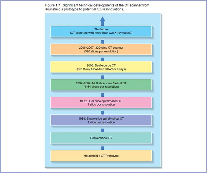
CT has experienced a number of significant technical innovations through the years as illustrated in Figure 1.7. In brief CT has evolved from a scanner dedicated to imaging the brain only to single-slice whole-body scanners and multislice scanners, and subsequently to scanners with two X-ray tubes coupled to two sets of detectors. These latter scanners are known as Dual Source CT (DSCT) scanners. Other notable technical innovations include the development of multislice detectors, iterative reconstruction algorithms, virtual reality imaging methods, dose optimization methods, and important quality control test tools and procedures.
These innovations are intended not only to improve image quality and reduce radiation dose, but also to play a role in the care and management of the patient, during CT imaging.