CT at a Glance gets readers quickly up to speed with the core knowledge and competencies required for computed tomography (CT) scanning, as established by the major radiography organizations around the world, including the ASRT and the CAMRT. This brand new title describes the basic science behind CT with an emphasis on the theory that is essential for practice. Featuring an abundance of illustrations, succinct, straightforward explanations and clear, step-by-step guidance, it includes the fundamental physics, technical principles, and imaging strategies and procedures involved in CT scanning.
Over the course of twenty four, concise modular chapters, CT at a Glance covers all the bases for entry-to-practice students, including:
The basic physics underlying CT scanning
State-of-the-art multi-slice technologies
Data acquisition strategies
Equipment components—their functions and applications
Image reconstruction and image quality control
CT dose and dose optimization procedures
Quality control fundamentals
CT at a Glance is an indispensable learning resource for students in medical imaging technology courses, including those covering radiography, nuclear medicine, and radiation therapy, as well as for biomedical engineering technology students.
Domande frequenti
Come faccio ad annullare l'abbonamento?
È semplicissimo: basta accedere alla sezione Account nelle Impostazioni e cliccare su "Annulla abbonamento". Dopo la cancellazione, l'abbonamento rimarrà attivo per il periodo rimanente già pagato. Per maggiori informazioni, clicca qui
È possibile scaricare libri? Se sì, come?
Al momento è possibile scaricare tramite l'app tutti i nostri libri ePub mobile-friendly. Anche la maggior parte dei nostri PDF è scaricabile e stiamo lavorando per rendere disponibile quanto prima il download di tutti gli altri file. Per maggiori informazioni, clicca qui
Che differenza c'è tra i piani?
Entrambi i piani ti danno accesso illimitato alla libreria e a tutte le funzionalità di Perlego. Le uniche differenze sono il prezzo e il periodo di abbonamento: con il piano annuale risparmierai circa il 30% rispetto a 12 rate con quello mensile.
Cos'è Perlego?
Perlego è un servizio di abbonamento a testi accademici, che ti permette di accedere a un'intera libreria online a un prezzo inferiore rispetto a quello che pagheresti per acquistare un singolo libro al mese. Con oltre 1 milione di testi suddivisi in più di 1.000 categorie, troverai sicuramente ciò che fa per te! Per maggiori informazioni, clicca qui.
Perlego supporta la sintesi vocale?
Cerca l'icona Sintesi vocale nel prossimo libro che leggerai per verificare se è possibile riprodurre l'audio. Questo strumento permette di leggere il testo a voce alta, evidenziandolo man mano che la lettura procede. Puoi aumentare o diminuire la velocità della sintesi vocale, oppure sospendere la riproduzione. Per maggiori informazioni, clicca qui.
CT at a Glance è disponibile online in formato PDF/ePub?
Sì, puoi accedere a CT at a Glance di Euclid Seeram in formato PDF e/o ePub, così come ad altri libri molto apprezzati nelle sezioni relative a Medicine e Medical Technology & Supplies. Scopri oltre 1 milione di libri disponibili nel nostro catalogo.
A significant and important technological innovation that has now become a popular tool for diagnostic imaging of patients is computed tomography (CT), an imaging technique that was first investigated as early as 1967. Later, in 1971, a prototype CT scanner for imaging the brain was developed by EMI Limited (Electric and Musical Industries [a manufacturer of records and electronics; the Beatles recorded under the EMI label], now Thorn EMI) in Middlesex, UK. This prototype resulted in the first patient being scanned in 1971, and this development earned two pioneers of CT, Godfrey Hounsfield and Allan Cormack, the Nobel Prize in Medicine in 1979.
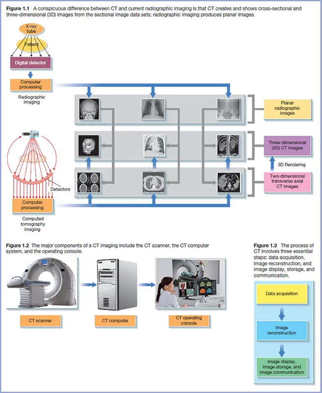
A striking fundamental difference between CT and current radiographic imaging is clearly illustrated in Figure 1.1 While CT creates and shows cross-sectional and three-dimensional (3D) images from the sectional image data sets, radiographic imaging produces planar images. There are other differences between these two imaging modalities, which will be described in later chapters. One such example is that CT uses much more sensitive electronic detectors, which can show very small differences in tissue attenuation compared to radiographic detectors. This characteristic results in CT providing much better tissue image contrast than radiography, and therefore the observer can see soft tissues much better than with radiography.
Radiographic imaging
The major components of radiographic imaging includes an X-ray tube and generator that provide the appropriate X-rays to image the patient, a detector that captures X-rays transmitted through the patient, a computer processing system, and an image display workstation (Figure 1.1). X-rays transmitted through the patient are converted into digital data for processing by the computer. The image output from the computer is subsequently displayed for viewing and interpretation by an observer. These radiographic images are usually referred to as planar images. The problems with these images are (i) superimposition of all structures on the detector (which makes it difficult and sometimes impossible to distinguish a particular detail) and (ii) the qualitative nature of radiographic imaging. The latter simply means that it is difficult to distinguish between a homogeneous object (one tissue type) of non-uniform thickness and a heterogeneous object (bone, soft tissue, and air) of uniform thickness. Finally, the beam used in radiography is an open beam (wide beam) and this creates more scattered rays, which get to the image and essentially destroy the image contrast.
CT imaging
CT overcomes these limitations by removing the superimposition of structures, improving image contrast, and imaging very small differences in tissue contrast.
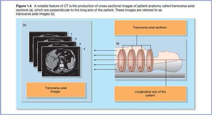
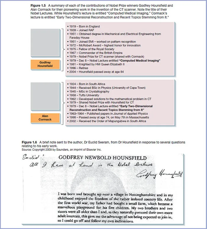
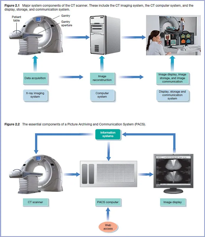
The major components of a CT imaging are shown in Figure 1.2 and include the CT scanner, the CT computer, and the CT operating console. Furthermore, the process of acquiring images of the patient involves three steps shown in Figure 1.3, data acquisition, image reconstruction, and image display, storage, and communication.
The CT scanner contains the X-ray tube and detectors, which rotate around the patient to collect attenuation data. These data are subsequently sent to the CT computer, which produces images using image reconstruction algorithms (computer programs that build up the image using the attenuation data). Furthermore, CT imaging now creates several 3D image types (Figure 1.1) using what is referred to as 3D rendering algorithms. These image types are intended to enhance diagnostic interpretation. Finally images are displayed for viewing and interpretation, after which they are stored for retrospective analysis, and sent to another location using computer network communications technology. One such popular technology is a Picture Archiving and Communication System (PACS).
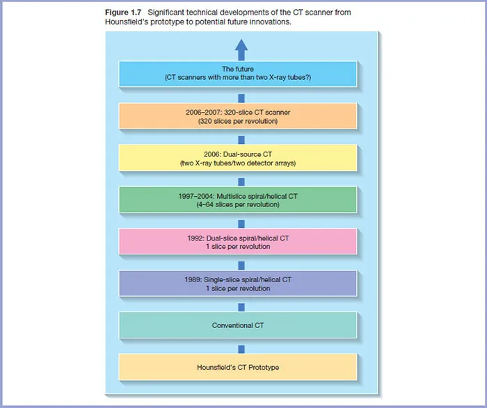
As noted earlier, CT produces cross-sectional images of patient anatomy, which are transverse axial sections (Figure 1.4a). These images are referred to as transverse axial images (Figure 1.4b).These sections are perpendicular to the long axis of the patient as illustrated in Figure 1.4a.
Nobel prize for the invention of the CT scanner
The invention of the CT scanner is credited to two individuals (Godfrey Hounsfield and Alan Cormack) working in two separate countries and who shared the Nobel Prize in Medicine in 1979 for their contributions to the development of the scanner. Photographs and detailed notes of their work are published by the Nobel Foundation (http://www.nobelprize.org). A summary of each of their contributions is shown in Figure 1.5. It is interesting to note the title of their Nobel Lectures. While Hounsfield’s lecture is entitled “Computed Medical Imaging,” Cormack’s lecture is entitled “Early Two-Dimensional Reconstruction and Recent Topics Stemming from It.” Both of these pioneers worked out the mathematical solutions to the problem in CT, but Hounsfield developed the first useful clinical CT scanner. Figure 1.6 shows a note sent to the author, Dr Euclid Seeram, from Dr Hounsfield in response to several questions relating to his early work.
The technical evolution of CT
CT has experienced a number of significant technical innovations through the years as illustrated in Figure 1.7. In brief CT has evolved from a scanner dedicated to imaging the brain only to single-slice whole-body scanners and multislice scanners, and subsequently to scanners with two X-ray tubes coupled to two sets of detectors. These latter scanners are known as Dual Source CT (DSCT) scanners. Other notable technical innovations include the development of multislice detectors, iterative reconstruction algorithms, virtual reality imaging methods, dose optimization methods, and important quality control test tools and procedures.
These innovations are intended not only to improve image quality and reduce radiation dose, but also to play a role in the care and management of the patient, during CT imaging.