Topographical and Pathotopographical Medical Atlas of the Pelvis, Spine, and Limbs
eBook - ePub

Topographical and Pathotopographical Medical Atlas of the Pelvis, Spine, and Limbs

Z. M. Seagal

  1. English
  2. ePUB (apto para móviles)
  3. Disponible en iOS y Android
eBook - ePub

Topographical and Pathotopographical Medical Atlas of the Pelvis, Spine, and Limbs

Z. M. Seagal

Detalles del libro
Vista previa del libro
Índice
Citas

Información del libro

The fourth medical atlas in this new series on the human body and filled with detailed pictures, this atlas details the topographical and pathotopographical anatomy of the pelvis, spine, and limbs, a useful reference for medical professionals and students alike.

Written by an experienced and well-respected physician and professor, this new volume, building on the previous volume, Ultrasonic Topographical and Pathotopographical Anatomy, and its sequels, also available from Wiley-Scrivener, presents the ultrasonic topographical and pathotopographical anatomy of the pelvis, spine, and limbs, offering further detail into these important areas for use by medical professionals.

This series of atlases of topographic and pathotopographic human anatomy is a fundamental and practically important series designed for doctors of all specializations and students of medical schools. Here you can find almost everything that is connected with the topographic and pathotopographic human anatomy, including original graphs of logical structures of topographic anatomy and development of congenital abnormalities, topography of different areas in layers, pathotopography, computer and magnetic resonance imaging (MRI) of topographic and pathotopographic anatomy. Also you can find here new theoretical and practical sections of topographic anatomy developed by the author himself which are published for the first time. They are practically important for mastering the technique of operative interventions and denying possibility of iatrogenic complications during operations.

This important new volume will be valuable to physicians, junior physicians, medical residents, lecturers in medicine, and medical students alike, either as a textbook or as a reference. It is a must-have for any physician's library.

Preguntas frecuentes

¿Cómo cancelo mi suscripción?
Simplemente, dirígete a la sección ajustes de la cuenta y haz clic en «Cancelar suscripción». Así de sencillo. Después de cancelar tu suscripción, esta permanecerá activa el tiempo restante que hayas pagado. Obtén más información aquí.
¿Cómo descargo los libros?
Por el momento, todos nuestros libros ePub adaptables a dispositivos móviles se pueden descargar a través de la aplicación. La mayor parte de nuestros PDF también se puede descargar y ya estamos trabajando para que el resto también sea descargable. Obtén más información aquí.
¿En qué se diferencian los planes de precios?
Ambos planes te permiten acceder por completo a la biblioteca y a todas las funciones de Perlego. Las únicas diferencias son el precio y el período de suscripción: con el plan anual ahorrarás en torno a un 30 % en comparación con 12 meses de un plan mensual.
¿Qué es Perlego?
Somos un servicio de suscripción de libros de texto en línea que te permite acceder a toda una biblioteca en línea por menos de lo que cuesta un libro al mes. Con más de un millón de libros sobre más de 1000 categorías, ¡tenemos todo lo que necesitas! Obtén más información aquí.
¿Perlego ofrece la función de texto a voz?
Busca el símbolo de lectura en voz alta en tu próximo libro para ver si puedes escucharlo. La herramienta de lectura en voz alta lee el texto en voz alta por ti, resaltando el texto a medida que se lee. Puedes pausarla, acelerarla y ralentizarla. Obtén más información aquí.
¿Es Topographical and Pathotopographical Medical Atlas of the Pelvis, Spine, and Limbs un PDF/ePUB en línea?
Sí, puedes acceder a Topographical and Pathotopographical Medical Atlas of the Pelvis, Spine, and Limbs de Z. M. Seagal en formato PDF o ePUB, así como a otros libros populares de Medicina y Anatomía. Tenemos más de un millón de libros disponibles en nuestro catálogo para que explores.

Información

Año
2019
ISBN
9781119618546
Edición
1
Categoría
Medicina
Categoría
Anatomía

Part 1:
The Pelvis

Topographic Anatomy of the Pelvis

Limits. The pelvis is a part of the body located between the abdomen and lower limbs. It is limited outside by the pelvic bones, sacrum, coccyx and perineum at the bottom.
External reference points: the iliac crests (crista iliaca), anterior superior iliac spines (spina iliaca anterior superior), pubic tubercle (tuberculum pubicum), pubic symphysis, (symphysis pubica), dorsal surface of the sacrum (facies dorsalis os sacrum), tailbone (os coccyges); ischial tuberosity (tuber ischiadicum), the greater trochanter of the femur (trochanter major ossis femoralis), subpubicalis angle (angulus subpubicus).

Individual, Gender and Age Differences:

  1. The individual features of the pelvis consist of different ratios of the longitudinal and transverse diameters of the pelvic ring. At one extreme type of variability, the longitudinal diameter is greater than the transverse one, the pelvis is “squeezed” from the sides, and the axes of the pelvic organs are often inclined to the sacrum. With a different type of variability, the longitudinal diameter is less than the transverse one, the pelvis is “compressed” in the anteroposterior direction, and the axes of the pelvic organs are inclined toward the pubic symphysis.
  2. The gender differences. The wings of the iliac bones in women are more horizontal, therefore in women the pelvis is wider and lower than that of men. The inferior branches of pubic bones in women are located at an obtuse angle and form a pubic arch (arcus pubis); in men they are located at an acute angle and form a subpubic angle (angulus subpubicus). The pelvic cavity in women has the shape of a curved cylinder; in men it has the shape of a curved cone. The incline of the pelvis (inclination pelvis) is the angle between the horizontal plane and the plane of the superior aperture of the pelvis. In women it’s 55–60°, in men – 50–55°.
  3. Age differences. By the time of birth, the pelvic consists of three parts, each of which has ossification centers. All three parts – iliac, ischial and pubic – are connected inter-layer cartilage in the area of the acetabulum. The iliac fossa is almost absent. By the age of seven, the size of the pelvic bone doubles, then the growth slows to 12 years. By the age of 13–18, parts of the pelvic bone coalesce, and their ossification ends. The synostosis of all elements of the hipbone is completed by the age of 25.
The walls of the pelvis. The pelvis is restricted to the pubic, iliac, ischium, sacrum and coccyx.
The pelvic floor is formed by the diaphragm of the pelvis (diaphragma pelvi) and partly by the urogenital diaphragm (diaphragma urogenitale).
The genitourinary diaphragm, diaphragma urogenitale (Figures 15) is formed by two muscles: the deep perineal transverse muscle and the external sphincter of the urethra.
Figure illustrates the topographic anatomy of the female pelvic region in terms of urogenital diaphragm formed by two muscles, the deep perineal transverse muscle and the external sphincter of the urethra and the figure parts are marked as 1 – diaphragm urogenitalis; 2 – nn. et vasa obturatorii; 3 – m. obturatorius internus; 4 – arcus tendineus m. levatoris ani; 5 – m. levator ani; 6 – anus; 7 – nn. et vasa glutei superiores; 8 – n. ischiadicus et fasc. gluteus inferior; 9 – m. piriformis; 10 – m. coccygeus; 11 – lig. anococcygeus; 12 – centrum tendineum perinei; 13 – vagina; 14 – urethra.
Figure 1 Urogenital diaphragm in women.
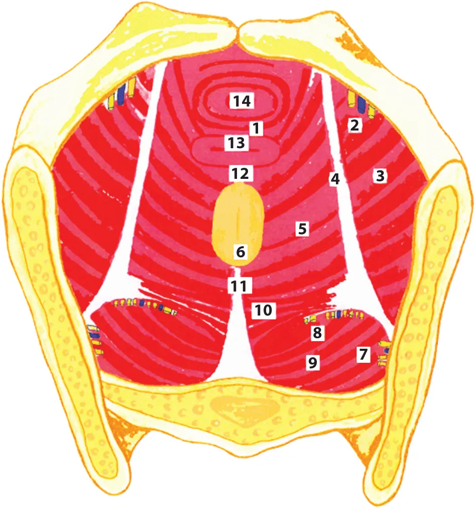
1 – diaphragma urogenitalis; 2 – nn. et vasa obturatorii; 3 – m. obturatorius internus; 4 – arcus tendineus m. levatoris ani; 5 – m. levator ani; 6 – anus; 7 – nn. et vasa glutei superiores; 8 – n. ischiadicus et fasc. gluteus inferior; 9 – m. piriformis; 10 – m. coccygeus; 11 – lig. anococcygeus; 12 – centrum tendineum perinei; 13 – vagina; 14 – urethra
Figure illustrates the front section of the male pelvic region where the peritoneum from the anterior wall of the abdomen passes to the bladder, covering the upper wall and partly the lateral and posterior ones and the other parts are marked as peritoneum; 5 – the visceral peritoneum; 6 – the deep perineal fascia; 7 – fascia m. obturatorii; 8 – the superficial perineal fascia; 9 – anus; 10 – m. sphincter ani externi; 11 – canalis pro vasa pudendi et n.; 12 – m. obduratorius internus; 13 – m. levator ani; 14 – rectum; 15 – derma perinei.
Figure 2 The front section of the pelvis.
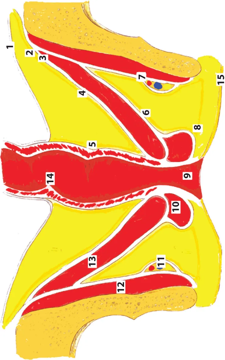
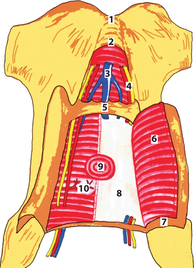
1 – peritoneum; 2 – fascia intraperitonealis; 3 – arcus tendneus; 4 – the parietal peritoneum; 5 – the visceral peritoneum; 6 – the deep perineal fascia; 7 – fascia m. obturatorii; 8 – the superficial perineal fascia; 9 – anus; 10 – m. sphincter ani externi; 11 – canalis pro vasa pudendi et n.; 12 – m. obduratorius internus; 13 – m. levator ani; 14 – rectum; 15 – derma perinei
Figure shows the innervation of the hip joint where (1) N. gluteus superior goes to the suprapiriform foramen with the same artery and vein and (4) N. gluteus inferior and (3) n. pudendus leave the cavity of the small pelvis through the infrapiriform foramen; in addition, n. pudendus, internal arteries and veins enter the ischioanal fossa through the lesser sciatic foramen and together with them, the long branches of the sacral plexus (2) (n. ischiadicus и n.cutaneus femoris posterior) enter the gluteal region and are directed into the infrapiriform foramen with the inferior gluteal vessels.
Figure 3 Te innervation of the hip joint.
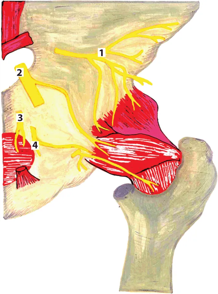
1 – n. gluteus superior; 2 – n. ischiadicus; 3 – n. pudendus; 4 – n. gluteus inferior
Figure shows the innervation of the hip joint where short branches of (1) n. femoralis and (2) n. obturatorius are guided along the side wall of the pelvis to the obturator foramen.
Figure 4 The innervation of the hip joint.
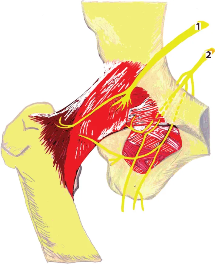
1 – n. femoralis; 2 – n. obturatorius
Figure illustrates the topographic anatomy of the male pelvic region in terms of urogenital diaphragm formed by two muscles, the deep perineal transverse muscle and the external sphincter of the urethra and the figure parts are marked as 1 – symphisis ossium pubis; 2 – lig. arcuaturn pubis; 3 – v. penis; 4 – a. et n. penis; 5 – lig. transversum perinea; 6 – m. transversus perinei profundus; 7 – folium superficialis aponeurosis urogenitalis; 8 – folium profundum aponeurosis urogenitalis; 9 – urethra et m. sphincter urethrae; 10 – glandula bulbourethralis.
Figure 5 Urogenital diaphragm in men.
1 – symphisis ossium pubis; 2 – lig. arcuaturn pubis; 3 – v. penis; 4 – a. et n. penis; 5 – lig. transversum perinea; 6 – m. transversus perinei profundus; 7 – folium superficialis aponeurosis urogenitalis; 8 – folium profundum aponeurosis urogenitalis; 9 – urethra et m. sphincter urethrae; 10 – glandula bulbourethralis
The diaphragm of the pelvis is formed by two muscles: a muscle lifting the anus (m. levator ani) and the coccygeal muscle (m. soccygeus).
The floors of the cavity of the small pelvis. The cavity of the small pelvis is divided into three floors: upper one – peritoneum (cavum pelvis peritoneale), middle one – subperitoneal (cavum pelvis subperitoneale) and lower one – subcutaneous or perineal (cavum pelvis subcutaneum s. perineale).
Peritoneum in the male pelvis. In the male pelvis, the peritoneum from the anterior wall of the abdomen passes to the bladder, covering the upper wall and partly the lateral and posterior ones. On the sides of the bladder the peritoneum forms fossae paravesicales. At the symphysis level, the peritoneum forms plica vesicalis transversa. Going down the back wall of the bladder, the peritoneum covers the medial edges of the vas deferens and the apex of the seminal glands, and passes to the rectum, forming the recto-vesical pouch (excavatio rectovesicalis). The ampulla is covered on three sides, and the lower part of the rectum is not covered with the peritoneum at all.
Peritoneum in the female pelvis. In the female pelvis, the peritoneum also passes from the anterior abdominal wall to the bladder, forming a transverse fold, and then covers its superior and posterior walls. After this, it passes to the anterior surface of the uterus at the level of the isthmus, where it forms a shallow vesicouterine pouch (excavatio vesicouterina). On the anterior surface of the uterus, the peritoneum covers only the body of the uterus. On the posterior surface of the uterus, the peritoneum covers the body of the uterus, the supramarginal part of the cervix and the posterior vaginal vault and passes to the rectum, forming a deep recto-uterine pounch (the excavatio rectouterine) or Douglas’ pouch. It is limited by folds of the peritoneum – plicae rectouterinae, which extend to the anterior surface of the sacrum.
Fascia and cellular spaces, their connection with the cellular spaces adjacent regions. The parietal pelvic fascia (fascia pelvis parietalis) covers the anterior surface of the sacrum behind the back and is called the presacral fascia (fascia presacralis), on the sides it covers the parietal muscles (m. piriformis, m. obturatorius internus), the fascia of which have corresponding names (fascia obturatoria, fascia m. piriformis) in front – the posterior surface of the symphysis and the superior branches of the pubic bones, from below – the upper surface m. levator ani. The visceral pelvic fascia (fascia pelvis visceralis) lining the extraperitoneal parts of the pelvic organs in men and forms two sagittal branches extending from the pubic bones to the sacrum. Part of the visceral fascia that stretching from the pubic bone to the prostate gland in men or to the bladder in women is called the puboprostatic (lig. puboprostaticum) or pubovesical (lig. pubovesicale) ligament. These ligaments are accompanied by bundles of smooth muscle fibers of m. puboprostaticus and m. pubovesicalis.
The sagittal branches of the visceral fascia that located behind the bladder also...

Índice

  1. Cover
  2. Title Page
  3. Copyright
  4. Introduction
  5. Part 1: The Pelvis
  6. Part 2: The Spine
  7. Part 3: The Limbs
  8. Conclusion
  9. About the Author
  10. End User License Agreement
Estilos de citas para Topographical and Pathotopographical Medical Atlas of the Pelvis, Spine, and Limbs

APA 6 Citation

Seagal, Z. (2019). Topographical and Pathotopographical Medical Atlas of the Pelvis, Spine, and Limbs (1st ed.). Wiley. Retrieved from https://www.perlego.com/book/1148975/topographical-and-pathotopographical-medical-atlas-of-the-pelvis-spine-and-limbs-pdf (Original work published 2019)

Chicago Citation

Seagal, Z. (2019) 2019. Topographical and Pathotopographical Medical Atlas of the Pelvis, Spine, and Limbs. 1st ed. Wiley. https://www.perlego.com/book/1148975/topographical-and-pathotopographical-medical-atlas-of-the-pelvis-spine-and-limbs-pdf.

Harvard Citation

Seagal, Z. (2019) Topographical and Pathotopographical Medical Atlas of the Pelvis, Spine, and Limbs. 1st edn. Wiley. Available at: https://www.perlego.com/book/1148975/topographical-and-pathotopographical-medical-atlas-of-the-pelvis-spine-and-limbs-pdf (Accessed: 14 October 2022).

MLA 7 Citation

Seagal, Z. Topographical and Pathotopographical Medical Atlas of the Pelvis, Spine, and Limbs. 1st ed. Wiley, 2019. Web. 14 Oct. 2022.