eBook - ePub
Quantitative Functional Brain Imaging with Positron Emission Tomography
Richard E. Carson,Peter Herscovitch,Margaret E. Daube-Witherspoon
This is a test
- 503 páginas
- English
- ePUB (apto para móviles)
- Disponible en iOS y Android
eBook - ePub
Quantitative Functional Brain Imaging with Positron Emission Tomography
Richard E. Carson,Peter Herscovitch,Margaret E. Daube-Witherspoon
Detalles del libro
Vista previa del libro
Índice
Citas
Información del libro
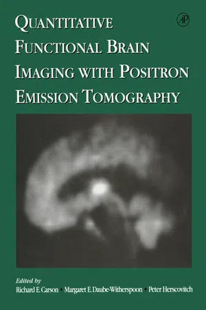
This book presents the latest scientific developments in the field of positron emission tomography (PET) dealing with data acquisition, image processing, applications, statistical analysis, tracer development, parameter estimation, and kinetic modeling. It covers improved methodology and the application of existing techniques to new areas. The text also describes new approaches in scanner design and image processing, and the latest techniques for modeling and statistical analyses. This volume will be a useful reference for the active brain PET scientist, as well as a valuable introduction for students and researchers who wish to take advantage of the capabilities of PET to study the normal and diseased brain.
- Authored by international authorities in PET
- Provides the latest up-to-date techniques and applications
- Covers all fundamental disciplines of PET in one volume
- A comprehensive resource for students, clinicians, and new PET researchers
Preguntas frecuentes
¿Cómo cancelo mi suscripción?
¿Cómo descargo los libros?
Por el momento, todos nuestros libros ePub adaptables a dispositivos móviles se pueden descargar a través de la aplicación. La mayor parte de nuestros PDF también se puede descargar y ya estamos trabajando para que el resto también sea descargable. Obtén más información aquí.
¿En qué se diferencian los planes de precios?
Ambos planes te permiten acceder por completo a la biblioteca y a todas las funciones de Perlego. Las únicas diferencias son el precio y el período de suscripción: con el plan anual ahorrarás en torno a un 30 % en comparación con 12 meses de un plan mensual.
¿Qué es Perlego?
Somos un servicio de suscripción de libros de texto en línea que te permite acceder a toda una biblioteca en línea por menos de lo que cuesta un libro al mes. Con más de un millón de libros sobre más de 1000 categorías, ¡tenemos todo lo que necesitas! Obtén más información aquí.
¿Perlego ofrece la función de texto a voz?
Busca el símbolo de lectura en voz alta en tu próximo libro para ver si puedes escucharlo. La herramienta de lectura en voz alta lee el texto en voz alta por ti, resaltando el texto a medida que se lee. Puedes pausarla, acelerarla y ralentizarla. Obtén más información aquí.
¿Es Quantitative Functional Brain Imaging with Positron Emission Tomography un PDF/ePUB en línea?
Sí, puedes acceder a Quantitative Functional Brain Imaging with Positron Emission Tomography de Richard E. Carson,Peter Herscovitch,Margaret E. Daube-Witherspoon en formato PDF o ePUB, así como a otros libros populares de Scienze biologiche y Biofisica. Tenemos más de un millón de libros disponibles en nuestro catálogo para que explores.
Información
Categoría
Scienze biologicheCategoría
BiofisicaSECTION VII
KINETIC MODELING
CHAPTER 54
Temporally Overlapping Dual-Tracer PET Studies1
R.A. KOEPPE, E.P. FICARO, D.M. RAFFEL, S. MINOSHIMA and M.R. KILBOURN, Division of Nuclear Medicine, University of Michigan Ann Arbor, Michigan 48109
There has been increased interest in studying multiple neurotransmitter–neuroreceptor systems within the same subject. Currently, examination of two distinct neuropharmacologic measures with positron emission tomography (PET) involves performing two independent scans. This chapter proposes a dual-tracer protocol with a single overlapping PET scan and analysis using a combined compartmental model. Advantages include (1) reduction in scan time by 1.5–2 hr, (2) neuropharmacology measures would be obtained over nearly the same interval, and (3) interventional protocols involving a pair of dual-tracer scans could be performed in a single session. Simulations were performed for various combinations of three tracers: [11C]flumazenil (FMZ), [11C]dihydrotetrabenazine (DTBZ), and N-[11C]methylpiperidinylpropionate (PMP). Noisy time-activity curves were generated simulating injection separations of 10–30 min. Model parameters were estimated for both tracers simultaneously using the combined model. For FMZ/DTBZ, two parallel two-compartment configurations were used, estimating K1 and distribution volume (DV) for each ligand. For tracer pairs involving PMP, a parallel two- and three-compartment combination was used, estimating K1 and DV for FMZ or DTBZ and K1 and k3 for PMP. Parameter estimation accuracy is strongly dependent on the choice of tracers, the brain region, the parameter of interest, the injection order, and, obviously, the injection spacing. Examination of the covariance among parameters is important for interpreting model performance. Making specific conclusions that can be generalized to any tracer pair is difficult; however, the use of tracers with rapid kinetics will yield greater success. The authors conclude that dual-tracer single-scan PET is feasible and can be implemented with a number of different radiotracers.
I INTRODUCTION
The authors’ goal is to develop methodology for use with positron emission tomography (PET) that will yield neuropharmacologic information related to two different biochemical systems or two different aspects of the same system of the brain at a single point in time. Currently, if one is interested in examining two distinct neuropharmacologic measures with PET, a scan session would involve injecting the first radioligand, scanning for about 1 hr, waiting for the first ligand to decay, injecting the second radioligand, and then scanning for approximately another hour. There are at least two obvious limitations of such a procedure: (1) the long study duration, making it difficult to scan certain patient groups; and (2) obtaining neuropharmacologic measures at two different points in time separated by about 2 hr when the physiologic/pharmacologic state of the subject may have changed. Furthermore, if one is interested in how two neuropharmacologic systems interact, a research study involving a pharmacologic challenge would require four PET scans, two baseline scans (one for each ligand), and two challenge scans. Such a protocol would require multiple days to perform and, in addition, the radiation dose to the subject may be excessive. A dual-tracer protocol with two radioligands injected 10–30 min apart and overlapping scanning offers a means of addressing these problems. The scan time required for a dual-tracer study would be reduced by 1.5–2 hr. The neuropharmacologic measures estimated from the two radioligands would be obtained over nearly the same time window. The lower doses afforded by three-dimensional (3D) imaging would allow interventional protocols involving a pair of dual-tracer studies (dual-baseline and dual-intervention scans) and could be performed in a single morning or afternoon scan session.
The rationale for a dual-tracer protocol stems from studies that indicate that many synaptic neurochemical indices by themselves may be insufficient to fully characterize neurologic diseases. As an example, multiple markers for striatal dopaminergic synapses are now available, including D1 (measured with [11C]SCH-23390) and D2 ([11C]raclopride; RAC) receptors, the presynaptic dopamine reuptake site ([11C]WIN35,428), the vesicular monoamine transporter ([11C]dihydrotetrabenazine; DTBZ), and DOPA decarboxylase activity ([18F]fluoroDOPA; FDOPA). In diseases involving both the number and the activity of dopaminergic nerve terminals, any or all of these markers have potential applications. Furthermore, there is mounting evidence that endogenous neurotransmitters and neuromodulators may influence in vivo measures with some of these agents. Finally, there is interest in examining the effects of pharmacologic treatments or test interventions with these markers. Experimental designs can now be envisioned in which two or more markers are studied under different conditions, permitting the testing of hypotheses regarding altered numbers and function of specific neurons.
An example of multitracer dopaminergic characterization is the investigation of altered presynaptic terminals and D2 receptors in unmedicated Parkinson’s disease (PD). It has been hypothesized that the observation of reduced DOPA decarboxylase activity in PD may be confounded by compensatory upregulation of enzymatic activity, whereas the density of synaptic vesicles as reflected by VMAT2 binding is a stable marker of presynaptic terminal integrity. This could be directly investigated by performing FDOPA, DTBZ, and RAC studies in unmedicated patients at baseline, followed by repeat characterizations after administering a pharmacologic dose of a D2 agonist drug. If the hypothesis were correct, FDOPA accumulation would be reduced in proportion to reduced RAC binding in the second scans due to D2 receptor-mediated downregulation of DOPA decarboxylase activity. DTBZ binding ...
Índice
- Cover image
- Title page
- Table of Contents
- Inside Front Cover
- Copyright
- Contributors
- Preface
- Acknowledgments
- SECTION I: DATA ACQUISITION AND QUANTIFICATION
- SECTION II: IMAGE PROCESSING
- SECTION III: APPLICATIONS
- SECTION IV: STATISTICAL ANALYSIS
- SECTION V: TRACER DEVELOPMENT
- SECTION VI: PARAMETER ESTIMATION
- SECTION VII: KINETIC MODELING
- SECTION VIII: BRAINPET97 DISCUSSION
- Index
- Color Plates
Estilos de citas para Quantitative Functional Brain Imaging with Positron Emission Tomography
APA 6 Citation
[author missing]. (1998). Quantitative Functional Brain Imaging with Positron Emission Tomography ([edition unavailable]). Elsevier Science. Retrieved from https://www.perlego.com/book/1810423/quantitative-functional-brain-imaging-with-positron-emission-tomography-pdf (Original work published 1998)
Chicago Citation
[author missing]. (1998) 1998. Quantitative Functional Brain Imaging with Positron Emission Tomography. [Edition unavailable]. Elsevier Science. https://www.perlego.com/book/1810423/quantitative-functional-brain-imaging-with-positron-emission-tomography-pdf.
Harvard Citation
[author missing] (1998) Quantitative Functional Brain Imaging with Positron Emission Tomography. [edition unavailable]. Elsevier Science. Available at: https://www.perlego.com/book/1810423/quantitative-functional-brain-imaging-with-positron-emission-tomography-pdf (Accessed: 15 October 2022).
MLA 7 Citation
[author missing]. Quantitative Functional Brain Imaging with Positron Emission Tomography. [edition unavailable]. Elsevier Science, 1998. Web. 15 Oct. 2022.