Biological Sciences
CT Scan vs MRI
CT scans and MRIs are both medical imaging techniques used to visualize the internal structures of the body. CT scans use X-rays to create detailed cross-sectional images, providing excellent visualization of bone and dense tissues. MRIs use magnetic fields and radio waves to produce detailed images of soft tissues, organs, and the brain. Each imaging modality has its own strengths and is used based on the specific clinical needs of the patient.
Written by Perlego with AI-assistance
Related key terms
1 of 5
10 Key excerpts on "CT Scan vs MRI"
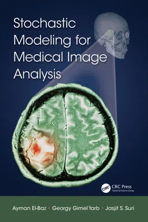
- Ayman El-Baz, Georgy Gimel’farb, Jasjit S. Suri, Georgy Gimel'farb(Authors)
- 2015(Publication Date)
- CRC Press(Publisher)
The functional CT is separated into the contrast-enhanced CT (CE-CT) and the CT angiography (CTA). Pros and cons, selected applications, and anatomical and/or functional information of the basic preoperative modalities, such as the MRI, CT, USI, and nuclear medical imaging (NMI), are briefly discussed below. However, their instrumental design, as well as the intraoperative imaging modalities are out of our scope. 1.1 Magnetic Resonance Imaging The MRI has become the most powerful and central noninvasive tool for clinical diagnostics. The human body to be imaged is entirely or partially 1 2 Stochastic Modeling for Medical Image Analysis Brain MRI Fetus US image Kidney CT image Lung PET image Liver SPECT image FIGURE 1.1 Most popular types of medical images. placed into a strong external static magnetic field to aligns parallel all hydro-gen (single-proton) nuclear magnetic spins in water molecules containing in different tissues, such as muscles, fat, and cerebral spinal fluid. Applied to the field radio frequency pulses form tissue-specific electromagnetic signals that encode spatial hydrogen distribution in the tissues and are measured to form an MR image. The MRI contrast strongly depends on how the image is acquired, because a preselected gradient of the external field and strengths, shapes, and tim-ing of sequences of the pulses highlight different components of the scanned areas. Generally, the MRI produces planar 2D slices, 3D volumes (spatial sequences of the slices), or 4D spatiotemporal images (temporal sequences of the 3D volumes) exemplified in Figures 1.2 and 1.3. (a) (b) (c) (d) FIGURE 1.2 Typical 2D MR slices of a knee (a) and a brain in sagittal (b), coronal (c), and axial (d) 3D planes.- Harcourt-Brown, Frances, Chitty, John, Harcourt-Brown, Frances, Chitty, John(Authors)
- 2013(Publication Date)
- British Small Animal Veterinary Association(Publisher)
CT and MRI are both tomographic techniques, tomos PeaninJ ¶slice’ in *reeN ,n ERtK tecKniTXes the object is displayed as multiple images or pic-tures, with every image representing one slice of the object. Every picture consists of pixels (picture elements) that have a thickness equal to the slice tKicNness 7Kese tKreediPensiRnal 3' ¶pi[els’ are called voxels (volume elements) (Bushburg et al ., 2002a). Creating CT and MR images is a complex process and therefore only some of the technical aspects will be highlighted in this section. Computed tomography Computed tomography (CT), or computed axial tomography (CAT) as it was previously named, was introduced in 1972. In the early period it took about 5 minutes to create one slice that was 13 mm thick and had pixels of 3 mm × 3 mm. The images were of low resolution with many artefacts (Bushburg et al ., 2002a). Many developments, such as helical scan-ning and multi-slice techniques, have improved the imaging process such that, within milliseconds, high-resolution images can be created that have a slice thickness of <1 mm. Helical scanning, consist-ing of continuous movement of the X-ray tube around the patient and simultaneous table move-ment, was introduced in 1989 and has improved scanning speed. The multi-slice technique, in which multiple slices are created in one rotation of the X-ray tube, was introduced in 1992 (Bushburg et al ., 2002a). The development and availability of advanced software programs has made image reconstruction and rendering possible and, as slice thickness decreases, high-resolution 3D image reconstruction or multi-planar reconstruction (MPR) become available. The 3D image reconstruction enables evaluation of the shape of anatomical struc-tures such as the skull or blood vessels (CT angio-gram). In MPR images, all views in the x -, y - and z -axes are available for reviewing, and all the struc-tures in the reconstructed slice can be evaluated without superposition of other structures. The equip-ment available in veterinary medicine currently con-sists mostly of 1 to 64 multi-slice, helical- eBook - PDF
- Ji-qian Fang, Ying Lu(Authors)
- 2003(Publication Date)
- World Scientific(Publisher)
380 J. S. Jin spatial resolution have made MRI the investigation of choice in many neurologic and orthopaedic diseases. X-rays are generated by the interaction of accelerated electrons with a target material (usually tungsten). X-rays are deflected and absorbed to different degrees by the various tissues and bones in the patient's body. The amount of absorption depends on the tissue composition. For example, dense bone matter will absorb many more X-rays than soft tissues, such as muscle, fat and blood. The amount of deflection depends on the density of electrons in the tissues. Tissues with high electron densities cause more X-ray scattering than those of lower density. Thus, since less photons reach the X-ray film after encountering bone or metal rather than tissue, the X-ray will look brighter for bone or metal. CT became generally available in the mid 1970s and is considered one of the major technological advances of medical science. X-ray CT gives anatomical information on the positions of air, soft tissues, and bone. Three-dimensional imaging is achieved by rotating an X-ray emitter around the patient, and measuring the intensity of transmitted rays from different angles. Ultrasound, as currently practiced in medicine, is a real-time tomo-graphic imaging modality. Not only does it produce real-time tomograms of the position of reflecting surfaces (internal organs and structures), but it can be used to produce real-time images of tissue and blood motion. The history of PET can be traced to the early 1950s, when workers in Boston first realized the medical imaging possibilities of a particular class of radioactive isotopes. Whereas most radioactive isotopes decay by release of a gamma ray and electrons, some decay by the release of a positron. A positron can be thought of as a positive electron. - No longer available |Learn more
- (Author)
- 2014(Publication Date)
- Learning Press(Publisher)
___________________________ WORLD TECHNOLOGIES ___________________________ Chapter- 5 X-ray Computed Tomography A patient is receiving a CT scan for cancer. Outside the scanning room is the imaging computers that reveal a 3D image of inside the body. Computed tomography (CT) is a medical imaging method employing tomography created by computer processing. Digital geometry processing is used to generate a three-dimensional image of the inside of an object from a large series of two-dimensional X-ray images taken around a single axis of rotation. CT produces a volume of data which can be manipulated, through a process known as windowing, in order to demonstrate various bodily structures based on their ability to block the X-ray beam. Although historically the images generated were in the axial or transverse plane, orthogonal to the long axis of the body, modern scanners allow this volume of data to be reformatted in various planes or even as volumetric (3D) rep-resentations of structures. Although most common in medicine, CT is also used in other fields, such as nondestructive materials testing. Another example is archaeological uses such as imaging the contents of sarcophagi or the DigiMorph project at the University of Texas at Austin which uses a CT scanner to study biological and paleontological specimens. ___________________________ WORLD TECHNOLOGIES ___________________________ Usage of CT has increased dramatically over the last two decades. An estimated 72 million scans were performed in the United States in 2007. Terminology The word tomography is derived from the Greek tomos (slice) and graphein (to write). Computed tomography was originally known as the EMI scan as it was developed at a research branch of EMI, a company best known today for its music and recording business. It was later known as computed axial tomography (CAT or CT scan) and body section röntgenography . - eBook - PDF
Introductory Biomedical Imaging
Principles and Practice from Microscopy to MRI
- Bethe A. Scalettar, James R. Abney(Authors)
- 2022(Publication Date)
- CRC Press(Publisher)
2 Image courtesy of Modus Medical Devices: www.modusqa.com/ products/deskcat-optical-ct-scanner-for-biophysics-education/. 271 DOI: 10.1201/b22076-15 13 Magnetic Resonance Imaging This chapter covers an unusual and exceptionally powerful technique – magnetic resonance imaging (MRI). MRI differs from the other methods that we have encountered, which use “ray trajectories” to achieve localization. This difference allows MRI to bypass the diffraction-dictated link between wave- length and resolution and thereby generate high (submillimeter) resolution images despite using EM radiation with wavelengths on the order of meters! MRI is intricate, as well as unusual, and has foun- dations in concepts from magnetism and quantum mechanics that have not yet arisen in the text. Thus, this chapter includes, of necessity, a very succinct review of select topics from these areas of physics. 1 13.1 Essence of the Technique An image is created by correlating a signal attribute (e.g., a ray trajectory or an echo time) with the location of the source. What makes MRI unusual is its use of signal frequencies to encode and map the location of the sources. In MRI, the sources typically are hydro- gen nuclei (protons) from water in living tissue. “Frequency encoding” is based on a resonance phe- nomenon exhibited by protons residing in a static magnetic field of strength B, typically specified using the unit tesla (T). The protons will undergo transi- tions between quantum energy states when exposed to an oscillating radiofrequency (RF) magnetic field, but only if the frequency of the oscillating field satis- fies the resonance condition B ν γ = ( γ is a property of the proton). 2,3 This link between ν and B means that spatially separated protons will absorb differently, 1 If time is limited, the material in Sections 13.1–13.6 can be used as the basis for a more introductory treatment of MRI. 2 For the moment, we ignore the difference between γ and γ - = γ/2π (Section 13.5.2). - No longer available |Learn more
- (Author)
- 2014(Publication Date)
- College Publishing House(Publisher)
An estimated 72 million scans were performed in the United States in 2007. It is estimated that 0.4% of current cancers in the United States are due to CTs performed in the past and that this may increase to as high as 1.5-2% with 2007 rates of CT usage. Terminology The word tomography is derived from the Greek tomos (slice) and graphein (to write). Computed tomography was originally known as the EMI scan as it was developed at a research branch of EMI, a company best known today for its music and recording busi-ness. It was later known as computed axial tomography (CAT or CT scan) and body section röntgenography . Although the term computed tomography could be used to describe positron emission tomography and single photon emission computed tomography, in practice it usually refers to the computation of tomography from X-ray images, especially in older medical literature and smaller medical facilities. In MeSH, computed axial tomography was used from 1977–79, but the current ind-exing explicitly includes X-ray in the title. Diagnostic use Since its introduction in the 1970s, CT has become an important tool in medical imaging to supplement X-rays and medical ultrasonography. It has more recently been used for preventive medicine or screening for disease, for example CT colonography for patients with a high risk of colon cancer, or full-motion heart scans for patients with high risk of heart disease. A number of institutions offer full-body scans for the general population. This is however a controversial practice, given its cost, significant radiation exposure, lack of proven benefit, and the risk of finding 'incidental' abnormalities that may trigger additional investigations. Head CT scanning of the head is typically used to detect infarction, tumours, calcifications, haemorrhage and bone trauma. - No longer available |Learn more
- (Author)
- 2014(Publication Date)
- Research World(Publisher)
________________________ WORLD TECHNOLOGIES ________________________ Chapter- 7 X-ray Computed Tomography A patient is receiving a CT scan for cancer. Outside of the scanning room is an imaging computer that reveals a 3D image of the body's interior. X-ray computed tomography (CT) is a medical imaging method employing tomography created by computer processing. Digital geometry processing is used to generate a three-dimensional image of the inside of an object from a large series of two-dimensional X-ray images taken around a single axis of rotation. CT produces a volume of data which can be manipulated, through a process known as windowing, in order to demonstrate various bodily structures based on their ability to block the X-ray beam. Although historically the images generated were in the axial or transverse plane, orthogonal to the long axis of the body, modern scanners allow this volume of data to be reformatted in various planes or even as volumetric (3D) representations of structures. Although most common in medicine, CT is also used in ________________________ WORLD TECHNOLOGIES ________________________ other fields, such as nondestructive materials testing. Another example is archaeological uses such as imaging the contents of sarcophagi or the DigiMorph project at the University of Texas at Austin which uses a CT scanner to study biological and paleontological specimens. Usage of CT has increased dramatically over the last two decades in many countries. An estimated 72 million scans were performed in the United States in 2007. It is estimated that 0.4% of current cancers in the United States are due to CTs performed in the past and that this may increase to as high as 1.5-2% with 2007 rates of CT usage. Terminology The word tomography is derived from the Greek tomos (slice) and graphein (to write). - eBook - PDF
- Karen M. Mudry, Robert Plonsey, Joseph D. Bronzino, Karen M. Mudry, Robert Plonsey, Joseph D. Bronzino(Authors)
- 2003(Publication Date)
- CRC Press(Publisher)
Systems that make use of x-rays have been designed to image anatomic structures, while others that make use of radioisotopes provide functional information. The fields of view that can be imaged range from the whole body obtained with nuclear medicine bone scans to images of cellular components using magnetic resonance (MR) microscopy. The design of transducers for the imaging devices to the postprocessing of the data to allow easier interpretation of the images by medical personnel are all aspects of the medical imaging devices field. Even with the sophisticated systems now available, challenges remain in the medical imaging field. With the increasing emphasis on health care costs, and with medical imaging systems often cited as not only an example of the investment that health care providers must make and consequently recover, but also as a factor contributing to escalating costs, there is increasing emphasis on lowering the costs of new systems. Researchers, for example, are trying to find alternatives to the high-cost superconducting mag-nets used for magnetic resonance systems. With the decreasing cost of the powerful computers that are currently contained within most imaging systems and with the intense competition among imaging companies, prices for these systems are bound to fall. Other challenges entail presentation of the imaging data. Often multiple modalities are used during a clinical evaluation. If both anatomic and functional information are required, methods to combine and present these data for medical interpretation need to be achieved. The use of medical image data to more effectively execute surgery is a field that is only starting to be explored. How can anatomic data obtained with a tomographic scan be correlated with the surgical field, given that movement of tissues and organs occurs during surgery? Virtual reality is likely to play an important role in this integration of imaging information with surgery. - eBook - ePub
- (Author)
- 2011(Publication Date)
- Academic Press(Publisher)
73 Magnetic Resonance ImagingJohn A. Detre, MDKeywords: brain • diffusion • magnetic resonance imaging • molecular imaging • perfusion • spinal cord • relaxationI. History of Magnetic Resonance ImagingII. Magnetic Resonance Imaging HardwareIII. Basic Principles of Magnetic Resonance ImagingIV. Image AnalysisV. Basic Clinical Magnetic Resonance ImagingVI. Perfusion and Diffusion Magnetic Resonance ImagingVII. Other Sources of Image Contrast in Proton Magnetic Resonance ImagingVIII. Exogenous Contrast AgentsIX. Imaging Nuclei Other Than ProtonsX. ConclusionReferencesHigh-quality imaging of the central nervous system (CNS) is more challenging than in other organs because the brain and spinal cord are soft tissue encased in dense bony structures. Although even routine x-ray can provide diagnostic information about organs in the torso, only limited inferences can be drawn about the brain from skull x-rays, primarily based on asymmetries or shifts in the location of a few possibly calcified structures. Computed tomography (CT) revolutionized the assessment of the CNS, providing sufficient dynamic range to visualize brain tissue within the skull—albeit with careful windowing with respect to bone, which is much denser than nervous tissue, dominates the signal, and often degrades image quality in the brainstem and spinal cord where the ratio of tissue to bone is reduced. CT scanning remains an important modality for rapid assessment of the CNS (e.g., head trauma), though tissue contrast in CT is limited to changes in tissue density and enhancement following exogenous contrast administration.Magnetic resonance imaging (MRI) currently is the most versatile and informative imaging modality for the CNS. There are a variety of reasons for this. First, MRI is detected as radio frequency (RF) signals that can penetrate the skull and spinal column without degradation. Second, the hardware used to generate MRI scans allows tremendous flexibility in imaging orientation and spatial resolution. Finally, there are numerous sources of image contrast that contribute to the characterization of structure, function, and lesions of the CNS. Some of these capabilities are covered in chapters on functional MRI and magnetic resonance spectroscopy (MRS) in this text. - eBook - PDF
- Angela Williams(Author)
- 2018(Publication Date)
- Omnigraphics(Publisher)
The risk of any harm is very slight, but repeated X-rays could cause cancer. The benefits nearly always outweigh the risk. Talk to your doctor about the need for each X-ray. Ask about shielding to protect parts of the body that are not in the picture. You should always let your doctor and the technician know if there is any chance that you are pregnant. 331 Chapter 30 Neurological Imaging Neurological Imaging in the Past Neurologists and neurosurgeons made clinical decisions based on first generation computed tomography (CT) scans. This was a quantum advance over the insensitive plain film X-ray techniques of previous generations. Early positron emission tomography (PET) and single photon emission computed tomography (SPECT) techniques utilized first generation radiographic tracers (or tags) to map brain function. Functional magnetic resonance imaging (fMRI) allowed researchers to measure blood oxygen level dependent (BOLD) changes in the brain of humans for the first time. fMRI enabled the noninvasive study of everything from finger movements to thoughts and emotions. Neurological Imaging Today Advanced magnetic resonance imaging (MRI) is revolutionizing the care of patients with neurologic disorders, as well as research in understanding the brain. Magnetic resonance This chapter includes text excerpted from Neurological Imaging, National Institutes of Health (NIH), March 29, 2013. Reviewed July 2018. 332 Medical Tests Sourcebook, Sixth Edition (MR) spectroscopy allows measurement of brain chemicals in living patients. PET imaging using compounds that bind to brain receptors now allows the study of molecular details not previously visualized. The resolution of brain and spinal cord imaging has increased tremendously. For example, modern techniques allow the neuroimaging of subtle abnormalities of neurological development that give rise to seizures and enable many more persons to benefit from a surgical treatment of epilepsy.
Index pages curate the most relevant extracts from our library of academic textbooks. They’ve been created using an in-house natural language model (NLM), each adding context and meaning to key research topics.
