Physics
X-rays
X-rays are a form of electromagnetic radiation with high energy and short wavelengths, lying between ultraviolet light and gamma rays on the electromagnetic spectrum. They are commonly used in medical imaging to visualize the internal structures of the body, as well as in industrial and scientific applications for their ability to penetrate materials.
Written by Perlego with AI-assistance
Related key terms
1 of 5
11 Key excerpts on "X-rays"
 eBook - PDF
- Rene Van Grieken, A. Markowicz, Rene Van Grieken, A. Markowicz(Authors)
- 2001(Publication Date)
- CRC Press(Publisher)
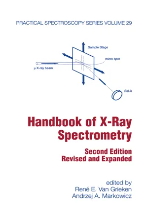
III. GENERAL FEATURES X-rays, or Rontgen rays, are electromagnetic radiations having wavelengths roughly within the range from 0.005 to lOnm. At the short-wavelength end, they overlap with y-rays, and at the long-wavelength end, they approach ultraviolet radiation. The properties of X-rays, some of which are discussed in detail in this chapter, are summarized as follows: Invisible Propagated in straight lines with a velocity of 3 x 10 8 m/s, as is light Unaffected by electrical and magnetic fields Differentially absorbed while passing through matter of varying composition, density, or thickness Reflected, diffracted, refracted, and polarized Capable of ionizing gases Capable of affecting electrical properties of liquids and solids Capable of blackening a photographic plate Able to liberate photoelectrons and recoil electrons Capable of producing biological reactions (e.g., to damage or kill living cells and to produce genetic mutations) X-ray Physics Emitted in a continuous spectrum whose short-wavelength limit is determined only by the voltage on the tube Emitted also with a line spectrum characteristic of the chemical elements Found to have absorption spectra characteristic of the chemical elements IV. EMISSION OF CONTINUOUS RADIATION Continuous X-rays are produced when electrons, or other high-energy charged particles, such as protons or a-particles, lose energy in passing through the Coulomb field of a nucleus. In this interaction, the radiant energy (photons) lost by the electron is called bremsstrahlung (from the German bremsen, to brake, and Strahlung, radiation; this term sometimes designates the interaction itself). The emission of continuous X-rays finds a simple explanation in terms of classic electromagnetic theory, because, according to this theory, the acceleration of charged particles should be accompanied by the emission of radiation. eBook - PDF
- K. Kirk Shung, Michael Smith, Benjamin M.W. Tsui(Authors)
- 2012(Publication Date)
- Academic Press(Publisher)
Equation (1-1) is a special form of the solution to the wave equation. The main difference between X-ray and light or radio waves is in their fre-quency or wavelength. Diagnostic X-rays typically have a wavelength from 100 nm to 0.01 nm, which is much shorter. Unlike light or radio waves, the propagation of X-ray radiation is sometimes difficult to interpret by treating the radiation as a wave alone. It is sometimes nec-essary to discuss X-ray radiation as if it possessed the dual characteristics of both particles and waves. I. Fundamentals of X -r a y 3 F i x e d , t X D i s t a n c e , χ (b) Fi u re1 Sinusoidal electromagnetic w a v e (a) as a function of time at fixed distance a n d (b) as a function of distance at a certain instant of time. The wave concept is useful in explaining such phenomena as the reflection, scattering, refraction, and diffraction characteristics of X-rays. However, in a number of situations, the particle concept has to be used. In discussing its particle properties, the X-ray radiation is thought to be parti-cles traveling at the speed of light and carrying an energy given by Ε = Λ/, where h is the Planck constant (4.13 x 10 1 8 keV-sec where 1 eV = 1.6 x 10 1 9 joules). These particles are called quanta, or photons. A photon having an energy level greater than a few electron volts is capable of ionizing atoms and molecules (i.e., knocking an electron out of its orbit), and it is called ionization radiation. Consider an X-ray photon with a wavelength of 1 nm. The energy of the pho-ton is then Ε = 4.13 x ΙΟ 1 5 x 3 x ΙΟΥΚΓ 9 = 1.2 x ΙΟ 3 eV Therefore, X-ray is an ionizing radiation. It can be easily shown that gamma rays and some ultraviolet rays are ionizing radiation as well. 4 C H A P T E R 1 X -r a y Incident X -r a y photon Scattered X -r a y photon A t o m Fi u re2 Coherent scattering of a n X -r a y photon by an atom. eBook - PDF
- Richards, Austin A.(Authors)
- 2011(Publication Date)
Chapter 4 X Rays and Gamma Rays: Crookes Tubes and Nuclear Light Light becomes something quite strange and powerful in the region of the electromagnetic spectrum in which wavelengths are shorter than in the near-UV ultraviolet waveband. This region, shown in Fig. 4.1, includes the extreme ultraviolet , x-ray and gamma-ray wavebands. X rays and gamma rays are electromagnetic waves with such short wavelengths (and correspondingly high energies) that they interact with matter very differently than do the longer wavelengths discussed previously. For the purposes of the following discussion I shall describe x rays and gamma rays in the context of photons , which are particles of light. Light has a dual nature: it can behave as a wave sometimes and a particle at other times. When it is a particle, or photon, it is best described by its energy; that is more appropriate than describing this light as a wave of a particular wavelength. This method of description reflects the way these high-energy photons interact with matter: when an x ray or a gamma ray does interact with an electron or a nucleus in material, there is a significant amount of energy transferred to a very localized area, like a bullet hitting a metal target. That is why x rays and gamma rays are considered radiation and are dangerous to living beings—the energy transfer often produces permanent chemical changes, which can lead to cellular mutation in living tissue. At the same time, when x rays and gamma rays traverse matter, the probability that they will interact with the matter is fairly low, and if the material is thin or has a low atomic number , then a significant fraction of the light can pass through with little or no loss. The thicker or denser the material, the more reduction in the intensity of a transmitted beam of x rays or gamma rays. eBook - PDF
- Anil Bharath(Author)
- 2022(Publication Date)
- Springer(Publisher)
As we shall see, there are many aspects which complicate the simplistic, ideal situation: 4 CHAPTER 2. DIAGNOSTIC X-RAY IMAGING • Statistical arrival of photons (Poisson process). • Photon scatter. • Lines of projection are not parallel i.e., one has beam divergence). • Photon detection is inefficient. • Beam hardening. • X-rays represent a form of ionizing radiation - there are health risks associated with prolonged or repetitive exposures. 2.2 RELEVANT PHYSICS 2.2.1 ATOMIC STRUCTURE All atoms have a similar structure, in that they consist of small, dense nuclei, which have a radius of ≈ 10 −14 m, with a positive charge given by Z × 1.6 × 10 −19 C, where Z is the atomic number of the atom i.e., the number of protons in the nucleus). In unionised form, the atom is electrically neutral: the nucleus is orbited by Z electrons, each of which has a negative charge of 1.6 × 10 −19 C, and a mass of 9 × 10 −31 kg, around 0.05% of the mass of each proton. Nuclei also contain uncharged particles, known as neutrons, of mass almost equal to that of protons, which provide short-range attractive forces that bind the protons together. The number of protons plus the number of neutrons is the mass of the atom. 2.2.2 NATURE OF X-rays X-rays represent electromagnetic radiation in the frequency range of about 10 18 -10 20 Hz. At this frequency range, the free space wavelength is of the order of λ = c/f = 3 × 10 8 /10 18 = 3 × 10 10 m (2.1) which is very small indeed. At such dimensions of wavelength, the quantum nature of electromagnetic phenomena becomes significant and, indeed, sets fundamental limits on imaging. The primary significance of this quantum nature is that the electromagnetic radiation is delivered in discrete lumps of energy, known as quanta, or photons. The quantity of energy in each photon is related to the wavelength of the radiation according to E = hf = hc/λ (2.2) where h is Planck’s constant, 6.626 × 10 −34 Js. eBook - PDF
- Patrick N. McDermott, Colin G. Orton(Authors)
- 2018(Publication Date)
- Medical Physics Publishing(Publisher)
5-1 X-ray Production II: Basic Physics and Properties of Resulting X-rays 5.1 Production of X-rays: Microscopic Physics 5.2 X-ray Spectrum 5.3 Efficiency of X-ray Production 5.4 Directional Dependence of Bremsstrahlung Emission 5.5 X-ray Attenuation 5.6 Half-value Layer (HVL) 5.7 Mass Attenuation Coefficient Appendix: Röntgen and the Discovery of X-rays Chapter Summary Problems Bibliography 5.1 Production of X-rays: Microscopic Physics When an electron strikes the target in an x-ray tube (or the target in a linear accelerator), it interacts with the matter in the target. On a microscopic level there are only two entities that the electron can interact with: atomic electrons or the atomic nucleus.* We will consider each of these two interactions in turn. *Very high-energy electrons can interact with individual nucleons, but that process does not play an important role in this context. 5 THE PHYSICS AND TECHNOLOGY OF RADIATION THERAPY 5-2 5.1.1 Characteristic X-rays When a high-energy electron interacts with atomic electrons in the target of an x-ray-producing device, there are two broad categories of subsequent events, depending upon whether the incoming electron interacts with an outer or an inner shell electron. Outer shell electrons can be excited to higher-level energy states. They will subsequently drop back to their original states and emit low-energy pho- tons in the process. These low-energy photons will be absorbed very quickly inside the target material. The energy of these photons will be converted into random motion of the atoms in the target; i.e., heat. Another possibility is that an outer shell electron can be ejected from the atom (i.e., the atom is ionized). The ejected electron will move through the target material; it may collide with other atoms, giving up some of its energy to the random motion of these atoms—heat again. eBook - PDF
Modern Diagnostic X-Ray Sources
Technology, Manufacturing, Reliability
- Rolf Behling(Author)
- 2015(Publication Date)
- CRC Press(Publisher)
The Interaction of X-rays with Matter 84 Medical imaging with X-rays is based on the generation of contrast by sending a “probe” of photons into a volume, capturing and analyzing the outgoing X-radiation. This analysis may comprise the simple recording of patterns of attenuation and also other characteristics such as distribu-tion of scattered radiation, spectral modulation, and phase shift. In any case, the result is spatially resolved information about the object. It is convenient to consider the X-ray probe as a spatially delimited shower of photons; a beam. The absence of proper lenses makes this assumption suspi-cious, however. As discussed below, there are no suitable lenses available for human imaging. Hence, an X-ray “beam” for human imaging is always divergent. It travels rectilinearly in air and nearly on straight paths in human tissue. Under the assumption that the dimensions of the source are small with respect to the distance r , the intensity I ( r ) obeys a square distance law, as shown in Figure 3.1, where I ( r → 0) denotes the intensity in an infinitesimal distance to a realistic source: ) ( = A I r I r ( 0) 2 . (3.1) However, the X-ray source is often extended, and the above law may deliver false expectations. When illuminating a patient with X-rays scattered radiation may emerge from wide areas close to the skin. Theoretically, an infinitely large planar source would cause constant intensity independent of the distance. Scattered radiation from a patient, which is illuminated with a delimited beam, decreases less rapidly with distance compared with the point source of Equation 3.1. eBook - PDF
Fundamentals Of Imaging, The: From Particles To Galaxies
From Particles to Galaxies
- Michael Mark Woolfson(Author)
- 2011(Publication Date)
- ICP(Publisher)
R¨ ontgen found that the glow was due to fluorescence of the barium-platinocyanide-covered cardboard strip. R¨ontgen concluded that some new form of radiation was respon-sible and he investigated its properties. He found that these rays, which he called X-rays to indicate their uncertain nature, blackened a photographic plate. Experiments that measured the extent of black-ening of a photographic plate showed that the transmission of X-rays depended on the type and thickness of the material through which they passed. Because bone and flesh absorb differently he found that the bones of a hand could be imaged photographically by pass-ing X-rays through it. A radiograph of a colleague’s hand is shown in Fig. 14.3. Medical radiography quickly became, and still is, an essential diagnostic technique. For the important discovery of X-rays R¨ontgen was awarded the first Nobel Prize for Physics in 1901. It took some time for the danger of overexposure to x-radiation to be discovered and x-ray generators were sold freely as a means of home entertainment to look at bones within the body; there must have been many premature deaths due to this cause. 14.2. X-ray Generators X-rays are a form of electromagnetic energy of extremely short wavelength, typically 0.1 nm. 1 In many applications of X-rays it has 1 1 nm (nanometre) is 10 − 9 m. 278 The Fundamentals of Imaging: From Particles to Galaxies Figure 14.3 A radiograph taken by R¨ontgen of the hand of Albert von Kolliker. been found convenient to express wavelengths in terms of another unit, the ˚ Angstrom unit ( ˚ A) that is 0 . 1 nm = 10 − 10 m. The simplest way to produce X-rays is to fire a beam of very high-energy elec-trons at a metal target, one preferably made of a heavy metal. The electrons, colliding with the metal atoms, lose energy in one or more interactions and each energy loss is converted into a photon, a packet of electromagnetic energy. No longer available |Learn more
- Maureen M. Julian(Author)
- 2014(Publication Date)
- CRC Press(Publisher)
287 C H A P T E R 6 Properties of X-rays Above is a picture of a modern x-ray tube. Most X-rays used in crystallography are produced by similar tubes. Synchrotrons also produce X-rays used in some protein studies. Nuclear bombs produce X-rays, which caused much damage to human tissue in Hiroshima. The sun produces X-rays, as do many other stars. X-ray astronomy is an important branch of star study. Certain radioactive materials decay to produce X-rays. For example, the low inten-sity X-rays from radioactive 55 Fe are used to test x-ray detectors. CONTENTS Chapter Objectives 288 6.1 Introduction 289 6.2 The Discovery of X-rays 289 6.3 Properties of Waves 290 6.3.1 Principle of Superposition 291 6.3.1.1 Constructive Interference 291 288 ◾ Properties of X-rays CHAPTER OBJECTIVES • Discovery of X-rays • Principle of superposition • Constructive, destructive, and partial interference • Young’s double-slit experiment • Properties of electromagnetic waves • X-ray spectrum • Bremsstrahlung and characteristic X-rays • Absorption • X-ray tubes and filters • X-ray diffraction • Bragg’s law • X-ray powder diffraction camera • Synchrotron as source of X-rays • Protein crystals and synchrotrons 6.3.1.2 Destructive Interference 292 6.3.1.3 Partial Interference 292 6.3.2 Young’s Double-Slit Experiment 292 6.3.3 Properties of Electromagnetic Waves 295 6.4 X-Ray Spectrum 297 6.4.1 Bremsstrahlung Radiation 297 6.4.2 Characteristic Radiation 299 6.4.3 Absorption 300 6.4.4 Electrons and X-rays 302 6.5 The X-Ray Tube 303 6.5.1 Filters 303 6.6 X-Ray Diffraction 305 6.6.1 First X-Ray Diffraction Photograph 305 6.6.2 Bragg’s Law 307 6.6.3 X-Ray Powder Diffraction Camera 309 6.7 Synchrotron X-rays 310 Definitions 312 Exercises 313 MATLAB Code: Starter Program for Chapter 6: Graphic of Powder Diffraction File 313 6.2 The Discovery of X-rays ◾ 289 6.1 INTRODUCTION Chapters 1 through 5 have provided an introduction to the principles of symmetry in crys-tals. eBook - PDF
- Anthony Wolbarst(Author)
- 2005(Publication Date)
- Medical Physics Publishing(Publisher)
Seen in cross section, the beam produced by an x-ray tube and incident on a patient is smooth and nearly uniform. But only 1% or so of the radiation entering any part of the body exits from the far side—and the beam that does emerge from it is spatially modulated. Some photons will have been either absorbed or scattered by atomic and molecular electrons, instead, and thereby removed from the beam. Imagine the beam as consisting of a bundle of separate geometric rays, as in Figures 3–1 and 2–1. Every one of these lines will encounter a unique set of tissues along its path. The properties of each such set of tissues determine how much of the transiting x-ray energy will be lost before emerging from the body to strike the film cassette. Frau Roentgen’s hand, for 3. X-RAY IMAGING I: OVERVIEW OF FILM RADIOGRAPHY 25 Figure 3–3. The energy spectra of x-ray radiation produced at two kVp settings. With a 60-kVp potential applied to the x-ray tube, only the continuous bremsstrahlung spectrum is evident. At 100 kVp, discrete peaks corre- sponding to tungsten characteristic X-rays also appear. Either way, the energy of the most energetic photons, in keV, is numerically equal to the kVp setting. Relative intensity Photon energy (keV) 20 100 80 60 40 100,000 V 60,000 V 26 PHYSICS OF RADIOLOGY, SECOND EDITION example, gave rise to a meaningful x-ray shadow because bone takes photon energy out of a beam more efficiently than do the surrounding soft tissues. It is the pattern of X-rays that have not interacted with the body’s tissues, and have not been absorbed or scattered, that comprises the primary x-ray image that exposes the radiographic cassette and thereby affects the film within. So, in a sense, an x-ray image is a negative: the thicker and denser parts of the body produce regions in the developed film that are lighter and more transparent. Conversely, the thinner or less dense the tissues in the beam path, the stronger the emerging beam and the darker the resulting film. eBook - PDF
- M. I. Pergament(Author)
- 2014(Publication Date)
- CRC Press(Publisher)
269 C H A P T E R 10 Measurements in the X-Ray Region of the Spectrum X -rays nowadays have applications in a wide range of investigations and measurements, from biology and medicine to crystallography and metallurgy, each area calling for its own detailed consideration of the techniques that are used. Naturally, one cannot possibly cover such a vast scope in a single discussion, and an attempt to do so would be counterpro-ductive. We will therefore concentrate our attention only on those X-ray measurement methods that are used in experimental physics and mainly confine the spectral interval considered to the so-called far (or vacuum) ultraviolet (VUV) and soft X-ray (SXR) regions of the spectrum. 10.1 SPECTRAL DOMAIN AND SOURCES OF X-RAY RADIATION More precisely, we are going to consider the wavelength range from 200 to about 0.25 nm (corresponding to photon energies ranging from some 6 to 5000 eV). * It is no accident that these wavelengths are so common in experimental physics applications. First, it is in this wavelength range that electromagnetic radiation actively interacts with atomic structures, thus * If wavelengths are given in nanometers and photon energy in electron volts, the transition between the two systems of units can be made using the relationship planckover2pi ωλ = × 1239.842 eV nm. 270 ◾ Methods of Experimental Physics becoming a tool for their exploration and analysis. * Second, the radiation emitted by very high-temperature objects is strongest in this range, whether it is from high-temperature plasma in a laboratory or from astro-physical objects in the universe. Third, the fact that these wavelengths are comparable with intra-atomic distances in a solid-state matter opens up a vast opportunity for various types of measurement. The wavelength range in question is arbitrarily divided into the VUV (200–10 nm) and the SXR (10–0.25 nm) regions. The boundary between the two regions is defined only for convenience. eBook - PDF
- L.H. Schwartz(Author)
- 2012(Publication Date)
- Academic Press(Publisher)
Chapter 4 Properties o f R a d i a t i o n U s e f u l f o r Studying the Structure o f M a t e r i a l s We have seen in Chapter 3 that a diffraction pattern can occur when the wavelength of radiation incident on a periodic array is the same order of magnitude as the period. As we now know from diffraction studies, these spacings are of the order of 1 A ( 1 0 -10 m) for atoms in solids, thus we wish to study the properties of radiation in this wavelength range. We shall confine our attention to the most commonly used radiations: χ rays, neutrons, and electrons. 4.1. PRODUCTION OF RADIATION—X RAYS Rontgen discovered, in the summer of 1895, that in an evacuated tube in which a voltage was applied across two metal plates, unknown or x rays emerged, penetrated matter, and exposed film. However, experiments to ex-amine the nature of these rays, to determine whether they were particles or electromagnetic radiation, were difficult. Ruled grating experiments which should give diffraction patterns for waves gave poor results that were hard to resolve and therefore not definitive. We know now that very fine gratings were needed because the x-ray wavelengths are so small, but such gratings were not available at that time. The results of these studies did suggest that if the rays were waves, they were of quite small wavelength, and these experiments helped to stimulate Laue's thoughts that χ rays might have the correct wave-lengths to diffract from crystals. As we shall see later, χ rays could be polarized like other electromagnetic waves, i.e., their amplitude of variation could be made to lie in only one direction perpendicular to their propagation direction. By contrast, χ rays could also eject electrons when they impinged on a material 118 4.1 PRODUCTION OF RADIATION—X RAYS 119 as in a particle collision. Experiments to reflect χ rays like waves also proved fruitless at first.
Index pages curate the most relevant extracts from our library of academic textbooks. They’ve been created using an in-house natural language model (NLM), each adding context and meaning to key research topics.