Biological Sciences
Insulin
Insulin is a hormone produced by the pancreas that plays a key role in regulating blood sugar levels. It helps cells in the body absorb glucose from the bloodstream to use as energy or store for later use. Insulin also helps to lower blood sugar levels by signaling the liver to store excess glucose. Its proper functioning is crucial for maintaining overall health and preventing conditions like diabetes.
Written by Perlego with AI-assistance
Related key terms
1 of 5
10 Key excerpts on "Insulin"
 No longer available |Learn more
- (Author)
- 2014(Publication Date)
- Orange Apple(Publisher)
As its level is a central metabolic control mechanism, its status is also used as a control signal to other body systems (such as amino acid uptake by body cells). In addition, it has several other anabolic effects throughout the body. When control of Insulin levels fails, diabetes mellitus will result. As a consequence, Insulin is used medically to treat some forms of diabetes mellitus. Patients with Type 1 diabetes mellitus depend on external Insulin (most commonly injected subcutaneously) for their survival because the hormone is no longer produced internally. Patients with Type 2 diabetes mellitus are often Insulin resistant, and because of such resistance, may suffer from a relative Insulin deficiency. Some patients with Type 2 diabetes may ________________________ WORLD TECHNOLOGIES ________________________ eventually require Insulin if other medications fail to control blood glucose levels adequately, though this is somewhat uncommon. Insulin also influences other body functions, such as vascular compliance and cognition. Once Insulin enters the human brain, it enhances learning and memory and in particular benefits verbal memory. Enhancing brain Insulin signaling by means of intranasal Insulin administration also enhances the acute thermoregulatory and glucoregulatory response to food intake, suggesting that central nervous Insulin contributes to the control of whole-body energy homeostasis in humans. Insulin is a peptide hormone composed of 51 amino acids and has a molecular weight of 5808 Da. It is produced in the islets of Langerhans in the pancreas. The name comes from the Latin insula for island. Insulin's structure varies slightly between species of animal. Insulin from animal sources differs somewhat in strength (in carbohydrate metabolism control effects) in humans because of those variations. Porcine Insulin is especially close to the human version. Gene The proInsulin precursor of Insulin is encoded by the INS gene.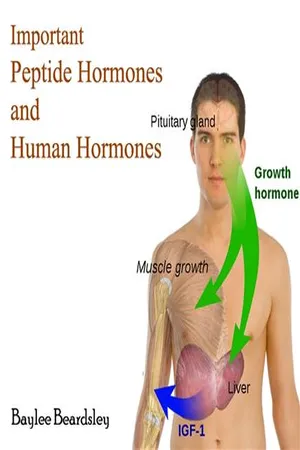 No longer available |Learn more
- (Author)
- 2014(Publication Date)
- University Publications(Publisher)
As its level is a central metabolic control mechanism, its status is also used as a control signal to other body systems (such as amino acid uptake by body cells). In addition, it has several other anabolic effects throughout the body. When control of Insulin levels fails, diabetes mellitus will result. As a consequence, Insulin is used medically to treat some forms of diabetes mellitus. Patients with Type 1 diabetes mellitus depend on external Insulin (most commonly injected subcutaneously) for their survival because the hormone is no longer produced internally. Patients with Type 2 diabetes mellitus are often Insulin resistant, and because of such resistance, may suffer from a relative Insulin deficiency. Some patients with Type 2 diabetes may eventually require Insulin if other medications fail to control blood glucose levels adequately, though this is somewhat uncommon. Insulin also influences other body functions, such as vascular compliance and cognition. Once Insulin enters the human brain, it enhances learning and memory and in particular benefits verbal memory. Enhancing brain Insulin signaling by means of intranasal Insulin administration also enhances the acute thermoregulatory and glucoregulatory response to food intake, suggesting that central nervous Insulin contributes to the control of whole-body energy homeostasis in humans. Insulin is a peptide hormone composed of 51 amino acids and has a molecular weight of 5808 Da. It is produced in the islets of Langerhans in the pancreas. The name comes from the Latin insula for island. Insulin's structure varies slightly between species of animal. Insulin from animal sources differs somewhat in strength (in carbohydrate metabolism control effects) in humans because of those variations. Porcine Insulin is especially close to the human version. Gene The proInsulin precursor of Insulin is encoded by the INS gene. Alleles No longer available |Learn more
- (Author)
- 2014(Publication Date)
- College Publishing House(Publisher)
As its level is a central metabolic control mechanism, its status is also used as a control signal to other body systems (such as amino acid uptake by body cells). In addition, it has several other anabolic effects throughout the body. When control of Insulin levels fails, diabetes mellitus will result. As a consequence, Insulin is used medically to treat some forms of diabetes mellitus. Patients with Type 1 diabetes mellitus depend on external Insulin (most commonly injected subcutaneously) for their survival because the hormone is no longer produced internally. Patients with Type 2 diabetes mellitus are often Insulin resistant, and because of such resistance, may suffer from a relative Insulin deficiency. Some patients with Type 2 diabetes may ________________________ WORLD TECHNOLOGIES ________________________ eventually require Insulin if other medications fail to control blood glucose levels adeq-uately, though this is somewhat uncommon. Insulin also influences other body functions, such as vascular compliance and cognition. Once Insulin enters the human brain, it enhances learning and memory and in particular benefits verbal memory. Enhancing brain Insulin signaling by means of intranasal Insulin administration also enhances the acute thermoregulatory and glucoregulatory response to food intake, suggesting that central nervous Insulin contributes to the control of whole-body energy homeostasis in humans. Insulin is a peptide hormone composed of 51 amino acids and has a molecular weight of 5808 Da. It is produced in the islets of Langerhans in the pancreas. The name comes from the Latin insula for island. Insulin's structure varies slightly between species of animal. Insulin from animal sources differs somewhat in strength (in carbohydrate metabolism control effects) in humans because of those variations. Porcine Insulin is especially close to the human version. Gene The proInsulin precursor of Insulin is encoded by the INS gene. eBook - ePub
- Robert H. Eckel(Author)
- 2011(Publication Date)
- Wiley-Blackwell(Publisher)
Chapter 1 Insulin action and beta-cell function: role in metabolic regulation Kristina M. Utzschneider and Steven E. Kahn Regulation of fuel utilization in health and diseaseThe normal processing and utilization of fuels is tightly regulated by hormonal, neural, and intracellular mechanisms so that carbohydrates, proteins, and fats supply energy to the brain, muscles, and other tissues, and excess fuel is stored efficiently for use during periods of fasting or increased energy needs. Two key players in the balance of hormones regulating these processes are Insulin and glucagon.Insulin is secreted by the islet beta-cell in response to glucose, amino acids, peptides, and fatty acids and then promotes tissue uptake of glucose and glyco-gen synthesis. Insulin also acts on lipid metabolism by promoting enzymes involved in de novo lipogenesis, while suppressing those enzymes involved in lipid oxidation and lipolysis, resulting in a decrease in circulating free fatty acids (FFAs). The net result is a shift towards utilization of glucose as the primary fuel. Insulin’s effects are mainly anabolic as Insulin levels increase when nutrient availability is high. During times when Insulin levels are low, such as during fasting, these processes reverse and fuel selection shifts to preferentially utilize fat. Insulin also acts centrally in the hypothalamus as a satiety signal by interacting with neural centers that regulate food intake.In contrast to Insulin, the hormone glucagon, which is secreted by the islet alpha-cell, acts as a catabolic hormone, stimulating production of glucose via glycogenolysis and gluconeogenesis, primarily in response to hypoglycemia. Glucagon is also important in the regulation of basal and postprandial glucose levels, with the balance of this hormone with Insulin being important. Thus, when Insulin levels rise, as occurs after nutrient ingestion, glucagon levels will normally decrease. eBook - ePub
- Anthony W. Norman, Gerald Litwack(Authors)
- 1997(Publication Date)
- Academic Press(Publisher)
CHAPTER 7Pancreatic Hormones: Insulin and Glucagon
I. INTRODUCTIONA. Background Information B. Regulation of Blood Glucose C. Nutritional and Metabolic InterrelationshipsII. ANATOMICAL, MORPHOLOGICAL, AND PHYSIOLOGICAL RELATIONSHIPSA. Introduction B. Development and Embryologic Origins C. Ultrastructure of Pancreatic Islets D. Vascularization and Innervation of Pancreatic IsletsIII. CHEMISTRYA. Insulin B. Glucagon C. Pancreatic Polypeptide D. Somatostatin E. Islet Amyloid Polypeptide F. LeptinIV. BIOCHEMISTRYA. Biosynthesis of Hormones B. Secretion of Pancreatic Hormones C. Pharmacological Agents Related to the Pancreas D. Metabolism of Insulin and GlucagonV. BIOLOGICAL AND MOLECULAR ACTIONSA. Interactions with Target Tissues B. Biological ActionsVI. CLINICAL ASPECTSA. Insulin B. GlucagonReferencesI INTRODUCTION
A Background InformationA feature essential for life of higher vertebrates is their ability to maintain a relatively constant blood glucose concentration. In the higher animals, glucose is essential as an energy source for all cells. Although some cells can utilize alternate “fuel metabolites,” such as amino acids or fatty acids, the brain and its neurons are dependent upon a continuous supply of blood-delivered glucose. Superimposed on the requirement for the maintenance of a constant blood glucose level are the perturbations in blood glucose that may occur naturally as a consequence of ongoing physiological and metabolic events. These can include (1) the intestinal absorption and concomitant systemic transport to storage depots (liver, muscle, and/or adipose tissue) of foodstuffs (e.g., carbohydrates, proteins, fat); (2) muscular activity; (3) thermogenesis and response to environmental extremes of heat and cold; (4) starvation; (5) pregnancy/lactation; and (6) disease or injury states. The endocrine gland largely responsible for the maintenance of blood glucose levels and the proper cellular uptake and exchange of “fuel metabolites” is the pancreas. The principal hormones secreted by the pancreas are Insulin and glucagon. In addition, the pancreas secretes a number of other peptide hormones (see Table 7-6 eBook - ePub
- Anthony W. Norman, Helen L. Henry(Authors)
- 2014(Publication Date)
- Academic Press(Publisher)
c Gastrin is predominantly secreted by the antrum of the stomach, the duodenum, and the pancreas. The antrum is at the bottom of the stomach.Insulin is a pluripotent hormone in that it has a wide sphere of influence; directly or indirectly it affects virtually every organ and tissue in the body. The main function of Insulin is to stimulate anabolic reactions to increase the amount of stored carbohydrates, proteins, and fats; this process will have the metabolic consequence of producing a lowered blood glucose level and the storing of energy.Glucagon can be thought of as an indirect antagonist of Insulin. Glucagon stimulates catabolic reactions that ultimately lead to the breakdown of glycogen, proteins, and fat and result in an elevation in blood glucose levels. Thus, the pancreas is continuously adjusting the relative amounts of glucagon and Insulin secreted, in response to the continuous perturbations of blood glucose and other fuel metabolites occurring as a consequence of changes in the relative amounts of anabolism and catabolism in the various tissues.B Regulation of Blood Glucose
The blood concentration of glucose normally lies within the range of 80–110 mg/100 mL (4.4–6.1 mM); see Table 6-2 . Reduction in blood glucose levels below 45–55 mg/100 mL (~2.5 mM) for a continued interval of time will lead to an impairment of brain function, tremors, and convulsions due to activation of the sympathetic nervous system, resulting ultimately in death. Conversely, a prolonged elevation of blood glucose above 130 mg/100 mL generates a state of hyperglycemia, due to a relative lack of Insulin. This then leads to a devastating wasting of metabolic energy, osmotic diuresis, and metabolic acidosis. Because glucose is a “small molecule,” all blood glucose is completely filtered into urine by the glomerulus of the kidney (see Figure 15-3B/C eBook - PDF
- Franklyn F. Bolander(Author)
- 2013(Publication Date)
- Academic Press(Publisher)
1. Insulin Although Insulin consists of two peptides linked by disulfide bonds, it is originally synthesized as an 81-amino-acid precursor, proInsulin. The entire molecule is required to approximate the appropriate cysteines, and the disul-fide bridges form while the prohormone is in the rough endoplasmic reticu-lum. Once this linkage occurs, the intervening piece, or C peptide, must be Table 2-3 Histochemical Characteristics of Some Pancreatic Hormones Hormone Location Structure Glucagon A (a) cells: Outermost rim of 29-Amino-acid linear poly-islets peptide Insulin Β (β) cells: Core of islet Two chains (21 and 30 amino acids) connected by disul-fide bonds SRIF D (δ) cells: Inner rim of islets 14-Amino-acid cyclic peptide (via disulfide bond) Pancreatic polypeptide F cells 36-Amino acid linear poly-peptide 54 2. Classical Endocrinology removed since proInsulin is only about 10% as active as Insulin. Cleavage is accomplished by a trypsin-like protease attracted to two pairs of basic amino acids (Fig. 2-16). Cleavage begins when the molecule is in the Golgi appa-ratus and continues in the secretory granules, although it is never complete: about 6% of Insulin is secreted as proInsulin. Finally, zinc is transported into the granules and triggers the crystallization of Insulin as dinners and hex-amers. To understand the control of Insulin secretion, it is necessary to discuss its actions briefly. Insulin is an anabolic hormone, that is, it is involved with energy storage. Primarily, this action is manifested as an increase in glucose and amino acid transport into cells and as the stimulation of conversion of these precursors into storage forms such as glycogen, protein, and triglycer-ides. Therefore, elevated blood levels of glucose, fatty acids, or amino acids will stimulate Insulin release. eBook - PDF
- Gerald Litwack(Author)
- 2012(Publication Date)
- Academic Press(Publisher)
BIOCHEMICAL ACTIONS OF HORMONES, VOL. VIII CHAPTER 5 Effects of Insulin on Intraoellular Functions Ira D. Goldfine I. Introduction 274 IL Intraoellular Effects of Insulin 274 A. Effects of Insulin on the Nucleus 274 B. Effects of Insulin on the Endoplasmic Reticulum 277 C. Effects of Insulin on Lysosomes 278 D. Effects of Insulin on Mitochondria 278 E. Effects of Insulin on Cytoplasmic Enzymes 279 F. Effects of Insulin on Phosphorylation 279 III. Mechanism of Insulin Action 280 IV. Cellular Binding Sites for Insulin 281 A. Plasma Membranes 281 B. Nucleus and Nuclear Envelope 282 C. Smooth and Rough Endoplasmic Reticulum 286 D. Golgi 287 E. Other Subcellular Fractions 288 V. The Entry of Insulin into Target Cells 288 A. Cell Fractionation Studies 288 B. Fluorescent-Labeled Insulin 290 C. Autoradiographic Analysis 290 D. Is Internalization Necessary for the Biological Actions of Insulin? 297 VI. Hypothesis, Speculation, and Conclusion 299 References 301 273 Copyright © 1981 by Academic Press, Inc. All rights of reproduction in any form reserved. ISBN 0-12-452808-2 274 Ira D. Goldfine I. INTRODUCTION Insulin is a major anabolic hormone that regulates the metabolism of most cells. The potency of Insulin is derived from its having short, in-termediate, and long-term effects on cellular functions. The short-term effects of Insulin (occurring within seconds) are the rapid regulation of plasma membrane activities including membrane potential and the transport of sugars, amino acids, and ions (Krahl, 1974; Pilkis and Park, 1974; Fain, 1974; Czech, 1977; Goldfine, 1977, 1978a). The in-termediate effects of Insulin (occurring within minutes) are the activation and inactivation of enzymes, stimulation of protein synthesis, and the in-hibition of protein degradation (Krahl, 1974; Pilkis and Park, 1974; Fain, 1974; Czech, 1977; Goldfine, 1977, 1978a). eBook - PDF
The Hormones
Physiology, Chemistry, and Applications
- Gregory Pincus, Kenneth V. Thimann, E. B. Astwood, Gregory Pincus, Kenneth V. Thimann, E. B. Astwood(Authors)
- 2013(Publication Date)
- Academic Press(Publisher)
The development of these immunological techniques has provided important methods for detecting structural dif-ferences in Insulins and promises to revolutionize the assay of Insulin in body fluids and studies of factors which regulate the storage and secre-tion of the hormone by the pancreatic ft cell. Advances of comparable importance have also taken place in the sphere of Insulin action. The membrane theory of Insulin action proposed by Levine and his colleagues (130) and reviewed extensively by Stetten and Bloom has been strength-ened, and an important early paper by Lundsgaard (134) published in 1939, which proposed a membrane theory of Insulin action and provided experimental evidence acceptable even today, has been rediscovered. More recent studies on the action of Insulin have been greatly facilitated by the development of satisfactory in vitro techniques, notably the per-fused rat heart by Fisher and colleagues (27) and Morgan and Park and colleagues (166), an improved rat diaphragm preparation by Kipnis and Cori (95), and in vitro preparations of adipose tissue by Krahl (106), Wertheimer and Shafrir (246), Gordon and Cherkes (67), White and Engel (247), and Winegrad and Renold (251). Studies with these preparations have led to the realization that the action of the hormone leads to seemingly independent changes in the metabolism of carbohy-drate, of protein, and, in some respects, of fat. In assessing current knowledge about Insulin, the amino acid sequence of the hormone stands alone as a seemingly unchallengeable fact. The particular steps in metabolism which are affected by the action of the hormone are probably largely defined, and something is known about storage of Insulin in /? cells and of factors which alter the rate of its secretion. eBook - PDF
- Richard I. G. Holt, Allan Flyvbjerg(Authors)
- 2024(Publication Date)
- Wiley-Blackwell(Publisher)
Am. J. Physiol. Endocrinol. Metab. 303 (2): E265–E271. 111 Textbook of Diabetes, Sixth Edition. Edited by Richard I.G. Holt and Allan Flyvbjerg. © 2024 John Wiley & Sons Ltd. Published 2024 by John Wiley & Sons Ltd. Insulin and Insulin-like growth factor (IGF) signalling integrates metabolism, growth, reproduction, and lifespan with environmen- tal signals [1]. Lower animals express many Insulin-like peptides – 38 in Caenorhabditis elegans and 7 in fruit flies – that bind to a single Insulin-like receptor tyrosine kinase [2], whereas the human genome encodes a smaller family of structurally related Insulin-like peptides, including Insulin itself, IGF-I, and IGF-II. Diabetes is a common metabolic disease of persistent hyperglycaemia owing to various mechanisms that cause deficiency of Insulin and/or Insulin action, including impaired glucose sensing or Insulin secretion by pancreatic β cells (maturity-onset diabetes of youth, MODY); autoimmune-mediated β-cell destruction (type 1 diabetes); or insufficient β-cell Insulin secretory capacity to compensate for peripheral Insulin resistance (type 2 diabetes) [3]. MODY is caused by mutations in at least 14 genes associated with β-cell function, notably hepatocyte nuclear factor-4α (HNF4α, MODY1), glucoki- nase (GCK, MODY2), and hepatocyte nuclear factor-1α (HNF1α, MODY3) [4]. During the past 50 years, advances in all areas of biomedical sci- ence led to a multidisciplinary understanding of Insulin receptor signalling. Cloning of the Insulin receptor cDNA and its gene revealed novel mechanisms of signal transduction. Dysregulated Insulin signalling causes Insulin resistance, which includes any state of diminished cellular or systemic Insulin action [5]. Mutations in 9 Mechanism of Insulin Action Morris F. White Division of Endocrinology, Children’s Hospital Boston, Harvard Medical School, Boston, MA, USA Key points • The Insulin receptor is a transmembrane protein.
Index pages curate the most relevant extracts from our library of academic textbooks. They’ve been created using an in-house natural language model (NLM), each adding context and meaning to key research topics.