Biological Sciences
Interferon
Interferon is a group of signaling proteins produced by the body in response to viral infections, tumors, and other immune challenges. It plays a crucial role in regulating the immune system and has antiviral, antiproliferative, and immunomodulatory effects. Interferon can be used therapeutically to treat certain diseases, including hepatitis C, multiple sclerosis, and certain cancers.
Written by Perlego with AI-assistance
Related key terms
1 of 5
8 Key excerpts on "Interferon"
- eBook - PDF
Biological Response Modifiers
New Approaches to Disease Intervention
- Paul Torrence(Author)
- 2012(Publication Date)
- Academic Press(Publisher)
3 Immunoregulatory Functions of Interferon JOHN J . HOOKS BARBARA DETRiCK Clinical Branch and Experimental Immunology Section National Eye Institute National Institutes of Health Bethesda, Maryland I. Introduction 57 II. Interferon Production by Lymphoid Cells 58 III. Interferon y and the Lymphokine Circuit 59 IV. Effects of Interferon on Immune Cells and Immune Responses 60 Α. Β Lymphocytes 61 Β. Τ Lymphocytes 61 C. Natural Killer Cells 61 D. Basophils 62 E. Macrophages 63 V. Interferon and Class II Antigen Expression 64 VI. Interferons in Immunologically Related Disorders 67 A. Interferon-Induced Disorders in Animals 68 B. Circulating Interferon in Human Immunologically Related Disorders 69 C. Production of Interferon in Human Immunologically Related Disorders 71 VII. Conclusions , 71 References 72 I. INTRODUCTION Over 25 years have passed since Interferon (IFN) was first described as an antiviral glycoprotein produced by cells in response to viruses (Isaacs and Lindenmann, 1957). Now, however, numerous studies show that the actions of IFN are not exclusively antiviral. In fact, the IFN proteins can modify a variety of biological activities and can be considered one of the body's regulatory proteins (Merigan and Friedman, 1982; Baron et al., 1982; Vilcek and De Maeyer, 1984). BIOLOGICAL RESPONSE MODIFIERS 57 Copyright © 1985 by Academic Press, Inc. All rights of reproduction in any form reserved. 58 John J. Hooks and Barbara Detrick Biological regulatory systems, frequently consisting of proteins interact-ing with cell receptors, are important in maintaining physiological integrity. The cellular production of the IFN proteins can be activated by viruses, immune responses, and other environmental factors. - eBook - PDF
Pharmaceutical Biotechnology
Fundamentals and Applications, Third Edition
- Daan J. A. Crommelin, Robert D. Sindelar, Bernd Meibohm, Daan J. A. Crommelin, Robert D. Sindelar, Bernd Meibohm(Authors)
- 2016(Publication Date)
- CRC Press(Publisher)
11 Interferons and Interleukins Jean-Charles Ryff Biotech Research and Innovation Network, Basel, Switzerland Ronald W. Bordens Schering-Plough Corporation, Kenilworth, New Jersey, U.S.A. Sidney Pestka Department of Molecular Genetics, Microbiology, and Immunology, University of Medicine and Dentistry of New Jersey–Robert Wood Johnson Medical School, Piscataway, New Jersey, U.S.A. INTRODUCTION In 1957 a substance was described (Isaacs and Lindenmann, 1957) that was produced by virus-infected cell cultures and “interfered” with infection by other viruses; it was called Interferon (IFN). Over the following decades it was realized that “IFN” comprises a family of related proteins with several additional properties. Starting in the 1960s various “factors” produced primarily by white blood cells (WBC) as well as other cell supernatants were described which acted in various ways on other WBCs or somatic cells. They were usually given a descriptive name either associated with their cell of origin or their activity on other cells resulting in a myriad of names. The application of molecular technology allowed us to determine that some cyto-kines had multiple activities and that different cyto-kines had similar overlapping activities. A systematic classification based on genetic structure and protein characterization has been effective. The interactive networks and cascades of cytokines, IFN, interleukins (IL), growth factors (GF), chemokines (CK), their receptors (r or R) and signaling pathways are highly complex and will be further explored in this chapter. Cytokine is a term coined in 1974 by Stanley Cohen in an attempt at a more systematic approach to the numerous regulatory proteins secreted by hemopoietic and nonhemopoietic cells. Cytokines play a critical role in modulating the innate and adaptive immune systems. They are multifunctional peptides that are now known to be produced by normal and neoplastic cells, apart from those of the immune system. - eBook - PDF
- Anthony R. Mire-Sluis, Robin Thorpe(Authors)
- 1998(Publication Date)
- Academic Press(Publisher)
Receptors 372 6.1 Characterization 372 6.1.1 General Features 372 6.1.2 Molecular Cloning of IFN Receptor Components 373 7. Signal Transduction 375 7.1 Signal Transduction 375 7.1.1 Molecular Mechanisms 375 7.2 IFN-inducible Genes 375 7.2.1 IFN-response Gene Sequences 375 7.2.2 Proteins Induced by IFN 375 8. Mouse IFN-~ and IFN-[3 376 9. Clinical Uses of IFNs 378 9.1 General Considerations 378 9.2 IFN Treatment of Malignant Diseases 379 9.3 IFN Treatment of Viral Diseases 379 9.4 IFN Treatment of Other Human Diseases 380 10. References 380 1. Introduction 1.1 OUTLINE OF DISCOVERY AND CHARACTERIZATION OF InterferonS (IFN) The phenomenon of viral interference was first described nearly 60 years ago when Hoskins (1935) described the protective action of a neurotropic yellow fever virus against a viserotropic strain of the same virus in monkeys. Although viral interference was further investigated in the 1940s and 1950s, the underlying mechanism was not discovered until 1957 when Isaacs and Lindemann, working at The National Institute for Medical Research (London, UK), isolated a biologically active substance from virally-infected chicken cell cultures that, on transfer to fresh chicken cell cultures, Cytokines ISBN 0-12-498340-5 Copyright 9 1998 Academic Press Limited All rights of reproduction in any form reserved. 362 A. MEAGER produced a protective antiviral effect (Isaacs and Lindemann, 1957). The word Interferon (IFN) was coined for this substance. Its discovery aroused considerable scientific and medical interest since by 1957 antibiotics were widely available to control bacterial infections, but, in stark contrast, viral diseases such as influenza, measles, polio, and smallpox were virtually untreatable. Interest was further heightened by many subsequent studies that demonstrated that IFN could be produced by human cells and was active against a broad spectrum of viruses (see Schlesinger, 1959, for an early review). - eBook - PDF
The Interferons
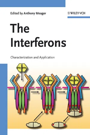
Characterization and Application
- Anthony Meager(Author)
- 2006(Publication Date)
- Wiley-Blackwell(Publisher)
The IFN family is composed of transcriptionally activated and secreted proteins with pleiotropic biological effects on the host. IFNs play a central role in the resis- tance of mammalian hosts to pathogens, and modulate antiviral and immune re- sponse [4, 5]. The IFN family is classified into two subgroups: type I IFNs (IFN-a, -b, -o and -l) and type II IFN (IFN-g), characterized mainly by the receptor complex used for signaling, the cell type from which they are secreted and their intrinsic biological properties (Tab. 2.1). Type I IFNs (IFN-b and multiple types of IFN-a), also referred to as viral IFNs, play an essential role in the host immune response against viruses. IFN-a, previously referred to as leukocyte IFN, is comprised of at least (1) 13 IFN-a species, whereas IFN-b, also known as fibroblast IFN, is a single species. Each subtype of IFN-a is encoded by its own gene and is regulated by its own promoter sequence, all lacking introns [6]. Type II IFN, also known as IFN-g, is encoded by one gene that contains four exons and three introns; expression is controlled by lymphoid restricted transcription factors such as nuclear factor of activated T cells (NF-AT), The first two authors contributed equally to the work 35 - eBook - PDF
- Wahid Ali Khan(Author)
- 2019(Publication Date)
- IntechOpen(Publisher)
It includes three main classes, designated as type I IFNs, type II IFN and type III IFNs. The two main type I IFNs includes IFN-α (further classified into 13 different subtypes such as IFN-α1, -α2, -α4, -α5, -α6, -α7, -α8, -α10, -α13, -α14, -α16, -α17 and -α21), and IFN-β. The term Interferon derives from the ability of these cytokines to interfere with viral replication. Type I IFNs present a potent anti-viral effect by inhibiting viral replication, increasing the lysis potential of natural killer (NK) cells and the expression of MHC class I molecules on virus-infected cells, and stimulating the development of Th1 cells. During an infectious process, this type of Interferon becomes abun-dant and is easily detectable in the blood. On the other hand, type II IFN has only one repre-sentative, IFN-γ. This cytokine plays a major role is macrophage activation both in innate and adaptive immune responses. Type III IFNs, also denoted IL-28/29, present similar biological effects to type I IFN, playing an important role in host defense against viral infections [5 – 8 ]. 2.1. History Interferon was the first described member of the class of protein molecules now known as cytokines. Nowadays, Interferons are well known to participate in innate immune system, mediating responses against viral infections. This role of the IFNs was first described in the 1930s, when a research conducted by Hoskins demonstrated that rabbits previously infected by the herpes simplex virus were protected against subsequent infections by the same type of virus. In 1937, a few years after Hoskins’ experiment, Findlay and MacCallum showed that the virus-infected animals were also resistant to infections caused by antigenically different viruses, corroborating and complementing the existing evidence regarding IFNs functions at that time. - eBook - PDF
Cytokines and Cytokine Receptors
Physiology and Pathological Disorders
- Constantin A. Bona, Jean-Pierre Revillard(Authors)
- 2001(Publication Date)
- CRC Press(Publisher)
19 TYPE I InterferonS Edward De Maeyer and Jaqueline De Maeyer-Guignard Institut Curie, Université Paris-Sud, Orsay, France Type I Interferons (IFNs) constitute a family of structurally related proteins that are derived from the same ancestral gene and act on a common cell-surface receptor. Contrary to many other cytokines, the production of type I IFNs is not a specialized function, and all cells in the organism can produce them, mainly, but not exclusively, as a result of induction by viruses, usually via the formation of double-stranded RNA. Type I IFNs are responsible for the first line of defense during virus infection and act through the induction of a great number of proteins. Of these, at least forty have been characterized, and there are probably many more. In addition to their direct antiviral effects, type I IFNs exert a variety of other activities, such as for example the induction of various cytokines and the stimulation of different effector cells of the immune system. Due to these pleiotropic effects, recombinant Interferons are used in the clinic to treat a variety of diseases, among which some forms of cancer, viral hepatitis and relapsing-remitting multiple sclerosis. INTRODUCTION The designation of Interferons as “Type I” originated more than 25 years ago, to distinguish one class of Interferons, characterized by lack of inactivation after exposure to pH2, from another antiviral protein that was acid-labile and was referred to as “Type II” Interferon. Later on, type II IFN was called immune IFN, and has since become IFN-γ. It is a lymphokine that displays no molecular homology with type I IFNs, but shares some biological activities. In fact, the activities of type I and type II IFNs are intimately related, in that type I IFN can up- or down-regulate the production of type II IFN, and type II IFN can induce the synthesis of type I IFN.Moreover, the signal transduction pathways of type I and type II IFN partly overlap. - Josef S. Smolen, Peter E. Lipsky, Josef S. Smolen, Peter E. Lipsky(Authors)
- 2007(Publication Date)
- CRC Press(Publisher)
19 The Interferons Lars Rönnblom, Maija-Leena Eloranta and Gunnar Alm Introduction • The IFN proteins and genes • Activation of type I IFN genes • Activation of the type II IFN (IFN-γ ) gene • Activation of type III IFN genes • Cellular basis of type I and III IFN production • Mode of activation of type I IFN production in immature PDCs • The IFN receptors and their signaling pathways • Negative regulation of IFN signaling pathways • Genes, proteins, and cell functions regulated by IFNs • Activation of the type I IFN system in autoimmune diseases • A causative role for type I IFN in autoimmunity • Type II IFN (IFN-γ ) in autoimmune diseases • An etiopathogenic mechanism for autoimmunity involving the type I IFN system • Therapeutic targets • Acknowledgments • References INTRODUCTION The Interferons (IFNs) were the first cytokines to be discovered, 1 evaluated as therapeutic agents in viral infections and cancers, and produced on a large scale as recombinant proteins for clinical use. 2 The IFNs are defined by their ability to interfere with replication of viruses in cells via induction of new mRNA and protein synthesis. They are grouped into type I IFN, type II IFN, and type III IFN and are encoded by no less than 21 different genes in man. These three major classes of IFNs (I–III) act on separate receptors. The type I IFNs have been most extensively studied and constitute an important part of innate immunity against viral infections, but also act as a link to adaptive immunity via many effects on key immune cells, such as T cells, B cells, and dendritic cells (DCs). The inducers of type I IFN production (typically virus), the cells producing type I IFNs, the type I IFN genes and proteins, as well as the targets cells affected by the type I IFNs, can be defined as the type I IFN system. The type III IFNs may be included in this system, because of many similarities to type I IFN.- eBook - ePub
- Abner Louis Notkins(Author)
- 2014(Publication Date)
- Academic Press(Publisher)
For many years antibody was considered the host’s major defense mechanism against viral infections. The interaction of antibody with extracellular virus neutralizes virus, thereby preventing infection of new cells. In the late 1950’s a second host defense mechanism, Interferon, was discovered. This substance acts at the level of the cell and prevents viral replication by inhibiting transcription and/or translation. In the 1960’s a third host defense mechanism, cellular immunity, was identified. Immune lymphocytes are thought to recognize viral induced antigens on the surface of infected cells, and reject these cells in much the same way that immune lymphocytes recognize and reject tumor cells. Over the last few years it has become apparent that a wide variety of biological mediators are released from immune lymphocytes when they are stimulated by specific antigens. One of these mediators is Interferon. The purpose of this review is to bring together information suggesting that the cellular immune response to certain viral infections may be mediated through Interferon.Immune Specific Induction of Interferon in Vitro
In 1965 Wheelock showed that human leukocyte cultures stimulated by phytohemagglutinin (PHA) not only underwent blastogenesis but also produced Interferon (1 ). When compared to standard Interferon induced by incubating leukocyte cultures with Newcastle disease virus (NDV), the PHA-induced Interferon differed in two respects; it was not stable at pH 2.0 and it lost biological activity at 56° C. Subsequently a variety of other mitogens including pokeweed mitogen (PWM) and concanavallin A were found to be capable of inducing the production of Interferon (2 –4 ). In addition, Falcoff et al. found that stimulation of lymphocytes by anti-lymphocyte immunoglobulin resulted in the production of Interferon (5 ).The specific induction of Interferon from immune leukocytes by non-viral antigens was first demonstrated by Green et al. (3
Index pages curate the most relevant extracts from our library of academic textbooks. They’ve been created using an in-house natural language model (NLM), each adding context and meaning to key research topics.
