Biological Sciences
Ribosomes
Ribosomes are cellular structures responsible for protein synthesis. They are composed of RNA and protein and can be found in the cytoplasm or attached to the endoplasmic reticulum. Ribosomes read the genetic information from messenger RNA and use it to assemble amino acids into proteins through a process called translation.
Written by Perlego with AI-assistance
Related key terms
1 of 5
8 Key excerpts on "Ribosomes"
- eBook - PDF
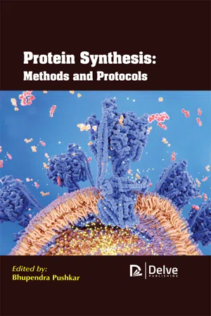
- Bhupendra Pushkar(Author)
- 2020(Publication Date)
- Delve Publishing(Publisher)
This chapter also provides knowledge about the function of a ribosome. 6.1. INTRODUCTION There have been often seen that there are many organelles which tend to work together to complete the cell activities while examining the animal as well as plant cell by using a microscope. This is one of the essential cell organelles which are Ribosomes. These are in charge of the protein synthesis. The ribosome is considered to be a complex made of protein and RNA. The ribosome tends to add up to numerous million Daltons in size and it is assumed to have an important part in the course of decoding the genetic message which is reserved in the genome into protein. There is an essential chemical step of protein synthesis and that is peptidyl transfer. In this, the movement of the developing or nascent peptide takes place from one tRNA molecule to the amino acid along with another tRNA. While developing the polypeptide, the amino acids are included along with the arrangement of codons of an mRNA. Therefore, the ribosome has important sites for one mRNA and not less than two tRNAs. A ribosome is said to be made up of two subunits. These include the big and the little subunit which contains a couple of ribosomal RNA (rRNA) molecules and a random number of ribosomal proteins. Several protein factors catalyze the distinct impressions of protein synthesis. It is important to consider the translation of the genetic code for the manufacturing of the protein which is useful and also, for the growth of the cells. Ribosomes are said to be the particles that are present in large numbers in all the living cells. It serves as the site where the protein synthesis takes place. Ribosomes can occur both as free particles in prokaryotic as well as eukaryotic and also, it acts as the particles which are attached to the membranes of the endoplasmic reticulum in eukaryotic cells. Ribosome Structure and Function 151 Ribosomes are the small particles which are remarkably abundant in cells. - eBook - PDF
The Plant Cell
A Comprehensive Treatise
- N. E. Tolbert(Author)
- 2013(Publication Date)
- Academic Press(Publisher)
Ribosomes ERIC DAVIES BRIAN A. LARKINS 11 I. Introduction 413 II. Ribosome Structure and Biogenesis 414 A. Ribosomal RNA 414 B. Ribosomal Proteins 417 C. Ribosome Assembly 419 D. Ribosome Structure 419 Ε. Control of Ribosome Content in Eukaryotes 420 F. Interaction of Ribosomes with Other Subcellular Components 423 III. PolyRibosomes 424 A. Polyribosome Function 424 B. Polyribosome Isolation 424 C. Free and Membrane-Bound Polysomes 428 D. Changes in Polysome Aggregation 432 References 433 I. INTRODUCTION The term ribosome was first introduced about 20 years ago (Roberts, 1958) to describe a particle made up of approximately equal amounts of RNA and protein that was intimately involved in protein synthesis. At that time, more was known about the Ribosomes from animals and plants than about those from bacteria, but that situation has changed considerably, as exemplified by the selection of articles in the most comprehensive review on the subject (Nomura^/ aL, 1974). At least part of the reason for the emergence ofE. coli as a system for studies on Ribosomes is its comparative simplicity. Bacteria (prokaryotes) have only one genome and produce only one type of ribosome The Biochemistry of Plants, Vol. 1 Copyright © 1980 by Academic Press, Inc. All rights of reproduction in any form reserved. ISBN 0-12-675401-2 413 414 Eric Davies and Brian A. Larkins II. RIBOSOME STRUCTURE AND BIOGENESIS Perhaps the major difference between eukaryotes and prokaryotes in re-gard to ribosome biogenesis is that the latter have no apparent subcellular compartmentalization of the sites of synthesis and assembly of their various ribosomal components. In contrast, strict compartmentaUzation exists in eukaryotes. The RNA component of Ribosomes is synthesized directly from a DNA template in the fibrillar region of the nucleolus, whereas the ribosomal proteins are made on cytoplasmic polyRibosomes and must be transported into the nucleolus for assembly (Warner^/ Λ /., 1973). - eBook - ePub
- Anders Liljas, Lars Liljas;Miriam-Rose Ash;G?ran Lindblom;Poul Nissen;Morten Kjeldgaard(Authors)
- 2016(Publication Date)
- WSPC(Publisher)
11Protein Synthesis â Translation
11.1Evolution of the Translation System
The translation of genetic information into functional protein molecules is a central process for life; and the genetic code, tRNA molecules and the machinery for protein synthesis are all highly conserved. The ribosome on which translation occurs is composed of protein and rRNA molecules. Carl Woese showed that sequenced fragments of ribosomal rRNA molecules from a large variety of species were related. Thus, the ribosomal RNAs could be used to analyze the relationship between species. By 1977, he could show that it was not correct to divide the present living organisms into prokaryotes and eukaryotes. A unique new kingdom had to be introduced: archaea. Living organisms, according to Woese, had to be organized into bacteria, archaea and eukaryotes.In comparisons of completely sequenced genomes, the molecules of the translation apparatus stand out as dominating the group of universally conserved molecules. The genetic code, tRNAs, ribosomal rRNA, ribosomal proteins and translation factors must have coevolved at a very early phase of biological evolution and have subsequently gone through only limited further changes.An important aspect of protein synthesis is that nucleic acid molecules have central roles, in contrast to most other processes in cells where proteins dominate. Central components are the mRNA, the tRNA and the ribosomal rRNA molecules. An mRNA molecule contains a copy of the gene sequence and binds to the ribosome. The tRNA molecules, the adapters suggested by Francis Crick, decode the gene sequence and link the amino acid into the growing peptide on the ribosome.Fig. 11.1 ▪ - eBook - ePub
Cell Biology E-Book
Cell Biology E-Book
- Thomas D. Pollard, William C. Earnshaw, Jennifer Lippincott-Schwartz, Graham Johnson(Authors)
- 2022(Publication Date)
- Elsevier(Publisher)
Assembly factors consisting of snoRNAs and numerous proteins then orchestrate the stepwise assembly of rRNAs and ribosomal proteins into the small and large subunits and guide their export from the nucleus into the cytoplasm. Ribosomes are stable for days, but starving cells may use them as a source of nutrients, after selection by autophagy and delivery to lysosomes for degradation (see Fig. 24.9). Although the genes for many ribosomal proteins are essential for viability, mutations in some cause remarkably specific defects. For example, humans with just one functional gene for ribosomal protein RPSA are missing their spleen but are otherwise normal. Mutations in genes for other subunits cause anemia and mutations in genes for certain assembly proteins cause liver disease. One explanation for these defects in specific organs is that translation of particular mRNAs in specialized cells may be sensitive to the concentration or total activity of Ribosomes. Mechanism of Protein Synthesis Organisms in all three domains of life use homologous components and similar biochemical reactions catalyzed by Ribosomes to synthesize proteins in four steps: initiation, elongation, termination, and subunit recycling (Fig. 12.1). Conformational changes move a ribosome along a mRNA as the gene sequence is read out. Ribosomes make few errors thanks to precise pairing between mRNA codons and tRNAs with their amino acids and to guanosine triphosphatase (GTPase) proteins that regulate the fidelity of each step (see Fig. 4.6 for details on GTPase cycles). As expected after 3 billion years of evolutionary divergence, some details differ across the phylogenetic tree. Initiation Phase The goal of initiation is to bring together an initiator tRNA carrying methionine (or N -formylmethionine, fMet, in Bacteria) and the AUG initiator codon of a mRNA on the ribosome ready for elongation of the polypeptide (Fig. 12.8) - eBook - ePub
- (Author)
- 2003(Publication Date)
- Elsevier Science(Publisher)
8Ribosome Biogenesis in Yeast: rRNA Processing and Quality Control
Ross N. Nazar [email protected] Department of Molecular Biology and Genetics, University of Guelph, Guelph, Ontario, Canada N1G 2W1.The ribosome, being a cell’s factory for protein synthesis, represents a cellular substructure that is critical, both for cell growth and survival. In the eukaryotes, the cytoplasmic ribosome is synthesized, almost exclusively, in a specialized subnuclear compartment called the nucleolus. Biogenesis begins with the transcription of rRNA precursors (pre-rRNAs) from rRNA genes (rDNA) that are localized in the nucleolus and ends with the transport of pre-ribosomal subunits to the cytoplasm where the final steps in the maturation process occur. In the course of ribosome biogenesis, the pre-rRNAs are cleaved and covalently modified while assembling with ribosomal proteins to form the mature subunits. These assembly and maturation processes are dependent on both cis-acting elements and many non-ribosomal protein or RNA trans-acting factors. The mature ribosome is the result of numerous macromolecule interactions involving many RNA and protein molecules which are actual ribosomal constituents and other molecules which ultimately are excluded from the mature particle. Together, the maturation processes provide a cell, and perhaps biotechnologists, with a means to control cell growth and even a mechanism for quality control.1 INTRODUCTION
A great variety of genetic and biochemical approaches has been used extensively to study ribosome biogenesis over a 40 year period. In general, the maturation of the pre-rRNA and the basic processes in the biogenesis of the ribosomal subunits appear to be largely conserved among the eukaryotes, but differences in many species-specific details also have been reported. In all the organisms that have been examined, the nascent pre-rRNA is first assembled into an 80-90S nucleolar particle. Structural rearrangements and nucleotide modifications occur as the ribosomal proteins are recruited, followed with cleavages which ultimately result in the mature cytoplasmic 40S and 60S ribosomal subunits. Since yeasts can be manipulated easily both genetically and biochemically, a majority of these studies has occurred in yeast cells, initially in Sacharomyces cerevisiae (Grivell and Planta, 1990 ) but, more recently, also in Schizosacharomyces pombe - eBook - PDF
- Edward Bittar(Author)
- 1995(Publication Date)
- Elsevier Science(Publisher)
Chapter 10 The Ribosome RICHARD BRIMACOMBE Introduction Components of the E. Coli Ribosome Function Initiation Elongation Termination Structure Electron Microscopy Neutron Scattering Protein-Protein Cross-Linking, and Amino Acid Sequences RNA Primary Sequences and Secondary Structure Intra-RNA and RNA-Protein Cross-Linking Foot-Printing Three-Dimensional Models of the Ribosomal RNA Structure-Function Correlation Location of Functionally Important Sites in the 3OS and 50S Subunits Site-Directed Cross-Linking with Functional Ligands Conclusions And Prospects Site-Directed Mutagenesis Crystallization of Ribosomal Proteins and Whole Ribosomal Subunits H , ,,, 254 255 256 256 256 259 259 259 260 261 261 262 263 263 264 264 267 269 270 270 Principles of Medical Biology, Volume 2 Cellular Organelles, pages 253-273 Copyright 9 1995 by JAI Press Inc. All rights of reproduction in any form reserved. ISBN:l-55938-803-X 253 254 RICHARD BRIMACOMBE INTRODUCTION The flow of genetic information from DNA to RNA to protein is a universal process in all cellular organisms, and the final stage in this process-namely, the translation of a messenger RNA sequence into a protein molecule~takes place on the ribosome. Each consecutive triplet sequence of three bases on the mRNA corresponds to a single amino acid according to the genetic code, and the amino acids to be incorporated into the protein chain are brought to the ribosome in the form of covalent aminoacyl-tRNA complexes. A molecule of tRNA consists of about 80 nucleotides, which are folded into a rather rigid L- shape (Kim et al., 1973). The amino acid is attached to the short arm of the L, whereas at the end of the long arm of the L there is a triplet anti-codon sequence, which is able to bind to a coding triplet on the mRNA by Watson-Crick base-pairing. - eBook - PDF
Conformational Proteomics Of Macromolecular Architecture: Approaching The Structure Of Large Molecular Assemblies And Their Mechanisms Of Action (With Cd-rom)
Approaching the Structure of Large Molecular Assemblies and Their Mechanisms of Action(With CD-ROM)
- R Holland Cheng, Lena Hammar(Authors)
- 2004(Publication Date)
- World Scientific(Publisher)
CONCLUDING REMARKS Ribosomal crystallography, initiated two decades ago, yielded exciting structural and clinical information. We found that both the decoding cen- ter and the peptidyl transferase centers are formed of RNA. Proteins seem to serve ancillary functions such as stabilizing required conforma- 280 Ada Yonath tion, binding of non-ribosomal factors, assisting the directionality of the translocation and gating of the ribosomal tunnel. The ribosome is an accurate and intricate machine, and as such it has ample of mobile regions. These include the head and the shoulder of the small subunit, the features lining the mRNA path; the peptidyl trans- ferase; the intersubunit bridges; the exit tunnel that control the release of nascent chains; the L1 stalk that provides the door for exiting tRNA. The studies presented here show that the peptidyl transferase center tolerates various binding modes, but precise positioning appears to be crucial for the biosynthesis of protein chains. This precise positioning is determined by the tRNA helical stem, rather than by its 3' end, and the ribosome provides the structural frame for it. Once properly positioned, the peptide bond can be formed spontaneously. Ribosomal components appear not participate directly in the catalytic event. They may, however, be of major importance for cell vitality, as they may increase the effi- ciency or enhance the rate of the reaction.. Ribosomes are a major target for antibiotics. The therapeutic use of antibiotics has been severely hampered by the emergence of drug resis- tance in many pathogenic bacteria. With the increased popularity of anti- biotics to treat bacterial infections, pathogenic strains have acquired anti- biotic resistance, thus became ineffective. Resistance posed extremely serious medical problems that have prompted extensive effort in the de- sign of modified or new antibacterial agents. - eBook - ePub
The Liver
Biology and Pathobiology
- Irwin M. Arias, Harvey J. Alter, James L. Boyer, David E. Cohen, David A. Shafritz, Snorri S. Thorgeirsson, Allan W. Wolkoff(Authors)
- 2020(Publication Date)
- Wiley-Blackwell(Publisher)
7 ]). It is intriguing that dysregulation of such an essential process like ribosome biogenesis would result in a viable organism. Indeed, how changes in ribosome biogenesis, a process that is required for every cell type, would create tissue‐specific pathologies is an outstanding question in the field.Much of the current knowledge of the steps in ribosome biogenesis comes from studies in the budding yeast, Saccharomyces cerevisiae. Only recently have scientists made advances into understanding how this process works in humans. Additionally, how ribosome biogenesis changes among different tissues is largely unknown. One of the best models for understanding ribosome biogenesis at the tissue level, however, has been the liver. Foundational studies on the liver have provided insights into how Ribosomes are made and how the cell responds to various stimuli to regulate the production of Ribosomes. Indeed, nucleoli, the non‐membrane‐bound organelles responsible for ribosome production, were first biochemically isolated from liver cells in 1956 [8 ]. It is likely that in the future, the liver will continue to play a large role in our understanding of ribosome biogenesis and its relation to human disease.OVERVIEW OF RIBOSOME BIOGENESIS
Ribosome biogenesis begins in a non‐membrane‐bound organelle inside the cell nucleus called the nucleolus. It starts with transcription of the tandemly repeated ribosomal DNA (rDNA) by RNA polymerase I (RNAPI). As the rDNA is being transcribed, the nucleolus forms around it. Thus, the repeats of rDNA on the chromosomes are called nucleolar organizing regions, or NORs. The NORs are located on five different chromosomes in humans (13, 14, 15, 21, and 22). Therefore, human cells have the potential to form 10 nucleoli per cell, although far fewer nucleoli are often observed [9 ]. The number of nucleoli in a diploid mouse hepatocyte ranges from 3 to 6 (mice have the potential to form up to 12 nucleoli per cell), with 3 nucleoli per nucleus being seen most often [10
Index pages curate the most relevant extracts from our library of academic textbooks. They’ve been created using an in-house natural language model (NLM), each adding context and meaning to key research topics.
