Chemistry
transfer RNA
Transfer RNA (tRNA) is a type of RNA molecule that plays a crucial role in protein synthesis. It carries specific amino acids to the ribosome, where they are added to the growing protein chain during translation. Each tRNA molecule has an anticodon that pairs with a complementary codon on the messenger RNA, ensuring the correct amino acid is incorporated into the protein.
Written by Perlego with AI-assistance
Related key terms
1 of 5
7 Key excerpts on "transfer RNA"
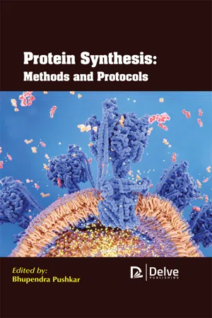 eBook - PDF
- Bhupendra Pushkar(Author)
- 2020(Publication Date)
- Delve Publishing(Publisher)
t-RNA and Aminoacyl-tRNA Synthetases 57 Until the year 1973, there was no firm information that was available in the three - dimensional structure of the molecule. In early 1973, the polynucleotide chain of yeast tRNA phe has been traced in a 4--X ray diffraction analysis. Structural work is noticed to progress rapidly, from then to the point where the atomic coordinates is now available. As these coordinates are derived from 2.5.-X ray diffraction analyses from the two different crystal forms of the same molecule. Knowledge about the detailed three-dimensional structure of the molecule that makes a change that occur separately in the type of research that can be also carried out. The main aim behind describing some of the details in the manner to obtain knowledge about the three -dimensional structure of tRNA species. they also want to discuss the extent to which it can be explained and can make various aspects understandable that are involved in chemistry and solution behavior of the same and tRNA species. The major biological function of the tRNA is related to the role of the tRNA in protein synthesis. The existence of a molecule like tRNA is a sense that is made necessary by the fact that nature encodes the information about the genetics in the nucleotide sequence in the nucleic acid. This helps in expressing the biological information in the ordered sequence in the polypeptide structure of amino acids. transfer RNA is said to have a fundamental biological role which is being used in acting as an interface between the polypeptide and polynucleotides. tRNA works by interacting with the messenger RNA in the ribosome at one end. While at the other end it is containing the growing peptide chain. tRNA is often seen to be involved in a large number of biological processes. Aminoacyl-tRNAs are the substrates that are used for the translation process and they are considered as an essential for determining how the genetic code is being interpreted as an amino acid.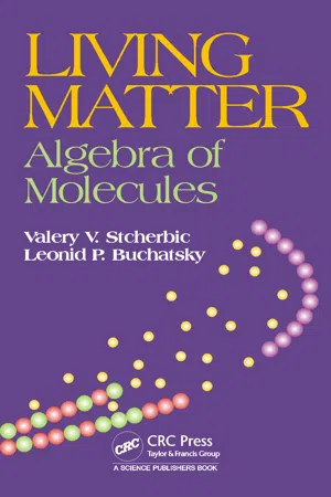 eBook - PDF
Living Matter
Algebra of Molecules
- Valery V. Stcherbic, Leonid P. Buchatsky(Authors)
- 2016(Publication Date)
- CRC Press(Publisher)
2 tRNA Molecule in Af fi ne Space Transport RNA (tRNA) are intermediate links, molecular adapters in the chain of protein synthesis on ribosome. These small molecules, composed of 74–95 nucleotides, covalently unite anticodons of the genetic code and amino acids. tRNA structure in the form of a clover-leaf (Figure 2.1) is well-known; its tertiary structure resembles the Latin character L. All tRNA molecules contain many modified nucleotides (frequently— inosine I , ribothimidine T , pseudouridine Ψ , dihydrouridine D ), which are basically intended for creating standard three-dimensional surface (especially in the anticodon region) necessary for ribosome to recognize them. tRNA molecules contain four regions, each possessing invariant properties irrespective of the amino acid linked. 3’-End of acceptor stem always contains CCA triplet; anticodon is located at the opposite end in position 34–36 of tRNA nucleotides. tRNA tertiary structure is stabilized by hydrogen bonds in the stems. Standard tRNA structure includes 76 nucleotides. However, D-loop often has an expanded structure; there is also a variable loop with variable number of nucleotides. tRNA molecule contains approximately 20 paired nucleotides, which do not always form canonical pairs. Often, acceptors of the same amino acid may be several different, isoaccepting tRNA with different anticodons; however, there may be tRNAs with identical anticodons but different structures. The functions of such tRNA are not clear. Nevertheless, surface shape and volume of tRNA depend little on the primary structure. 32 Living Matter: Algebra of Molecules To perform adapter functions in the process of mRNA translation, tRNA molecule should be attached to the amino acid, which corresponds to mRNA codon, since nonloaded tRNA is not perceived by ribosome. Joining of amino acids to tRNA is realized by highly specific enzymes, aminoacyl-tRNA synthetases. eBook - PDF
- Gerald Karp, Janet Iwasa, Wallace Marshall(Authors)
- 2021(Publication Date)
- Wiley(Publisher)
Decoding the information in an mRNA is accomplished by transfer RNAs, which act as adaptors. On the one hand, each tRNA is linked to a specific amino acid (as an aa-tRNA), but on the other hand, that same tRNA is able to recognize a particular codon in the mRNA. The inter- action between successive codons in the mRNA and specific aa-tRNAs leads to the synthesis of a polypeptide with an ordered sequence of amino acids. To understand how this occurs, you must first understand the structure of tRNAs. The Structure of tRNAs In 1965, after seven years of work, Robert Holley of Cornell Uni- versity reported the first base sequence of an RNA molecule, that of a yeast transfer RNA that carries the amino acid alanine (Figure 11.40a). This tRNA is composed of 77 nucleotides, 10 of which are modified from the standard 4 nucleotides of RNA (A, G, C, U), as indicated by the shading in the figure. Over the following years, other tRNA species were puri- fied and sequenced, and a number of distinct similarities pres- ent in all the different tRNAs became evident (Figure 11.40b). All tRNAs were roughly the same length—between 73 and 93 nucleotides—and all had a significant percentage of unusual bases that were found to result from enzymatic modifications of one of the four standard bases after it had been incorpo- rated into the RNA chain, that is, posttranscriptionally. In addi- tion, all tRNAs had sequences of nucleotides in one part of the molecule that were complementary to sequences located in other parts of the molecule. Because of these complementary sequences, the various tRNAs become folded in a similar way to form a structure that can be drawn in two dimensions as a cloverleaf. The base-paired stems and unpaired loops of the tRNA cloverleaf are shown in Figures 11.42 and 11.43. The unusual bases, which are concentrated in the loops, disrupt hydrogen bond formation in these regions and serve as poten- tial recognition sites for various proteins.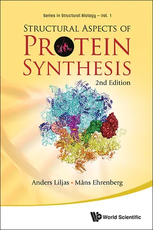
- Anders Liljas, Mans Ehrenberg(Authors)
- 2013(Publication Date)
- World Scientific(Publisher)
41 5 The Adaptor — tRNA The transfer RNAs (tRNAs) are central molecules in protein syn-thesis. Historically, when the structure of DNA and the basics of protein synthesis were clarified, the existence of tRNAs was still unknown. In 1956, Crick drew attention to the problem of assem-bling a polypeptide from an RNA template (Crick, 1958; Woese, 2001). A stereochemical complementarity between the mRNA codons and amino acids seemed impossible. He suggested that small RNA molecules could function as “adaptors.” These could be charged with specific amino acids by enzymes, specifically recognizing both the RNA adaptor and its cognate amino acid. Subsequently, the adaptors would decode the mRNA by Watson–Crick base pairing, thereby ensuring that the amino acids were incorporated into polypeptide chains according to the pre-scription of the genetic code. Indeed, such adaptors were identified experimentally (Hoagland et al. , 1957; Hoagland, 2003). They were initially called soluble RNAs (sRNAs) but are now known as trans-fer RNA molecules, or tRNAs, normally containing about 75 nucleotides. Each tRNA is specific for one amino acid, but one amino acid can be specific for several tRNAs. The latter are often referred to as “isoacceptors,” i.e. accepting the same amino acid in the reaction where tRNA is aminoacylated. For instance, there are six codons for the amino acid leucine. In E. coli these are read by 42 Structural Aspects of Protein Synthesis five leucine-specific tRNAs, all charged with leucine by the same aminoacyl-tRNA synthetase: LeuRS. 5.1 THE tRNAs Noncoding RNA molecules, i.e. micro RNAs, which are not messenger RNAs, exist in all kingdoms of life. They are often involved in regulation of gene expression (Vogel & Wagner, 2007). The tRNAs have a different and well-understood role in transla-tion and form the earliest known group of small noncoding RNAs. The tRNA genes are dispersed throughout the genomes and are frequently found in clusters. eBook - PDF
- S Bresler(Author)
- 2012(Publication Date)
- Academic Press(Publisher)
2. transfer RNA and the Activation of Amino Acids Since all types of RNA are implicated in protein synthesis, let us begin our discus-sion with transfer RNA (tRNA), actually a class of similar compounds that transport different amino acids to the sites of protein synthesis and there participate in the very first stages of polypeptide chain formation (1). transfer RNA is highly soluble and is further distinguished by a rather low molecular weight, on the order of 25,000 to 30,000 daltons. Most tRNA molecules in the cell are attached to amino acids which they acquire after the amino acid is first raised to a higher energy level, or activated, in a reaction with ATP. This corresponds to the observation that polypeptide chain syn-thesis requires the presence of covalently bonded complexes between amino acids and tRNA, not free amino acids. Without doubt, tRNA structure must be specially suited for the transfer of amino acids to growing polypeptide chains. In fact, tRNA appa-rently serves as an adaptor for the proper positioning of the amino acid during the assembly of proteins on a nucleic acid template. One of the principal functions of tRNA is to incorporate cellular amino acids into a chemically labile compound from which they can then be transferred to the growing polypeptide on the ribosomes under energetically favorable conditions. The role of tRNA exemplifies a difficulty with which the cell must continually cope. Many essential metabolites, which either penetrate the cell membrane from the external medium or are elaborated inside the cell itself, must be protected against undesired side reactions that could prevent them from fulfilling more useful ends. Therefore, amino acids are activated specifically for protein synthesis when they are temporarily sequestered by tRNA molecules and are not simply burned up in the cell as an energy source. eBook - PDF
DNA and RNA Modification Enzymes
Structure, Mechanism, Function and Evolution
- Henri Grosjean(Author)
- 2009(Publication Date)
- CRC Press(Publisher)
References 1. Chapeville F, Lipmann F, Ehrenstein GV ct al. On the role of soluble ribonucleic acid in coding for amino acids. Proc Acad Sci USA 1962; 48:1086-1092. 2. Ibba M, Francklyn C, Cusack S. The Aminoacyl-tRNA Synthetases. Georgetown: Landes Bioscience 2005. 3. Sheppard K, Yuan J, Hohn MJ et al. From one amino acid to another: tRNA-dependent amino acid biosynthesis. Nucleic Acids Res 2008; 36:1813-1825. 4. Jiihling F, Mori M, Hartmann R et al. tRNAdb 2009: compilation of tRNA sequences and tRNA genes. Nucleic Acids Res 2009; 37(Database issue) :in press. 5. Hou Y-M, Schimmel P. A simple structural feature is a major determinant of the identity of a transfer RNA. Nature 1988; 333:140-145. 6 . McClain WH, Foss K. Changing the identity of a tRNA by introducing a G-U wobble pair near the 3' acceptor end. Science 1988; 240:793-796. 7. Tamura K, Himeno H, Asahara H et al. In vitro study of E. coli tRNAArgand tRNALy*identity. Nucleic Acids Res 1992; 20:2335-2339. 8 . Zeevi M, Daniel V. Aminoacylation and nucleoside modification of in vitro synthesised transfer RNA. Nature 1975; 260:72-74. 9. Samuelsson T, Boren T, Johansen TI et al. Properties of a transfer RNA lacking modified nucleosides. J Biol Chem 1988; 27:13692-13699. 10. Sampson JR, DiRenzo AB, Behlen LS et al. Nucleotides in yeast tRNAplie required for the specific recognition by its cognate synthetase. Science 1989; 243:1363-1366. 11. Perret V, Garcia A, Grosjean H et al. Relaxation of transfer RNA specificity by removal of modified nucleotides. Nature 1990; 344:787-789. 12. Piitz J, Florentz C, Benseler F et al. A single methyl group prevents the mischarging of a tRNA. Nature Struct Mol Biol 1994; 1:580-582. 13. Grosjean H, de Crecy-Lagard V, Bjork GR. Amino acylation of the anticodon stem by a tRNA-synthetase paralog: Relic of an ancient code? Trends Biochem Sci 2004; 29:519-522. 14. Ibba M, Francklyn C. Turning tRNA upside down: When aminoacylation is not a prerequisite to protein synthesis. eBook - PDF
- D. S. Eggleston, Catherine D. Prescott, Neil D. Pearson(Authors)
- 1997(Publication Date)
- Academic Press(Publisher)
These examples suggest that the type of additional RNA binding domain coupled with the class II synthetase catalytic domain is corre-lated with the number of anti-codon nucleotides that have to be recognized to discriminate tRNAs. 1 Introduction The fidelity of protein synthesis depends to a large extent on the high specificity with which aminoacyl-tRNA synthetases charge their cognate tRNAs with the correct amino acid. Synthetases catalyse the aminoacylation reaction in two steps, firstly the activation of the amino acid using ATP to form the enzyme bound aminoacyl-adenylate and secondly, the transfer of the amino acid to the 2' or y hydroxyl of the ribose of the 3^ terminal A-76 of the tRNA. In Esherichia coli. *A11 images for this chapter appear in the colour plate section between p. 112 and p. 113. THE MANY FACES OF RNA Copyright © 1998 Academic Press Ltd ISBN 0-12-233210-5 All rights of reproduction in any form reserved 56 S. Cusack et al. there are at least 46 different tRNA molecules with anticodons corresponding to the various amino acids, and the seryl-tRNA synthetase, for instance, has to selectively charge the six serine isoacceptors and ignore the others. Given that tRNAs superficially have similar secondary and tertiary structures, what is the structural basis for the specific recognition between aminoacyl-tRNA synthetases and tRNAs? 2 tRNA identity In order to analyse this question, the concept of tRNA identity elements has been introduced (reviewed in Saks etal., 1994). This refers to those structural elements, usually a limited number of nucleotides or base pairs, that are in general conserved amongst tRNA iso-acceptors for an amino acid X and are essential for specific recognition by the cognate synthetase. In the ideal case, when these identity elements are transplanted into a tRNA for a different amino acid Y, they switch the identity of this tRNA so that it becomes an X iso-acceptor.
Index pages curate the most relevant extracts from our library of academic textbooks. They’ve been created using an in-house natural language model (NLM), each adding context and meaning to key research topics.