Psychology
Motor Area of Brain
The motor area of the brain is a region in the cerebral cortex responsible for planning, control, and execution of voluntary movements. It is located in the frontal lobe and is divided into the primary motor cortex and the premotor cortex. The primary motor cortex directly controls muscle movements, while the premotor cortex is involved in coordinating complex movements and integrating sensory information.
Written by Perlego with AI-assistance
Related key terms
1 of 5
12 Key excerpts on "Motor Area of Brain"

- Ian McMillan, Gail Carin-Levy(Authors)
- 2013(Publication Date)
- Wiley-Blackwell(Publisher)
The larger anterior part of the lobe, which lies above the orbit of the eyes (supraorbital area), is involved in planning and problem-solving aspects of both movement and behaviour. This part of the frontal lobe is also called the prefrontal lobe. Practice note-pad 3A: stroke Stroke is brain ti ssue damage that results from disrup ti on of the blood supply to a localised area of the brain. It may be caused by haemorrhage from a blood vessel, but is more com-monly due to arterial occlusion by a thrombus or an embolism. The disrup ti on of the blood supply results in infarct ( ti ssue death) of the a ff ected area, giving rise to a lesion. The symptoms and prognosis for each pa ti ent will be determined primarily by the loca ti on, extent and causal mechanism. A stroke can occur in any area of the brain, but usually a ff ects one cerebral hemisphere, giving rise to di ffi cul ti es associated with the func ti ons of that hemisphere. See prac ti ce note pads 3B to 3E. The band of grey ma tt er lying immediately in front of the central sulcus (precentral gyrus) is the primary motor area , which is concerned with the genera ti on of movement in the whole of the opposite side of the body (Figure 3.7a). Adjacent to this area are the premotor area and the supplementary motor area which are discussed in more detail below. The cell bodies of the neurones in the motor cortex project not to individual muscles, but to func ti onal groups of muscles. Direct links to the small muscles of the hands, the feet and the face are par ti cularly important, and damage to the primary motor area o ft en results in loss of precision movements. There is representa ti on of half of the body in an ‘upside-down’ posi ti on in each primary motor cortex. The head is represented in the lower cortex on the lateral side, then the upper limb and trunk above, and fi nally the lower leg and feet in the cortex on the medial surface of the lobe. eBook - ePub
Muscles, Nerves and Movement
In Human Occupation
- Barbara Tyldesley, June Grieve(Authors)
- 2013(Publication Date)
- Wiley-Blackwell(Publisher)
Fig. 3.8b ). Neuroimaging studies have recorded an increase in cerebral blood flow in the supplementary motor area immediately before the execution of a complex sequence of movements of the fingers, and when both hands are involved. These studies suggest a role in the planning of movement that is internally generated.The motor speech area identified by Broca lies in the lower part of the frontal lobe in the lip of the lateral sulcus (see Fig. 3.8a ). The function of this area, usually only found in the dominant hemisphere, is in the production of fluent speech.The prefrontal area (supraorbital area) occupies the large anterior area of the frontal lobe and connects with all the other lobes of the cerebral hemispheres, the thalamus, the limbic system and many other brain areas. Interaction with the limbic system is concerned with the emotional aspects of movement. The prefrontal area is also concerned with the planning of goal-directed movement and behaviour and in modifying the plan in response to changes in the environment. These are known as the executive functions.Fig. 3.8Main functional areas of the cerebral hemispheres: (a) lateral view; (b) medial view. MC = primary motor cortex; SMA = supplementary motor area; SC = somatosensory area.Fig. 3.9Primary motor cortex seen in frontal section to show the representation of body parts.Clinical note-pad 3B: Frontal lobe lesion- Primary and premotor cortex. Lesion on one side leads to muscle weakness in the muscles of the opposite side of the body, known as hemiplegia. Muscle tone may be low (flaccid) or high (spastic). Fine skilled movements of the extremities are particularly affected.
- Prefrontal cortex. Lesions in this area lead to problems in planning movement and in reviewing it during progress. This has implications for safety. Loss of insight into movement performance may be a major factor in the poor prospect of successful rehabilitation. There may be inability to monitor social behaviour. The inability to plan and monitor movement and behaviour resulting from lesions in the prefrontal cortex is known as dysexecutive syndrome (DES).
 eBook - PDF
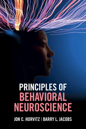
- Jon C. Horvitz, Barry L. Jacobs(Authors)
- 2022(Publication Date)
- Cambridge University Press(Publisher)
Some of these neurons may have been far from the site of electrical stimulation but pos- sessed strong synaptic connections with the directly stimulated neurons. In either case, these data suggest that activation of a particular area of the motor cortex may generate meaningful behaviors, not just movement of individual or nearby body parts. KEY CONCEPTS • The left primary motor cortex produces right-side body movements, and vice versa. • The motor cortex contains a general map of body parts. However, the neurons that control particular body parts spread around the motor cortex in a patchy way rather than group together in distinct regions. 4.3 PREMOTOR AREAS 167 4.3 PREMOTOR AREAS The premotor areas include a number of frontal lobe regions anterior to the primary motor cortex (Figure 4.15), including both the dorsal and ventral premotor areas and the supplementary motor area (SMA). The primary motor cortex receives much of its input from the premotor areas. Therefore, as we explore the functions of the premotor areas, we are revealing some of the kinds of information that may be relayed to corti- cospinal neurons to influence movement. 4.3.1 The Supplementary Motor Area Is Active during the Conscious Desire to Move Move your right arm sometime in the next 30 seconds. You can decide the direction and moment to move the arm. Wait until you wish to do it. It is unclear exactly how brain activity produces the conscious decision to move. However, activity within the SMA, a premotor area on the med- ial surface of the brain, coincides with the feeling of wanting to move. Individuals who elected to undergo brain surgery to treat epilepsy agreed to participate in studies involving stimulation of movement-related brain areas. When the SMA was stimulated, they reported an “urge” or “need” to move a body part (Fried et al., 1991).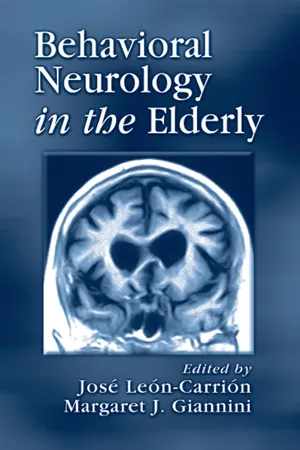 eBook - ePub
- Jose Leon-Carrion, Margaret Giannini, Jose Leon-Carrion, Margaret Giannini(Authors)
- 2001(Publication Date)
- CRC Press(Publisher)
2 The performance of voluntary movements depends on the participation of several anatomical structures in the nervous system and periphery. In addition to the motor neurons, the striated muscles, the specific body parts involved in the motion, and the central nervous system (CNS) are involved in the organization of the behavior and participate in the execution of voluntary movement. This complex process requires the coactivation of motor, planning, and organizational structures together with sensory structures in the CNS. Because of the multiple components involved in the execution of voluntary movement, it is difficult to identify the cerebral structures that are exclusively responsible for motor behavior. This chapter describes the cortical and subcortical structures involved in the execution of voluntary movement, and reviews some of the current theories on motor systems.A complete review of the evolution of all motor activities is beyond the scope of this chapter. The review here is limited to human motor functions that affect activities of daily living when the quality of the movement changes. Reviewed are gait, crucial in independent functioning; swallowing and oral-motor skills because of their importance in feeding and verbal communication; studies of normal elderly subjects in the execution of upper limb movements, including manual motor skills; and the effects of aging on learning new motor skills. Finally, this chapter reviews some of the most relevant studies in apraxia, defined as a neurological disorder of learned purposive behavior movement skill that is not explained by deficits in elemental motor or sensory systems3 and describes praxis in normal elderly subjects that were used as control groups in research.7.2 ANATOMY OF MOTOR FUNCTIONS
Among the cortical structures involved in voluntary movements, the frontal cortex contains the primary motor cortex (MI), which corresponds to area 4 of Brodman, the supplementary motor area (SMA), which conforms to the lateral and medial premotor cortex in the medial part of area 6, and the premotor cortex (PM), also called the dorsal prefrontal cortex, above the frontal eye field (areas 9 and 46). The basal ganglia and the cerebellum are the two major noncortical components of the motor system.7.2.1 CORTICAL AREAS
7.2.1.1 Primary Motor Area
The neurons of the MI connect to spinal and bulbar motor neurons and receive afferent impulses from the premotor area (PM), SMA, and cerebellum. Studies in cerebral blood flow (CBF) and cerebral metabolism (CMR) show that, except for voluntary eye movements, all other types of voluntary activity of the limbs, head, and face increase regional cerebral blood flow (rCBF), regional cerebral metabolism (rCMR), or open regional calcium channels (Ca2+ ) in the somatotopical field of the MI.4 These signs of increased energy utilization of areas in MI during voluntary motor activities of the limbs is mostly contralateral, although in the early phases of motor learning small activation can appear in the ipsilateral MI areas.4 eBook - ePub
Regulatory Functions of the CNS Principles of Motion and Organization
Proceedings of the 28th International Congress of Physiological Sciences, Budapest, 1980
- J. Szentágothai, M. Palkovits, J. Hámori(Authors)
- 2013(Publication Date)
- Pergamon(Publisher)
Neural mechanisms of voluntary movements and precentral motor area Passage contains an image
OPENING REMARKS ON THE NEURAL MECHANISMS OF VOLUNTARY MOVEMENTS AND PRECENTRAL MOTOR AREA
C.G. Phillips, Department of Anatomy, South Park Road, Oxford OX1, EnglandPublisher Summary
This chapter presents remarks on the neural mechanisms of voluntary movements and precentral motor area. The precentral motor area has been studied extensively in primates. It lies far downstream on the pathways that project the motor output from the central nervous system. It contains corticospinal neurons in lamina V, which make monosynaptic connections with alpha motoneurons, and are thus separated by only one synapse from the neuromuscular junctions. The number of neurons contained in any radially-orientated cylinder of cortex is similar to the number contained in cylinders of equal diameter from any other area of neocortex except area 17. Between the widely spaced neurons of area 4 is a bulky neuropil that provides a richness of synaptic connectivity unequaled in other areas. In the arm area, which has been the most studied, microstimulation finds that the motor outputs to different joints of the arm, forearm, and hand are located in multiple hot spots for flexion or extension of each joint, widely scattered, and interspersed with scattered hot spots for other joints.A century ago it would have been generally supposed that the Rolandic area of the human cortex was the actual locus of initiation of voluntary movements. But in the eighteen eighties there were two dissentient voices, those of François-Franck and Sherrington. The first concluded that this area contained ‘points of departure’ and not centres for movement. The second tried, unsuccessfully, to introduce the term ‘cord area’ in order to escape the theoretical obligations of the term ‘motor area’, and to designate merely that area of the cortex which is so intimately connected with the spinal cord that cortical lesion is followed by degeneration of a spinal tract.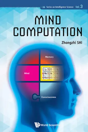 eBook - ePub
- Zhongzhi Shi(Author)
- 2017(Publication Date)
- WSPC(Publisher)
The thalamus is the organ of the synthesis and release of the sense of thalamus. It is the main body of the “I”. The brain association area is the activity place of the sense of thalamus. Consciousness in the brain association area is yet to be achieved. The outputs of motion samples, analyzed by the brain, the basal ganglia and the cerebellum activate the thalamus. According to the motion samples, the thalamus synthesizes and delivers to the brain association area, causes the brain to produce the awareness of motion and also produces the motion intention. Motion intention is consciousness and is divided into three categories: one is from the brain’s motor intention, one is from the basal ganglia, the cerebellum’s motor sensory and one kind is from sensory neurons after the motion sense.The main function of the brain is to analyze the portion that produces samples. The frontal lobe is the most advanced and important organ, including the association area, the premotor and motor areas. Frontal, occipital and temporal association zones are the main areas of activity of consciousness. According to the needs of the external environment, they can produce motor intention, clear movement direction or a mode of behavior. The brain is not motor-specific control, command and does not analyze the program and instructions of motor, which are executed by the basal ganglia and cerebellum, so that people can concentrate on all kinds of thinking activity. The brain frontal lobe motor area administers the movement order and at the end of the procedure executes its release. The motor area will exercise the program, and the instruction is issued to produce the movement. The motor area obeys the association area and the consciousness. Consciousness may suspend the motor procedure, and the instruction release at any time, and thus the movement can be interrupted.The basal ganglia are the main organs of motor control and command. Analysis of output samples of basal ganglia include control, campaign director, procedures and instructions. Motor sample analysis and production obey the motor intention. The basal ganglia and cerebellum analyze and produce motion samples according to the motor intention when brain association area forms the motor intention. Functions of the cerebellum are in many aspects, and there may participate in the consciousness, experiences and motor activities. In the process of movement, cerebellum analyzes output motion parameters, motion control details for motion accuracy and precision work. eBook - PDF
Concise Learning and Memory
The Editor's Selection
- (Author)
- 2010(Publication Date)
- Academic Press(Publisher)
The relationship of single-unit activity to behavioral aspects of motor tasks in awake, freely moving animals has also been an important approach to dis-tinguishing motor fields. Finally, motor areas have been characterized based on functional differences in ablation-behavior studies in nonhuman primates and functional imaging studies in humans. The nomenclature used for subdivisions of pri-mate motor areas has varied across laboratories. The generalized current scheme includes M1 (or Brodmann’s area 4); four subdivisions of the lateral premotor cortex (PMd-c, PMd-r, PMv-c, and PMv-r, or F2, F7, F4, and F5, respectively); two premotor subdivisions on the mesial surface of the hemisphere (SMA and pre-SMA, or F3 and F6), and three subdivi-sions of the cingulate motor area within regions lining the cingulate sulcus (CMAr, CMAd, and CMAv, or area 24c, area 6c, and area 23c). M1 is the most easily recognizable area in histological stains, as it contains very large pyramidal neurons in layer V, the so-called Betz cells. Also, it contains a greatly reduced layer IV, compared with sensory cortex that contains a thick layer IV with substantial numbers of granule cells. For this reason, M1 is sometimes referred to as agra-nular cortex. Figure 1 illustrates the location of the main cortical motor areas. 25.3.2 Cortical Motor Areas in Rodents Because rodents are often used to study the role of motor cortex in motor skill learning, a brief compara-tive account of cortical motor areas in rodents is instructive. (While motor cortex in cats is also fre-quently a focus of motor learning experiments, this species will not be discussed in this review.) Intracortical microstimulation studies of sensorimo-tor cortex in the rat have shown a complete motor representation that is cytoarchitectonically defined as agranular cortex (Hall and Lindholm, 1974; Donoghue and Wise, 1982) and commonly called M1 based on its similarities to primate M1. eBook - ePub
Motor Cortex in Voluntary Movements
A Distributed System for Distributed Functions
- Alexa Riehle, Eilon Vaadia(Authors)
- 2004(Publication Date)
- CRC Press(Publisher)
Why does the frontal lobe contain all of these premotor areas? Although there is as yet no definitive answer to this question, current data from anatomical, physiological, behavioral, and imaging studies suggest that each premotor area is concerned with a specific aspect of movement planning, preparation, and execution. Thus, the task of generating and controlling movement appears to be broken up into a number of subtasks that are accomplished through parallel distributed processing in multiple motor areas. Multiple motor areas may thereby decrease response time and increase response flexibility. In any event, the cortical control of movement is achieved by multiple motor areas, all of which send signals to the spinal cord.ACKNOWLEDGMENTS
This work was supported by the Veterans Affairs Medical Research Service, and by U.S. Public Health Service grant #24328 (PLS).REFERENCES
1 . Kuypers, H.G.J.M., Anatomy of the descending pathways, in Handbook of Physiology, Section I: The Nervous System, Vol. II: Motor Control, Part I, Brooks, V.B., Ed., American Physiological Society, Bethesda, MD, 1981, 567.2 . Wise, S.P., The primate premotor cortex fifty years after Fulton, Behav. Brain Res., 18, 79, 1985.3 . Wiesendanger, M., Recent developments in studies of the supplementary motor area of primates, Rev. Physiol. Biochem. Pharmacol., 103, 1, 1986.4 . Hepp-Reymond, M.C., Functional organization of motor cortex and its participation in voluntary movements, in Comparative Primate Biology, Vol. 4: Neurosciences, Alan R.Liss, 1988, 501.5 . Asanuma, H., The Motor Cortex, New York, Raven Press, 1989.6 . Cheney, P.D., Fetz, E.E., and Mewes, K., Neural mechanisms underlying corticospinal and rubrospinal control of limb movements, Prog. Brain Res., 87, 213, 1991.7. Georgopoulos, A., Higher order motor control, Annu. Rev. Neurosci., 14, 361, 1991.8 . Tanji, J., The supplementary motor area in the cerebral cortex, Neurosci. Res., 19, 251, 1994.9 . Dum, R.P. and Strick, P.L., The corticospinal system, a structural framework for the central control of movement, in Handbook of Physiology, Section 12: Exercise, Regulation and Integration of Multiple Systems, Rowell, L.B. and Shepard, J.T., Eds., American Physiological Society, New York, 1996, 217.10. Geyer, S. et al., Functional neuroanatomy of the primate isocortical motor system, Anat. Embryol., eBook - PDF
- Marie T. Banich, Rebecca J. Compton(Authors)
- 2018(Publication Date)
- Cambridge University Press(Publisher)
It is thought to create a forward model that helps to predict the sensory consequences of a motor plan. • Via its connections through a series of loops through the thalamus and up into the cortex, the basal ganglia can mod- ulate the initiation and cessation of movements. They also play a role in motor planning and learning. • Primary motor cortex generates movement most likely by controlling the force or other parameters of muscle movement. • The supplementary motor complex is thought to be involved in specifying, preparing, and initiating a motor plan for an action, which is an abstract representation of an intended movement that is preprogrammed before the motor act is initiated. • Premotor regions are thought to specify the type of motor action (such as a grasp) that is necessary to perform a task. A portion of the premotor area, known as the frontal eye field, programs voluntary eye movements such as those involved in scanning visual space. • The anterior cingulate cortex plays an important role in the selection of motor responses, especially when they are novel or atypical. It also plays a role in the evaluation of the out- come of such movements, such as whether or not they lead to an error. • The right inferior frontal cortex has been suggested to play a specific role in the inhibition of motor responses. • The parietal lobe links movements with sensory informa- tion, including visual, proprioceptive, and kinesthetic information. It is thought to estimate what motor actions are required to meet a particular end state, and can aid in the on-line modulation of actions. It is also important for link- ing motoric actions to their conceptual significance, such as occurs when saluting or making the sign of the cross. • Complex action requires the coordinated effort of all these regions in an integrated manner. eBook - ePub
- Larry Squire, Darwin Berg, Floyd E. Bloom, Sascha du Lac, Anirvan Ghosh, Nicholas C. Spitzer, Larry R. Squire, Floyd E. Bloom, Nicholas C. Spitzer(Authors)
- 2008(Publication Date)
- Academic Press(Publisher)
Axons from the cerebral cortex and from the red nucleus in the brain stem descend in pathways located dorsolaterally in the spinal cord to control voluntary movements. In addition to corticospinal and rubrospinal pathways, the motor cortex also sends projections to the red nucleus and to the pontomedullary reticular formation, providing indirect pathways for voluntary control. Multiple cortical motor areas—distinguished by their inputs, their cytoarchitecture, and their interconnections with other cortical areas and with the thalamus—make different contributions to the control of voluntary movements. The most detailed motor map is found in the primary motor cortex, M1, but the somatotopic organization is limited by convergence and divergence in the corticospinal projection. Populations of M1 neurons are also in most direct control of movement kinematics and dynamics. Other cortical motor areas participate differentially in the selection of, and preparation for, voluntary movements based on a variety of internal and external cues.SUMMARY
The motor cortex, the red nucleus, the pontomedullary reticular formation, and the vestibular nuclei each send major axonal projections descending from the brain to the spinal cord to control bodily movements. Although these pathways normally provide seamless movement control, different contributions of the medial and lateral descending pathways have been identified experimentally.The medial system—vestibulospinal and reticulospinal—receives sensory input from the vestibular apparatus and mediates postural responses that keep the head and body stabilized for stance and gait. Additional visual and proprioceptive inputs that reach the vestibular and reticular nuclei via brain stem pathways or after processing in the cerebellum (Chapter 32 ) also influence postural control. Context-dependent strategies established via centers including the cerebellum and motor cortex further adapt postural responses to the needs of particular complex situations.The lateral system—corticospinal and rubrospinal—receives, in addition to somatosensory inputs, information processed by the cerebellum (Chapter 32 ), the basal ganglia (Chapter 31 eBook - PDF
- Gregory R. Bock, Maeve O'Connor, Joan Marsh, Gregory R. Bock, Maeve O'Connor, Joan Marsh(Authors)
- 2008(Publication Date)
- Wiley(Publisher)
The principal sulcal cortex is additionally interconnected with the primary somatosensory area and the somatosensory association areas, in the frontoparietal operculum, with area PF of von Bonin and Bailey in the posterior parietal cortex, and with parts of the ‘motor’ thalamus. Recent be- havioural and electrophysiological studies in monkeys (Macaca mulatta) demon- strate that the principal sulcus can influence delayed-responding, whether the response is a hand or an eye movement. The anatomical and functional evidence supports the thesis that prefrontal cortex has access to and can direct the output of several motor centres. 1987 Motor areas of the cerebral cortex. Wiley, Chichester (Ciba Foundation Symposium 132) p 187-200 The role of cerebral cortex in motor control is generally considered to involve three major ‘motor’ areas-primary motor cortex (Brodmann’s area 4), the premotor cortex (Brodmann’s area 6) and the supplementary motor cortex (medial area 6). Although motor, premotor and supplementary motor areas are involved in the highest order control over voluntary behaviour, none of them are thought of as the ‘prime mover’ in the chain of command. In this paper I will review recent evidence on the role that prefrontal cortex may play in the initiation, facilitation and inhibition of motor responses. Although it has been proposed that prefrontal cortex performs command functions, the necessary functional and anatomical relationships between prefrontal cortex and motor centres have proved difficult to specify precisely and are usually not provided. However, recent behavioural, anatomical and physiological 187 188 Goldman-Rakic studies of the so-called motorically ‘silent’ areas of the frontal lobe indicate that such relationships exist and may provide new ways of conceptualizing the cortical and subcortical interactions underlying the regulation of voluntary motor behaviour. eBook - PDF
- Donald W Pfaff(Author)
- 2015(Publication Date)
- World Scientific(Publisher)
The easiest way to think about this subject is to assume that cortical controls over movement evolved after brainstem controls, and that the former are effectively superimposed on the latter. Voluntary movements guided by the cerebral cortex may be regarded as the most complicated of all movements to explain. This is because some scientists feel that motor control systems in the brain contain a kind of internal map — like a three-dimensional Google map — of all the movements it could make, and, in fact, that it rep-resents the movement to itself (in the form of a parallel recurrent pathway) just thousandths of a second before the actual movement occurs. Many neurons whose firing will make muscles contract have axonal branches that inform the relevant sensory systems as to what is about to happen. These are movements that result from an “intention” to act. In that part of the cerebral cortex primarily devoted to movement, different zones of neurons regulate movements in different parts of the body. Unusually large zones of neurons regulate muscle groups that produce the most finely graded movements, and the reverse is also true. Damaging these cerebral cortex neurons leads to loss of the abil-ity to make precise movements whose force and timing must be con-trolled within narrow limits. Once the command for any movement is sent out from the cerebral cortex, signaling from any given fore-brain motor control neuron is regulated by feedback control from the various senses (for example, pressure on the skin) and, indeed, from the relevant muscles themselves in order to guide that movement to a successful conclusion. It is exciting to think about how the initiation of movement plays into neuronal circuitry required for human consciousness, which, as defined by neurologists, is the ability not only to be aware of one’s environment but also to make purposeful, voluntary movements and to communicate with other humans.
Index pages curate the most relevant extracts from our library of academic textbooks. They’ve been created using an in-house natural language model (NLM), each adding context and meaning to key research topics.