Psychology
The Cerebral Cortex
The cerebral cortex is the outer layer of the brain responsible for higher cognitive functions such as thinking, perceiving, and decision-making. It is divided into four lobes—frontal, parietal, temporal, and occipital—each with specific roles in processing sensory information and controlling motor functions. The cerebral cortex plays a crucial role in human behavior, personality, and consciousness.
Written by Perlego with AI-assistance
Related key terms
1 of 5
9 Key excerpts on "The Cerebral Cortex"
 eBook - ePub
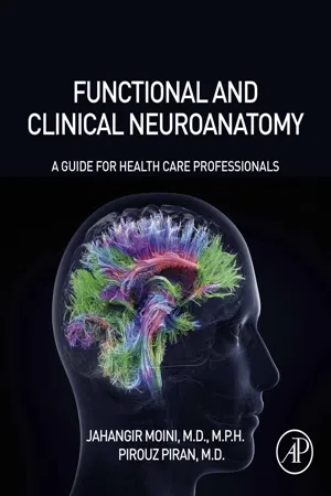
Functional and Clinical Neuroanatomy
A Guide for Health Care Professionals
- Jahangir Moini, Pirouz Piran(Authors)
- 2020(Publication Date)
- Academic Press(Publisher)
Chapter 6Cerebral cortex
Abstract
The Cerebral Cortex is a layer of neurons and synapses (gray matter) on the surface of the cerebral hemispheres. This is folded into gyri, and about two-thirds of the cortex's area is buried inside fissures. The Cerebral Cortex integrates higher mental functions, general movements, functions of the viscera, perceptions, and behavioral reactions. It has many different classifications. The Cerebral Cortex has 47 separate function areas, with differing cellular designs.Keywords
Cerebrum; Gross anatomy; Cerebral cortex; Neocortex; Electroencephalogram; Split-brain syndromeThe Cerebral Cortex is a layer of neurons and synapses (gray matter) on the surface of the cerebral hemispheres. This is folded into gyri, and about two-thirds of the cortex's area is buried inside fissures. The Cerebral Cortex integrates higher mental functions, general movements, functions of the viscera, perceptions, and behavioral reactions. It has many different classifications. The Cerebral Cortex has 47 separate function areas, with differing cellular designs.Stimulation of the precentral cortex or motor area, using electrodes, causes contractions of the voluntary muscles. If the motor speech area in the inferior frontal gyrus is destroyed, this causes motor aphasia or speech defects, even though the vocal organs are healthy and intact. If the brain cortex is stimulated as in case of a seizure, this stimulation will affect circulation, respiration, reactions of the pupils, and other visceral activities.Cerebrum
The cerebrum is the largest and most obvious portion of the human brain. It forms from the embryonic structure called the telencephalon . The cerebrum is the center of voluntary motor control and complex mental processes.Gross anatomy
The cerebrum is much larger than the other portions of the brain. It is divided into two cerebral hemispheres that are separated by the longitudinal fissure . A prominent tract of fibers called the corpus callosum also connects the hemispheres. Each hemisphere has conspicuous gyri , which appear as “wrinkles” that are separated by grooves known as sulci . The surface of the cerebrum folds into the gyri in a way that allows for a larger amount of cortex to fit inside the cranium and because of the gyri, the cerebrum has about 2500 cm2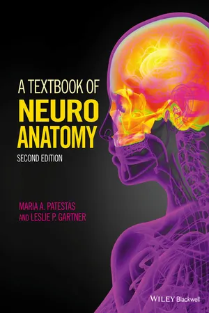 No longer available |Learn more
- Maria A. Patestas, Leslie P. Gartner(Authors)
- 2016(Publication Date)
- Wiley-Blackwell(Publisher)
A 45-year-old woman, who was previously in perfect health, had two seizures in the past week. Both occurred suddenly and each lasted less than a minute. The first seizure started as twitching of the right side of the face, which progressed to twitching of her right hand and then her right leg. She felt some perioral numbness as well. There was no loss of consciousness or other symptoms, and she was aware of what had occurred. This happened while in bed. The second seizure, as described by her husband, started with facial twitching but she rapidly lost consciousness and then had a generalized convulsion. She bit her tongue and had blood in her mouth. She also had urinary incontinence. She began responding a few minutes after the convulsion, but she was still confused and somewhat agitated for about 30 minutes after that. She had never had a seizure before. In between these spells she had been normal and examination was normal.The cerebral cortex is the most complex component of the human brain, as a result of its complex and widespread connections. It functions in the planning and initiation of motor activity, perception and conscious awareness of sensory information, learning, cognition, comprehension, memory, conceptual thinking, and awareness of emotions.The Cerebral Cortex (L., cortex, “bark”) is a multilayered sheet of nerve cell bodies and associated cell processes that covers the paired and prominent cerebral hemispheres of the cerebrum, forming a superficial layer much like bark covers a tree. The majority of The Cerebral Cortex consists of phylogenetically the most highly evolved and complex neural tissue of the human brain.The central nervous system (CNS) is comprised of white and gray matter. White matter consists mostly of nerve cell axons, whereas gray matter consists mostly of nerve cell bodies. Gray matter is arranged into nuclei or cortex. Nuclei are aggregations of nerve cell bodies embedded deep within the cerebrum or in the spinal cord. The cerebral cortex consists of 50–100 billion nerve cell bodies arranged into a three- to six-layered sheet that laminates the brain surface.The Cerebral Cortex overlies the subcortical white matter of the cerebral hemispheres. In other animals, the brain's cortical surface appears smooth, whereas in humans the brain surface is convoluted displaying prominent, alternating grooves and elevations as a result of the folding of The Cerebral Cortex, which occurs during development. The elevations are referred to as gyri, whereas the grooves are referred to as sulci or fissures (Fig. 25.1 ). Sulci are shallow, short grooves, whereas fissures are deeper and more constant grooves, with a consistent location on the brain surface. The cortex forming the gyri dips down into the pit of the adjacent sulci or fissures to line them. Certain gyri, sulci, and fissures are similar in all normal human brains. Others, however, may vary in different brains and in the two cerebral hemispheres of the same brain. The gyri and sulci greatly increase the total surface area of The Cerebral Cortex. If The Cerebral Cortex of a normal human brain were spread out (that is, if the pleats formed by the sulci and fissures were stretched out), the cortex would extend over 0.23 m2 (2.5 ft2 eBook - PDF
Neuropsychology
A Textbook of Systems and Psychological Functions of the Human Brain
- Stuart J. Dimond(Author)
- 2013(Publication Date)
- Butterworth-Heinemann(Publisher)
According to this analysis, the mental world of man is divided up by the separateness of the mid-brain mechanisms into four realms of action: the intellect, emotion, motor functions, and sex and sleep. Each, with the exception of sleep, shares a massive cortical component, but each devolves upon a separate structure at the mid-brain region, and it is this which leads to the view that these are the four pillars of the mind given because the brain is constructed in the way that it is. The Cortex That vast confusing web of tissue that we call the cortex appears at first sight to defy description. The range of its functions is enormous and the complexity of its organization intricate, and yet there is a basic order in the cortex both in terms of the way in which functions are arranged and mapped on to the cortex and also in terms of the way in which the business of the cortex is conducted in order to fulfil these essential functions. The cortex is not a chaotic structure and there is a discernible order to it, as is apparent from the work described in this volume. We have argued previously that if we adopt an internal perspective for the functions of the brain, and we take a brain's eye view of its function rather than that, say, of the neurosurgeon who views the brain from without, then we come to a different interpretation not only of the respective functions of the internal parts, but also of the way in which they relate to the cortex itself. If we imagine ourselves to be an observer standing at some central important pivotal point in the brain and looking through the structures to the periphery bounded by the cranial wall, then the structures of the cortex form the distant extent of our vision. They are the furthest, most distant envelope, but envelope they are, containing and restricting the structures within. Essentially, therefore, the cortex forms the external lining for the brain.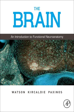 eBook - ePub
The Brain
An Introduction to Functional Neuroanatomy
- Charles Watson, Matthew Kirkcaldie, George Paxinos(Authors)
- 2010(Publication Date)
- Academic Press(Publisher)
The analytical abilities of the cortex are the product of its cellular architecture and the way it communicates with the rest of the nervous system. This chapter will deal with the cellular and anatomical properties of the cortex, as well as its functional abilities.The Cerebral Cortex–anatomy and histologyThe human nervous system shares its basic layout and functional arrangement with other mammals. However, the human cerebral cortex is huge and complicated compared with that of rodents, forming an overgrowth that bulges out and distorts the shape of structures lying inside the hemispheres, including the ventricles, the striatum, and the hippocampal complex. The human cerebrum expands forwards to form the frontal lobe, backwards to form the occipital lobe, and grows downwards and outwards into the temporal lobe. These expansions hide part of the cortex that does not grow as fast–a buried area called the insula, concerned with taste and gut control, which is found on the surface of the cortex in rodents.The word cortex means an outer layer, such as the bark of a tree or the skin of an orange; The Cerebral Cortex is a sheet of neurons and glia covering the rest of the forebrain. In rodents it is a flat sheet, but the human cortex expands so much that it is thrown into folds (called gyri) and grooves (called sulci). Beneath the cortex is the white matter, a dense mat of axon fibers joining cortical regions and communicating with the rest of the nervous system.Uniformity of structureOne of the most striking features of The Cerebral Cortex is how little its cellular structure varies from region to region, even between areas dealing with very different types of sensory input. This reflects the abstract nature of cortical processing, which is not so much concerned with the specifics of receptors and effectors, as with the meaning of sensory stimuli and their relevance to behavior. Most of the human cerebrum consists of neocortex, the part that has most recently evolved in mammals. The neocortex is also called isocortex, because a standard set of cellular arrangements is repeated across the entire sheet (‘iso’ means uniform or unchanging). As a consequence, neuroanatomists trying to distinguish one cortical area from another have to analyze the thickness of cellular layers and the sizes of cells found there. Even then the boundaries in many cases are not obvious, because each area merges with its neighbors. The clearest distinction is between granular (sensory) cortex and agranular (motor) cortex. In the sensory cortical areas, the spiny stellate cells form a dense granular layer, whereas the motor cortex is agranular because it lacks these cells. eBook - ePub
Vision Facts
Questions about the Human Eye
- Jason Yang, Charles Pidgeon(Authors)
- 2018(Publication Date)
- Universal-Publishers(Publisher)
great-grandmother cell hypothesis is a name for an early attempt to explain how the brain processes visual information. In this hypothesis, there is a neuron in the brain for every object that can be recognized, including one for your great-grandmother. However, this hypothesis has been refuted, as a neuron for every recognizable object means that the brain would have to be far larger than is realistic.Gross, C. G. “Genealogy of the ‘grandmother cell.’” Neuroscientist 8, no. 5 (October 2002): 512–18. Accessed September 21, 2017. https://www.ncbi.nlm.nih.gov/pubmed/12374433 .Q97 What is The Cerebral Cortex? A97 The cerebral cortex (or “cortex”) is a layer of neurons two to four millimeters thick on the surface of the brain. It is composed closely packed neurons forming gray matter. The Cerebral Cortex consists of four lobes: the occipital, parietal, temporal, and frontal lobes. It is divided into the left and right cerebral hemispheres.Some main functions of The Cerebral Cortex include perception, awareness, memory, consciousness, speech, and higher level thinking.“The Cerebral Cortex.” AP Psychology Community . Accessed September 14, 2017. http://www.appsychology.com/Book/Biological/cerebral_cortex.htm .The Cerebral Cortex consists of four lobes: the frontal lobe (red), parietal lobe (green), temporal lobe (blue), and occipital lobe (yellow).Q98 What is the function of each lobe of the cortex? A98 Each lobe in the cortex serves unique roles. The frontal lobe is considered to be the control center for emotions and personality. It is also involved in problem solving, motor skills, judgement, and social behaviour. The parietal lobe processes information related to several senses, including taste, temperature, touch, as well as helping out with vision. The temporal lobe is primarily involved in hearing and language. However, parts of the temporal lobe are also dedicated to vision. Finally, the occipital lobe eBook - PDF
- Karen R. Huffman, Katherine Dowdell, Catherine A. Sanderson(Authors)
- 2017(Publication Date)
- Wiley(Publisher)
Without a functioning cortex, we would be almost completely unaware of ourselves and our surroundings. Although The Cerebral Cortex is only about one-eighth of an inch thick, it’s made up of approximately 30 billion neurons and nine times as many glial cells. Its numerous wrinkles, called convolutions, significantly increase its surface area. Interestingly, the amount of “wrin- kling” or convolutions reflects the brain’s functional complexity and information-processing capacity. This means that having fewer convolutions (a “smooth” brain) is correlated with lower levels of cognitive functioning (Tallinen et al., 2014). Cerebral cortex The thin surface layer on the cerebral hemispheres that regulates most complex behavior, including sen- sations, motor control, and higher mental processes. Completing this self-test and connections section, and then checking your answers by clicking on the answer button or by looking in Appen- dix B, will provide immediate feedback and helpful practice for exams. Self-Test 1. Label the following structures/areas of the brain: a. corpus callosum b. amygdala c. cerebellum d. thalamus e. hippocampus f. cerebral cortex 2. Damage to the medulla can lead to loss of . a. vision b. respiration c. hearing d. smell 3. The pons, cerebellum, and the medulla are all . a. higher-level brain structures b. cortical areas c. association areas d. hindbrain structures 4. The brainstem is primarily involved with your . a. sense of smell and taste b. sense of touch and pain c. automatic survival functions d. emotional behavior 5. An interconnected group of forebrain structures particularly responsible for emotions is known as the . a. subcortical center b. homeostatic controller c. limbic system d. master endocrine gland Connections—Chapter to Chapter Answering the following question will help you “look back and look ahead” to see the important connections among the various subfields of psychology and chapters within this text. eBook - PDF
- Eddy Estlin, Stephen Lowis(Authors)
- 2005(Publication Date)
- Mac Keith Press(Publisher)
21 AN OVERVIEW OF CEREBRAL FUNCTIONING AND ITS APPLICATION IN THE DEVELOPING BRAIN Andrew Curran This chapter aims to provide a readily understandable overview of the mechanisms of how basic functions, such as memory and the emotional content of thoughts, are now believed to work. The aim is to build a picture of brain functioning that allows the reader to place a tumour in any position in the brain and be able to have some idea of the impact that the tumour, surgical removal and radiotherapy will have on cognitive and emotional functions. The first part outlines normal functioning of the brain, with particular reference to the key anatomical structures central to this functionality. Part two describes how structures in the developing brain gradually mature to integrate emotional perception into our memories. The penultimate section focuses on the effects of stress on the developing brain and reinforces the need for sensitive handling of children undergoing stressful experiences. The final section looks at the hemispheric lateralization of functionality. In all fields of medicine, but especially in the care of children, the understanding of how the brain works, and especially the importance of emotional health in allowing the individual to achieve their maximal potential must, I believe, always be paramount in our thoughts. The normal brain T HE R EPTILIAN B RAIN The control of sophisticated structures, such as the bodies of multicellular living organisms, requires coordination and control beyond that seen in simpler organisms. A unified ‘master control system’ to govern these activities in lesser systems is provided by the spinal cord, brainstem and corpus striatum (Fig. 21.1), the so-called reptilian brain, in higher organisms. MacLean (1990) has described this as the first step towards the “triune brain”, the ultimate expression of which is seen in modern man. eBook - PDF
Duus' Topical Diagnosis in Neurology
Anatomy, Physiology, Signs, Symptoms
- Mathias Baehr, Michael Frotscher(Authors)
- 2012(Publication Date)
- Thieme(Publisher)
9 9 Cerebrum Development . . . . . . . . . . . . . . . . . . . . . 226 Gross Anatomy and Subdivision of the Cerebrum . . . . . . . . . . . . . . . . . . 228 Histological Organization of The Cerebral Cortex . . . . . . . . . . . . . . . . . . . 231 Cerebral White Matter . . . . . . . . . . . . . 235 Functional Localization in The Cerebral Cortex . . . . . . . . . . . . . . . . . . . 238 9 226 9 Cerebrum Macroscopically, the cerebrum is made up of The Cerebral Cortex, the subcortical white matter , and the basal ganglia , which were discussed in Chapter 8. The gross structure of the cerebrum can be un-derstood best with reference to its embryological development. Its most impressive feature is the immense expansion of the cortex, which causes folding (gyration) of the brain surface. The in-dividual cortical areas are connected to each other, and to deeper brain structures, by the numerous fiber pathways that make up the subcortical white matter. Histologically, most of The Cerebral Cortex possesses a six-layered cellular architecture . This basic histological pattern undergoes characteristic variations from one location in the cortex to another, giving rise to numerous, cytoarchitectur-ally distinct cortical areas . The early neuroana-tomists proposed that the specific cellular struc-ture of each area corresponded to the specific task that it carried out. It has, indeed, been possible to assign a single, concrete function to a number of areas, the so-called primary cortical fields . Yet the greater part of The Cerebral Cortex consists of asso-ciation areas , whose function apparently consists of higher-level processing of information derived from, or traveling to, the primary fields. Higher cortical functions such as language, in particular, cannot be localized to a single cortical area but de-pend instead on a complex interaction of multiple areas. eBook - PDF
- Bradley R. Postle(Author)
- 2020(Publication Date)
- Wiley(Publisher)
And so, with these considerations in mind, we’ll stick with the label “cognitive neuroscience.” It’s not perfect, but if one is comfortable with a reasonably broad defini- tion of cognition as thinking, behaving, and the factors on which these depend, then this label will serve us rea- sonably well. CHAPTER 1 KEY THEMES ● Although the phenomenon of consciousness and the related construct of cognition (i.e., thinking) are the focus of many different scholarly disciplines, what distinguishes cognitive neuroscience is its grounding in the methods and traditions of neuroscience, and the primacy that it places on understanding the neurobiological bases of mental phenomena. ● There are two levels at which the term “cognitive neuroscience” is used: broadly, it has come to refer to the neuroscientific study of most domains of human behavior; narrowly, it refers to the study of neural bases of thinking – what influences it, what it consists of, and how it is controlled. ● The roots of cognitive neuroscience can be traced back to a nineteenth-century debate over two ways of thinking about brain function that both remain relevant today: localization of function vs. mass action. ● Mid-to-late nineteenth-century research, and the vigorous debate that accompanied it, led to models of localization of three functions: motor control (localized to posterior frontal lobes); vision (localized to occipital lobes); and speech production (localized to the left posterior inferior frontal gyrus). ● Motor control research introduced the principle of topographic representation, that is, adjacent parts of the body can be represented on adjacent parts of The Cerebral Cortex. ● Studying an aspect of cognition requires careful thought about the validity of the function to be studied; and not all aspects of human behavior can be studied with the same sets of assumptions, or even with the same methods.
Index pages curate the most relevant extracts from our library of academic textbooks. They’ve been created using an in-house natural language model (NLM), each adding context and meaning to key research topics.