eBook - ePub
Fundamental Mathematics and Physics of Medical Imaging
Jack Lancaster, Bruce Hasegawa
This is a test
- 322 Seiten
- English
- ePUB (handyfreundlich)
- Über iOS und Android verfügbar
eBook - ePub
Fundamental Mathematics and Physics of Medical Imaging
Jack Lancaster, Bruce Hasegawa
Angaben zum Buch
Buchvorschau
Inhaltsverzeichnis
Quellenangaben
Über dieses Buch
Authored by a leading educator, this book teaches the fundamental mathematics and physics concepts associated with medical imaging systems. Going beyond mere description of imaging modalities, this book delves into the mechanisms of image formation and image quality common to all imaging systems: contrast mechanisms, noise, and spatial and temporal resolution, making it an important reference for medical physicists and biomedical engineering students. This is an extensively revised new edition of The Physics of Medical X-Ray Imaging by Bruce Hasegawa (Medical Physics Publishing, 1991), and includes a wide range of modalities such as X-ray CT, MRI and SPECT.
Häufig gestellte Fragen
Wie kann ich mein Abo kündigen?
Gehe einfach zum Kontobereich in den Einstellungen und klicke auf „Abo kündigen“ – ganz einfach. Nachdem du gekündigt hast, bleibt deine Mitgliedschaft für den verbleibenden Abozeitraum, den du bereits bezahlt hast, aktiv. Mehr Informationen hier.
(Wie) Kann ich Bücher herunterladen?
Derzeit stehen all unsere auf Mobilgeräte reagierenden ePub-Bücher zum Download über die App zur Verfügung. Die meisten unserer PDFs stehen ebenfalls zum Download bereit; wir arbeiten daran, auch die übrigen PDFs zum Download anzubieten, bei denen dies aktuell noch nicht möglich ist. Weitere Informationen hier.
Welcher Unterschied besteht bei den Preisen zwischen den Aboplänen?
Mit beiden Aboplänen erhältst du vollen Zugang zur Bibliothek und allen Funktionen von Perlego. Die einzigen Unterschiede bestehen im Preis und dem Abozeitraum: Mit dem Jahresabo sparst du auf 12 Monate gerechnet im Vergleich zum Monatsabo rund 30 %.
Was ist Perlego?
Wir sind ein Online-Abodienst für Lehrbücher, bei dem du für weniger als den Preis eines einzelnen Buches pro Monat Zugang zu einer ganzen Online-Bibliothek erhältst. Mit über 1 Million Büchern zu über 1.000 verschiedenen Themen haben wir bestimmt alles, was du brauchst! Weitere Informationen hier.
Unterstützt Perlego Text-zu-Sprache?
Achte auf das Symbol zum Vorlesen in deinem nächsten Buch, um zu sehen, ob du es dir auch anhören kannst. Bei diesem Tool wird dir Text laut vorgelesen, wobei der Text beim Vorlesen auch grafisch hervorgehoben wird. Du kannst das Vorlesen jederzeit anhalten, beschleunigen und verlangsamen. Weitere Informationen hier.
Ist Fundamental Mathematics and Physics of Medical Imaging als Online-PDF/ePub verfügbar?
Ja, du hast Zugang zu Fundamental Mathematics and Physics of Medical Imaging von Jack Lancaster, Bruce Hasegawa im PDF- und/oder ePub-Format sowie zu anderen beliebten Büchern aus Medicina & Radiologia, radioterapia e medicina nucleare. Aus unserem Katalog stehen dir über 1 Million Bücher zur Verfügung.
Information
II
Intermediate Concepts
CHAPTER4
Physical Determinants of Contrast
A MEDICAL IMAGE CAN BE roughly described in terms of the three basic features given in Chapter 1: contrast, spatial resolution, and noise. Spatial resolution or clarity refers to the spatial detail of small objects within the image. Noise refers to the precision within the image; a noisy image will have large fluctuations in the signal across a uniform object, while a precise signal will have very small fluctuations. The subject of this chapter, contrast, relates to the difference in signals between a structure and its immediate surroundings. For example, if circles are displayed against a black background, a white circle will have larger contrast relative to the background when compared to gray circles (Figure 4.1). One uses the differences in gray shades to “visually” distinguish different tissue types, determine anatomical relationships, and sometimes assess their physiological functions. The larger the contrast between different tissue types, the easier it is to make distinctions clinically. It is often the objective of an imaging system to maximize the contrast in the image for a particular object or tissue of interest, although this is not always true since there may be design compromises where noise and spatial resolution are also very important. The contrast in an x-ray image depends on both physical characteristics of the object and properties of the device(s) used to image the object. The focus of this chapter is x-ray image contrast and we discuss the physical determinants of contrast, including material properties, x-ray spectra, detector response, and the role of perturbations such as scatter radiation and image intensifier veiling glare. We include physical determinants of contrast for several other medical imaging modalities, including those used in nuclear medicine, magnetic resonance imaging, and computed tomography to round off the discussion.
4.1COMPONENTS OF X-RAY IMAGE CONTRAST
Contrast can be quantified as the fractional difference in a measurable quantity between adjacent regions of an image. Usually, when we say “contrast,” we mean image contrast, which is the fractional difference in signals between two adjacent regions of an image. In conventional radiography, contrast can be separated into three components: (1) radiographic contrast, (2) detector contrast, and (3) display contrast (Table 4.1 and Figure 4.2).
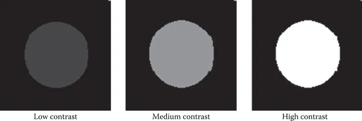
FIGURE 4.1Contrast relates to the relative difference between an object (circle here) and its surrounding background. Here the background gray level is set to black and the object gray level increased from a low-contrast ...
Inhaltsverzeichnis
- Cover
- Half Title
- Title
- Copyright
- Table of Contents
- Series Preface
- Preface
- Acknowledgments
- Author
- Introduction
- Section I Basic Concepts
- Section II Intermediate Concepts
- Section III Advanced Concepts
- Section IV Dynamic Imaging
- Section V Tomographic Imaging
- Index
Zitierstile für Fundamental Mathematics and Physics of Medical Imaging
APA 6 Citation
Lancaster, J., & Hasegawa, B. (2016). Fundamental Mathematics and Physics of Medical Imaging (1st ed.). CRC Press. Retrieved from https://www.perlego.com/book/2051498/fundamental-mathematics-and-physics-of-medical-imaging-pdf (Original work published 2016)
Chicago Citation
Lancaster, Jack, and Bruce Hasegawa. (2016) 2016. Fundamental Mathematics and Physics of Medical Imaging. 1st ed. CRC Press. https://www.perlego.com/book/2051498/fundamental-mathematics-and-physics-of-medical-imaging-pdf.
Harvard Citation
Lancaster, J. and Hasegawa, B. (2016) Fundamental Mathematics and Physics of Medical Imaging. 1st edn. CRC Press. Available at: https://www.perlego.com/book/2051498/fundamental-mathematics-and-physics-of-medical-imaging-pdf (Accessed: 15 October 2022).
MLA 7 Citation
Lancaster, Jack, and Bruce Hasegawa. Fundamental Mathematics and Physics of Medical Imaging. 1st ed. CRC Press, 2016. Web. 15 Oct. 2022.