eBook - ePub
Fundamental Mathematics and Physics of Medical Imaging
Jack Lancaster, Bruce Hasegawa
This is a test
- 322 pages
- English
- ePUB (adapté aux mobiles)
- Disponible sur iOS et Android
eBook - ePub
Fundamental Mathematics and Physics of Medical Imaging
Jack Lancaster, Bruce Hasegawa
Détails du livre
Aperçu du livre
Table des matières
Citations
À propos de ce livre
Authored by a leading educator, this book teaches the fundamental mathematics and physics concepts associated with medical imaging systems. Going beyond mere description of imaging modalities, this book delves into the mechanisms of image formation and image quality common to all imaging systems: contrast mechanisms, noise, and spatial and temporal resolution, making it an important reference for medical physicists and biomedical engineering students. This is an extensively revised new edition of The Physics of Medical X-Ray Imaging by Bruce Hasegawa (Medical Physics Publishing, 1991), and includes a wide range of modalities such as X-ray CT, MRI and SPECT.
Foire aux questions
Comment puis-je résilier mon abonnement ?
Il vous suffit de vous rendre dans la section compte dans paramètres et de cliquer sur « Résilier l’abonnement ». C’est aussi simple que cela ! Une fois que vous aurez résilié votre abonnement, il restera actif pour le reste de la période pour laquelle vous avez payé. Découvrez-en plus ici.
Puis-je / comment puis-je télécharger des livres ?
Pour le moment, tous nos livres en format ePub adaptés aux mobiles peuvent être téléchargés via l’application. La plupart de nos PDF sont également disponibles en téléchargement et les autres seront téléchargeables très prochainement. Découvrez-en plus ici.
Quelle est la différence entre les formules tarifaires ?
Les deux abonnements vous donnent un accès complet à la bibliothèque et à toutes les fonctionnalités de Perlego. Les seules différences sont les tarifs ainsi que la période d’abonnement : avec l’abonnement annuel, vous économiserez environ 30 % par rapport à 12 mois d’abonnement mensuel.
Qu’est-ce que Perlego ?
Nous sommes un service d’abonnement à des ouvrages universitaires en ligne, où vous pouvez accéder à toute une bibliothèque pour un prix inférieur à celui d’un seul livre par mois. Avec plus d’un million de livres sur plus de 1 000 sujets, nous avons ce qu’il vous faut ! Découvrez-en plus ici.
Prenez-vous en charge la synthèse vocale ?
Recherchez le symbole Écouter sur votre prochain livre pour voir si vous pouvez l’écouter. L’outil Écouter lit le texte à haute voix pour vous, en surlignant le passage qui est en cours de lecture. Vous pouvez le mettre sur pause, l’accélérer ou le ralentir. Découvrez-en plus ici.
Est-ce que Fundamental Mathematics and Physics of Medical Imaging est un PDF/ePUB en ligne ?
Oui, vous pouvez accéder à Fundamental Mathematics and Physics of Medical Imaging par Jack Lancaster, Bruce Hasegawa en format PDF et/ou ePUB ainsi qu’à d’autres livres populaires dans Medicina et Radiologia, radioterapia e medicina nucleare. Nous disposons de plus d’un million d’ouvrages à découvrir dans notre catalogue.
Informations
II
Intermediate Concepts
CHAPTER4
Physical Determinants of Contrast
A MEDICAL IMAGE CAN BE roughly described in terms of the three basic features given in Chapter 1: contrast, spatial resolution, and noise. Spatial resolution or clarity refers to the spatial detail of small objects within the image. Noise refers to the precision within the image; a noisy image will have large fluctuations in the signal across a uniform object, while a precise signal will have very small fluctuations. The subject of this chapter, contrast, relates to the difference in signals between a structure and its immediate surroundings. For example, if circles are displayed against a black background, a white circle will have larger contrast relative to the background when compared to gray circles (Figure 4.1). One uses the differences in gray shades to “visually” distinguish different tissue types, determine anatomical relationships, and sometimes assess their physiological functions. The larger the contrast between different tissue types, the easier it is to make distinctions clinically. It is often the objective of an imaging system to maximize the contrast in the image for a particular object or tissue of interest, although this is not always true since there may be design compromises where noise and spatial resolution are also very important. The contrast in an x-ray image depends on both physical characteristics of the object and properties of the device(s) used to image the object. The focus of this chapter is x-ray image contrast and we discuss the physical determinants of contrast, including material properties, x-ray spectra, detector response, and the role of perturbations such as scatter radiation and image intensifier veiling glare. We include physical determinants of contrast for several other medical imaging modalities, including those used in nuclear medicine, magnetic resonance imaging, and computed tomography to round off the discussion.
4.1COMPONENTS OF X-RAY IMAGE CONTRAST
Contrast can be quantified as the fractional difference in a measurable quantity between adjacent regions of an image. Usually, when we say “contrast,” we mean image contrast, which is the fractional difference in signals between two adjacent regions of an image. In conventional radiography, contrast can be separated into three components: (1) radiographic contrast, (2) detector contrast, and (3) display contrast (Table 4.1 and Figure 4.2).
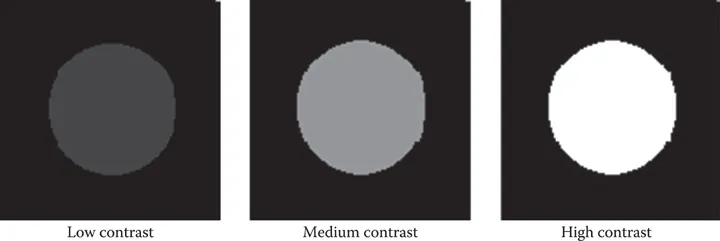
FIGURE 4.1Contrast relates to the relative difference between an object (circle here) and its surrounding background. Here the background gray level is set to black and the object gray level increased from a low-contrast ...
Table des matières
- Cover
- Half Title
- Title
- Copyright
- Table of Contents
- Series Preface
- Preface
- Acknowledgments
- Author
- Introduction
- Section I Basic Concepts
- Section II Intermediate Concepts
- Section III Advanced Concepts
- Section IV Dynamic Imaging
- Section V Tomographic Imaging
- Index
Normes de citation pour Fundamental Mathematics and Physics of Medical Imaging
APA 6 Citation
Lancaster, J., & Hasegawa, B. (2016). Fundamental Mathematics and Physics of Medical Imaging (1st ed.). CRC Press. Retrieved from https://www.perlego.com/book/2051498/fundamental-mathematics-and-physics-of-medical-imaging-pdf (Original work published 2016)
Chicago Citation
Lancaster, Jack, and Bruce Hasegawa. (2016) 2016. Fundamental Mathematics and Physics of Medical Imaging. 1st ed. CRC Press. https://www.perlego.com/book/2051498/fundamental-mathematics-and-physics-of-medical-imaging-pdf.
Harvard Citation
Lancaster, J. and Hasegawa, B. (2016) Fundamental Mathematics and Physics of Medical Imaging. 1st edn. CRC Press. Available at: https://www.perlego.com/book/2051498/fundamental-mathematics-and-physics-of-medical-imaging-pdf (Accessed: 15 October 2022).
MLA 7 Citation
Lancaster, Jack, and Bruce Hasegawa. Fundamental Mathematics and Physics of Medical Imaging. 1st ed. CRC Press, 2016. Web. 15 Oct. 2022.