eBook - ePub
Fundamental Mathematics and Physics of Medical Imaging
Jack Lancaster, Bruce Hasegawa
This is a test
- 322 pagine
- English
- ePUB (disponibile sull'app)
- Disponibile su iOS e Android
eBook - ePub
Fundamental Mathematics and Physics of Medical Imaging
Jack Lancaster, Bruce Hasegawa
Dettagli del libro
Anteprima del libro
Indice dei contenuti
Citazioni
Informazioni sul libro
Authored by a leading educator, this book teaches the fundamental mathematics and physics concepts associated with medical imaging systems. Going beyond mere description of imaging modalities, this book delves into the mechanisms of image formation and image quality common to all imaging systems: contrast mechanisms, noise, and spatial and temporal resolution, making it an important reference for medical physicists and biomedical engineering students. This is an extensively revised new edition of The Physics of Medical X-Ray Imaging by Bruce Hasegawa (Medical Physics Publishing, 1991), and includes a wide range of modalities such as X-ray CT, MRI and SPECT.
Domande frequenti
Come faccio ad annullare l'abbonamento?
È semplicissimo: basta accedere alla sezione Account nelle Impostazioni e cliccare su "Annulla abbonamento". Dopo la cancellazione, l'abbonamento rimarrà attivo per il periodo rimanente già pagato. Per maggiori informazioni, clicca qui
È possibile scaricare libri? Se sì, come?
Al momento è possibile scaricare tramite l'app tutti i nostri libri ePub mobile-friendly. Anche la maggior parte dei nostri PDF è scaricabile e stiamo lavorando per rendere disponibile quanto prima il download di tutti gli altri file. Per maggiori informazioni, clicca qui
Che differenza c'è tra i piani?
Entrambi i piani ti danno accesso illimitato alla libreria e a tutte le funzionalità di Perlego. Le uniche differenze sono il prezzo e il periodo di abbonamento: con il piano annuale risparmierai circa il 30% rispetto a 12 rate con quello mensile.
Cos'è Perlego?
Perlego è un servizio di abbonamento a testi accademici, che ti permette di accedere a un'intera libreria online a un prezzo inferiore rispetto a quello che pagheresti per acquistare un singolo libro al mese. Con oltre 1 milione di testi suddivisi in più di 1.000 categorie, troverai sicuramente ciò che fa per te! Per maggiori informazioni, clicca qui.
Perlego supporta la sintesi vocale?
Cerca l'icona Sintesi vocale nel prossimo libro che leggerai per verificare se è possibile riprodurre l'audio. Questo strumento permette di leggere il testo a voce alta, evidenziandolo man mano che la lettura procede. Puoi aumentare o diminuire la velocità della sintesi vocale, oppure sospendere la riproduzione. Per maggiori informazioni, clicca qui.
Fundamental Mathematics and Physics of Medical Imaging è disponibile online in formato PDF/ePub?
Sì, puoi accedere a Fundamental Mathematics and Physics of Medical Imaging di Jack Lancaster, Bruce Hasegawa in formato PDF e/o ePub, così come ad altri libri molto apprezzati nelle sezioni relative a Medicina e Radiologia, radioterapia e medicina nucleare. Scopri oltre 1 milione di libri disponibili nel nostro catalogo.
Informazioni
II
Intermediate Concepts
CHAPTER4
Physical Determinants of Contrast
A MEDICAL IMAGE CAN BE roughly described in terms of the three basic features given in Chapter 1: contrast, spatial resolution, and noise. Spatial resolution or clarity refers to the spatial detail of small objects within the image. Noise refers to the precision within the image; a noisy image will have large fluctuations in the signal across a uniform object, while a precise signal will have very small fluctuations. The subject of this chapter, contrast, relates to the difference in signals between a structure and its immediate surroundings. For example, if circles are displayed against a black background, a white circle will have larger contrast relative to the background when compared to gray circles (Figure 4.1). One uses the differences in gray shades to “visually” distinguish different tissue types, determine anatomical relationships, and sometimes assess their physiological functions. The larger the contrast between different tissue types, the easier it is to make distinctions clinically. It is often the objective of an imaging system to maximize the contrast in the image for a particular object or tissue of interest, although this is not always true since there may be design compromises where noise and spatial resolution are also very important. The contrast in an x-ray image depends on both physical characteristics of the object and properties of the device(s) used to image the object. The focus of this chapter is x-ray image contrast and we discuss the physical determinants of contrast, including material properties, x-ray spectra, detector response, and the role of perturbations such as scatter radiation and image intensifier veiling glare. We include physical determinants of contrast for several other medical imaging modalities, including those used in nuclear medicine, magnetic resonance imaging, and computed tomography to round off the discussion.
4.1COMPONENTS OF X-RAY IMAGE CONTRAST
Contrast can be quantified as the fractional difference in a measurable quantity between adjacent regions of an image. Usually, when we say “contrast,” we mean image contrast, which is the fractional difference in signals between two adjacent regions of an image. In conventional radiography, contrast can be separated into three components: (1) radiographic contrast, (2) detector contrast, and (3) display contrast (Table 4.1 and Figure 4.2).
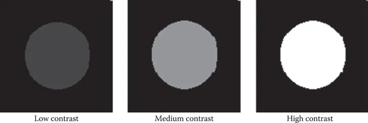
FIGURE 4.1Contrast relates to the relative difference between an object (circle here) and its surrounding background. Here the background gray level is set to black and the object gray level increased from a low-contrast ...
Indice dei contenuti
- Cover
- Half Title
- Title
- Copyright
- Table of Contents
- Series Preface
- Preface
- Acknowledgments
- Author
- Introduction
- Section I Basic Concepts
- Section II Intermediate Concepts
- Section III Advanced Concepts
- Section IV Dynamic Imaging
- Section V Tomographic Imaging
- Index
Stili delle citazioni per Fundamental Mathematics and Physics of Medical Imaging
APA 6 Citation
Lancaster, J., & Hasegawa, B. (2016). Fundamental Mathematics and Physics of Medical Imaging (1st ed.). CRC Press. Retrieved from https://www.perlego.com/book/2051498/fundamental-mathematics-and-physics-of-medical-imaging-pdf (Original work published 2016)
Chicago Citation
Lancaster, Jack, and Bruce Hasegawa. (2016) 2016. Fundamental Mathematics and Physics of Medical Imaging. 1st ed. CRC Press. https://www.perlego.com/book/2051498/fundamental-mathematics-and-physics-of-medical-imaging-pdf.
Harvard Citation
Lancaster, J. and Hasegawa, B. (2016) Fundamental Mathematics and Physics of Medical Imaging. 1st edn. CRC Press. Available at: https://www.perlego.com/book/2051498/fundamental-mathematics-and-physics-of-medical-imaging-pdf (Accessed: 15 October 2022).
MLA 7 Citation
Lancaster, Jack, and Bruce Hasegawa. Fundamental Mathematics and Physics of Medical Imaging. 1st ed. CRC Press, 2016. Web. 15 Oct. 2022.